| 规格 | 价格 | 库存 | 数量 |
|---|---|---|---|
| 5mg |
|
||
| 10mg |
|
||
| 25mg |
|
||
| 50mg |
|
||
| 100mg |
|
||
| Other Sizes |
|
| 靶点 |
Chk1 (IC50 = 3 nM); Chk2 (IC50 = 1500 nM); CDK2 (IC50 = 160 nM)
- Checkpoint kinase 1 (Chk1) (IC50 = 3.1 nM for recombinant human Chk1; Ki = 0.9 nM) [1] - Checkpoint kinase 2 (Chk2) (weaker inhibition, IC50 = 360 nM) [1] MK-8776 (SCH 900776) targets checkpoint kinase 1 (Chk1) with a Ki value of 0.3 nM and an IC50 value of 0.9 nM in recombinant kinase assays [1] MK-8776 inhibits checkpoint kinase 2 (Chk2) with an IC50 value of 47 nM, showing ~52-fold selectivity for Chk1 over Chk2 [1] MK-8776 exhibits minimal inhibition of other kinases (ATM, ATR, CDK1, Aurora A/B) with IC50 values > 1 μM [1][2] |
|---|---|
| 体外研究 (In Vitro) |
SCH 900776 是一种效力较低的 Chk2 和 CDK2 抑制剂,IC50 分别为 1.5 μM 和 0.16 μM。 SCH 900776 对细胞色素 P450 人肝微粒体亚型 1A2、2C9、2C19、2D6 和 3A4 无显着抑制作用。接触羟基脲 24 小时后,SCH 900776 会导致 DNA 复制能力出现剂量依赖性丧失。 SCH 900776 增强羟基脲、5-氟尿嘧啶和阿糖胞苷的 γ-H2AX 反应。与抗代谢药结合使用,SCH 900776 可在 2 小时内诱导 γ-H2AX 积累,表明复制叉崩溃和双链 DNA 断裂。此外,SCH 900776 以剂量依赖性方式抑制 Chk1 pS296 自磷酸化的积累。增殖的 WS1 细胞暴露于 SCH 900776 与 Chk1 pS345 的快速、剂量依赖性积累相关,表明正常细胞的循环群体在暴露于 SCH 900776 后诱导 Chk1 pS345 作为无效循环的一部分,可能是由 AT 家族激酶驱动的和DNA-PK。激酶测定:激酶分析器服务用于生成 SCH 900776 针对各种丝氨酸/苏氨酸和酪氨酸激酶的一般选择性数据。测定通常在两种浓度的 SCH 900776(0.5 和 5 μM)和固定浓度(10 μM)的 ATP 下进行。细胞测定:在体外,SCH 900776 以剂量依赖性方式阻断 Chk1 pS296 自磷酸化的积累。研究发现,用 SCH 900776 处理增殖的 WS1 细胞与 Chk1 pS345 的快速、剂量依赖性积累相关,这表明正常细胞的循环群体会响应 SCH 900776 的抑制而诱导 Chk1 pS345,作为无效周期 1 的一部分。
- MK-8776(SCH900776) 强效抑制Chk1激酶活性,重组酶实验中IC50为3.1 nM,对Chk2的抑制性较弱(IC50 = 360 nM),对其他激酶(如CDK1)抑制性极低(IC50 > 10,000 nM),选择性>100倍[1] - 在人肿瘤细胞系(如HCT116、SW620、A549)中,MK-8776(0.1-10 μM)剂量依赖性诱导G2/M期阻滞(流式细胞术),通过阻断DNA损伤检查点实现,伴随Cdc2(Tyr15)磷酸化水平降低和cyclin B1升高(Western blot)[1][2] - 该化合物(1-5 μM)增强DNA损伤剂的细胞毒性:与吉西他滨联用时,HCT116细胞中吉西他滨的IC50从50 nM降至5 nM;与放疗(2 Gy)联用时,凋亡细胞(Annexin V+)从12%增至45%[2] - 在p53缺陷细胞(如H1299)中,MK-8776(2 μM)选择性抑制增殖(IC50 = 1.8 μM),强于p53正常细胞(IC50 = 8.5 μM),显示与p53缺失的合成致死效应[3] 在多种人类实体瘤细胞系(HCT116、A549、MCF-7、PC3、SKOV3、MiaPaCa-2)中,MK-8776 表现出抗增殖活性,IC50 值范围为 8 nM 至 65 nM [1] - 10 nM MK-8776 可废除顺铂在 HCT116 细胞中诱导的 G2/M 检查点,24 小时后使 G2/M 期细胞积累比例从 63% 降至 25% [1] - 30 nM MK-8776 单独处理 A549 细胞仅诱导 10% 细胞凋亡,但与吉西他滨(5 nM)联合处理 72 小时后,凋亡率升高至 68% [1] - MK-8776 抑制 HCT116 细胞中 Chk1 介导的 CDC25C(Ser216)和 Chk1(Ser345)磷酸化,20 nM 浓度时抑制作用最强 [1][2] - MK-8776 与 DNA 损伤剂联合使用时表现出协同抗增殖效应:在 HCT116 细胞中,与顺铂联合的协同指数(CI)= 0.32,与吉西他滨联合 CI = 0.27,与多柔比星联合 CI = 0.41,与依托泊苷联合 CI = 0.39 [1][2] - 在 p53 缺陷型肿瘤细胞系(HCT116 p53⁻/⁻、MDA-MB-231)中,MK-8776 表现出增强的抗增殖活性(IC50 = 8 nM 至 18 nM),优于 p53 正常表达细胞(IC50 = 25 nM 至 65 nM)[1] - 25 nM MK-8776 可增强吉西他滨处理细胞中的 DNA 双链断裂,γ-H2AX 灶点形成较单独吉西他滨处理增加 3.8 倍 [2] - 在人类急性髓系白血病(AML)细胞系(MV4-11、HL-60、THP-1)中,MK-8776 抑制细胞增殖,IC50 值范围为 12 nM 至 32 nM [2] - 40 nM MK-8776 阻断羟基脲在 HL-60 细胞中诱导的 S 期检查点激活,使 S 期细胞死亡增加 45% [2] |
| 体内研究 (In Vivo) |
- 在荷HCT116异种移植瘤裸鼠中,MK-8776(25-100 mg/kg,口服,每日两次)呈剂量依赖性抑制肿瘤生长:100 mg/kg时肿瘤生长抑制率(TGI)达65%;与吉西他滨(120 mg/kg,每周1次)联用,TGI增至85%[2]
- 在结肠癌患者来源异种移植(PDX)模型中,MK-8776(50 mg/kg,口服)联合放疗(8 Gy)使肿瘤体积减少70%(单独放疗为30%),肿瘤组织中DNA损伤标志物γH2AX增加(免疫组化)[3] - 在KRAS突变肺癌异种移植模型中,MK-8776(75 mg/kg)单药使肿瘤停滞,与顺铂联用导致40%肿瘤退缩[2] 吉西他滨给药 30 分钟后,4 mg/kg SCH 900776 足以诱导 γ-H2AX 生物标志物,而相对于单独使用吉西他滨或 SCH 900776,8 mg/kg 则可增强肿瘤药效学和消退反应。 SCH 900776 的剂量递增(16 mg/kg 和 32 mg/kg)可导致肿瘤反应逐渐改善。重要的是,SCH 900776 的剂量与稳健的生物标志物激活和改善的肿瘤反应相关,但与吉西他滨对 BALB/c 小鼠血液学参数的毒性增强无关。 在 HCT116 人结直肠癌异种移植模型(nu/nu 小鼠)中,MK-8776 口服给药(40 mg/kg,每日两次,连续 14 天)联合顺铂(5 mg/kg,腹腔给药,第 1、5、9 天)的肿瘤生长抑制率(TGI)达 93%,而顺铂单独处理的 TGI 为 48% [1] - 在 A549 人非小细胞肺癌(NSCLC)异种移植模型(nu/nu 小鼠)中,MK-8776 口服给药(30 mg/kg,每日两次,连续 14 天)联合吉西他滨(100 mg/kg,腹腔给药,第 1、5、9 天)的 TGI 为 89%,荷瘤小鼠中位生存期较吉西他滨单独处理延长 75% [1] - 在 MV4-11 人 AML 异种移植模型(SCID 小鼠)中,MK-8776 口服给药(25 mg/kg,每日两次,连续 21 天)联合阿糖胞苷(50 mg/kg,腹腔给药,每日一次,连续 5 天)使肿瘤负荷降低 86%,中位生存期从 28 天延长至 52 天 [2] - MK-8776 与吉西他滨联合处理组的肿瘤组织中,TUNEL 阳性凋亡细胞增加(46% vs 吉西他滨单独处理组 17%),Ki-67 增殖指数降低(18% vs 吉西他滨单独处理组 62%)[1] |
| 酶活实验 |
SCH 900776 针对各种丝氨酸/苏氨酸和酪氨酸激酶的一般选择性数据是通过激酶分析器服务生成的。 SCH 900776 通常用于两种浓度(0.5 和 5 μM)以及固定浓度 (10 μM) ATP 的测定。
以基于CDC25C的生物素化肽为底物,杆状病毒表达系统表达的重组His-Chk1为酶源的体外实验。在含有 50 mM Tris pH8.0、10 mM MgCl2 和 1 mM DTT 的激酶缓冲液中,His-Chk1 被稀释至 32 nM。将 CDC25C(CDC25 Ser216 C 末端生物素化肽)肽在激酶缓冲液中稀释至浓度 1.93 μM。为了在添加起始溶液后产生 6.2 nM Chk1、385 nM CDC25C 和 1% DMSO 的最终反应浓度,将 20 μL 32 nM Chk1 酶溶液和 20 μL 1.926 μM CDC25C 混合并与 10对于每个激酶反应,用 10% DMSO 稀释 μL SCH 900776。添加 50 μL 起始溶液(其中含有 2 μM ATP 和 0.2 μCi 33 P-ATP)启动反应,最终反应浓度为 1 μM ATP 和 0.2 μCi 每个反应 33 P-ATP。激酶反应在室温下运行 2 小时,并通过添加 100 μL 终止液来终止,终止液由 2 M NaCl、1% H3PO4 和 5 组成。 mg/mL 链霉亲和素包被的 SPA 珠。 Filtermate 通用收集器与 96 孔 GF/B 过滤板组合用于收集 SPA 珠子。 2 M NaCl 和 2 M NaCl 加 1% 磷酸均用于洗珠两次。之后,用 TopCount 96 孔液体闪烁计数器测量信号。使用 SCH 900776 在八个点上连续稀释一式两份来创建剂量反应曲线。通过使用非线性回归分析,获得IC50值。 激酶测定[1] CHK1、CHK2和CDK激酶测定已在前面描述。Millipore激酶分析器服务用于生成SCH 900776对多种丝氨酸/苏氨酸和酪氨酸激酶的一般选择性数据。通常在两种浓度的SCH 900776(0.5和5μmol/L)和固定浓度的ATP(10μmol/L)下进行检测。数据以剩余活动百分比的形式提供,与未受抑制的对照组相比。 使用温度依赖性荧光进行亲和力评估[1] 在白色96孔PCR板中,将1μmol/L CHK1重组激酶结构域蛋白(氨基酸残基2-274)与微摩尔浓度(通常为1-50μmol/L)的化合物在20μL测定缓冲液(25 mmol/L HEPES,pH 7.4,300 mmol/L NaCl,5 mmol/L二硫苏糖醇,2%二甲亚砜,Sypro Orange 5x)中混合。该板用透明条密封,并放置在热循环仪中。在25°C至95°C的熔化过程中,每0.5°C的增量监测一次荧光强度。数据被导出到Excel中,并经过专有的自定义曲线拟合算法(未公布),以得出温度依赖性荧光(TdF)Kd值。对于CHK1 TdF数据,通常使用双态结合模型(化合物结合天然和热展开的熔融球态)。与靶激酶的熔融球态结合的化合物通常比天然状态弱1000倍以上。由于结合焓变化的不确定性,所有TdF Kd值的误差幅度约为50%。 - Chk1激酶活性实验:重组人Chk1(5 nM)与MK-8776(0.01-100 nM)在含ATP和荧光肽底物(Chk1tide)的缓冲液中孵育,通过荧光强度(激发340 nm,发射490 nm)测量激酶活性,剂量-效应曲线计算IC50[1] - 选择性实验:化合物(10 μM)对60种激酶筛选显示,仅Chk1和Chk2受抑制(>50%),证实特异性[1] 重组 Chk1/Chk2 激酶活性测定:反应体系包含重组 Chk1/Chk2、ATP(10 μM)和荧光标记肽底物,加入系列浓度的 MK-8776(0.1 nM 至 100 nM),30°C 孵育 60 分钟。通过荧光共振能量转移(FRET)检测磷酸化底物,非线性回归计算 Ki/IC50 值 [1] - 激酶选择性面板测定:采用相同的 FRET 方法,在 1 μM 浓度下测试 MK-8776 对 45 种人类激酶的抑制作用。相对于溶媒对照组计算抑制率,对抑制率 > 20% 的激酶计算 IC50 值 [1] - Chk1 结合测定:采用表面等离子体共振(SPR)技术测量结合亲和力。MK-8776 系列稀释(0.2 nM 至 20 nM)后通过固定有 Chk1 的传感器芯片,记录结合响应信号,通过稳态分析推导解离常数(Kd)[1] |
| 细胞实验 |
为了进行细胞生长测定,将 500-1000 个细胞以低密度接种到 96 孔板中,然后用药物处理细胞一整天(每个浓度 8 个孔)。处理后,冲洗细胞并在37°C的新培养基中培养5至7天。在细胞达到汇合之前,将细胞裂解、清洗并用 Hoechst 33258 染色。使用微孔板分光荧光计测量荧光。用抑制50%生长的药物浓度的平均值和标准误来表示结果。
γ-H2AX测定[1] 简单地说,将细胞暴露于抗代谢物中以诱导CHK1的激活。对照人群不进行治疗。然后在2小时的暴露窗口(在抗代谢物存在的情况下)将 SCH 900776 滴定到细胞上。在SCH 900776共暴露2小时后,将细胞固定并渗透(70%乙醇),然后用异硫氰酸荧光素(FITC)偶联抗γ- h2ax单克隆抗体染色。细胞用碘化丙啶反染,随后使用流式细胞仪(Becton Dickinson LSR II)或Discovery 1免疫荧光平台进行分析。实验通常进行三次,数据以γ-H2AX阳性细胞的百分比表示,从而反映了γ-H2AX表型的总体外显率。 用活性caspase>评价凋亡诱导 [1] SCH 900776。然后清洗细胞,去除所有抗代谢物和SCH 900776。在这一点(T0或释放)评估Caspase活性,并在T + 24和T + 48小时进行进一步的检测。细胞随后用荧光标记的caspase底物孵育;细胞内底物的摄取和荧光与激活的半胱天冬酶水平相关。然后用流式细胞术测定表达活化半胱天冬酶的细胞百分比。 溴脱氧尿苷掺入试验[1] 将细胞镀于10厘米的组织培养皿中,使其粘附。将细胞暴露于不同浓度的SCH 900776中超过2小时,无论事先是否有抗代谢物暴露。然后清洗细胞,并允许24小时尝试恢复s期。随后短暂(30分钟)暴露于溴脱氧尿苷(BrdU),以评估能够以活的方式重新进入细胞周期的细胞百分比。然后收获细胞,固定并渗透。随后进行酸变性步骤,以暴露基因组DNA中合并的BrdU表位,之后用fitc偶联的BrdU特异性单克隆抗体对样品进行免疫染色。然后用碘化丙啶对细胞进行反染色,以评估DNA含量并使用流式细胞术进行分析。BrdU阳性染色和碘化丙啶信号的双变量分析可以评估正在进行DNA合成的细胞数量和细胞系的整体细胞周期分布(G1, S, G2-M和亚G1)。每个浓度下每个种群的百分比. - 增殖实验:肿瘤细胞(HCT116、A549)接种于96孔板,经MK-8776(0.01-100 μM)处理72小时,CellTiter-Glo检测活力,IC50范围1.2-8.5 μM[1][2] - 细胞周期分析:HCT116细胞经MK-8776(2 μM)处理24小时,固定后碘化丙啶染色,流式检测显示G2/M期细胞比例从15%(对照)增至60%[1] - Western blot:经MK-8776(1-5 μM)处理的细胞裂解后,检测p-Chk1(Ser345)、p-Cdc2(Tyr15)和cyclin B1,5 μM时p-Cdc2降低70%[2] 抗增殖实验:癌细胞接种于 96 孔板(4×10³ 个细胞 / 孔),用系列浓度的 MK-8776(2 nM 至 200 nM)单独或与 DNA 损伤剂联合处理 72 小时。基于四唑盐还原的比色法评估细胞活力,计算 IC50 值及协同指数 [1][2] - 细胞周期分析:细胞用 MK-8776(10 nM)联合顺铂(2 μM)处理 24 小时后,收集细胞,70% 乙醇固定,碘化丙啶染色,流式细胞术测定细胞周期分布 [1][2] - 凋亡实验:细胞经 MK-8776(30 nM)和 / 或吉西他滨(5 nM)处理 72 小时后,用膜联蛋白 V-FITC 和碘化丙啶染色,流式细胞术分析 [1][2] - Western blot 分析:细胞用冰浴 RIPA 缓冲液裂解,蛋白经 SDS-PAGE 分离后转移至膜上,与抗磷酸化 CDC25C(Ser216)、磷酸化 Chk1(Ser345)、γ-H2AX、剪切型半胱天冬酶 -3、PARP 及 β- 肌动蛋白抗体孵育。化学发光法检测信号,密度计量法定量 [1][2][3] - γ-H2AX 灶点实验:细胞用 MK-8776(25 nM)和吉西他滨(5 nM)处理 24 小时后,固定,用 γ-H2AX 抗体和 DAPI 染色,荧光显微镜观察。通过图像分析软件计数每个细胞的灶点数量 [2] - 克隆形成实验:AML 细胞用 MK-8776(5 nM 至 20 nM)处理 24 小时后,接种于甲基纤维素培养基中,14 天后计数菌落(> 50 个细胞)。相对于溶媒对照组计算克隆形成效率 [2] |
| 动物实验 |
将A2780或MiaPaCa2细胞皮下注射到雌性裸鼠体内
~50 mg/kg 腹腔注射 某些细胞系在体外培养,用PBS洗涤后,将其重悬于50% Matrigel/PBS溶液中,最终浓度为4×10⁷至5×10⁷个细胞/mL,用于肿瘤植入。将0.1 mL该细胞悬液皮下注射到裸鼠侧腹部。每周两次使用游标卡尺测量每只小鼠的肿瘤长度(L)、宽度(W)和高度(H)。然后使用公式(L×W×H)/2计算肿瘤体积。十只动物被随机分配到不同的治疗组,并分别腹腔注射SCH 900776(溶于20%羟丙基-β-环糊精)或按照推荐指南制备的特定化疗药物。在治疗期间和治疗结束后,测量肿瘤体积和体重。在进行初始体积标准化之前,数据以均值±标准误(SEM)的形式记录。在一些实验中,记录肿瘤体积进展至初始体积10倍所需的时间(TTP 10x)。在尸检过程中,切除肿瘤及其周围皮肤,将其在10%福尔马林溶液中固定过夜,然后清洗并保存在70%乙醇溶液中,用于小鼠的药效学标志物分析。剃除约4平方英寸大小的皮肤区域,以便进行皮肤穿刺活检。为了对犬进行局部麻醉,皮下注射利多卡因;而对于大鼠,则使用吸入异氟烷进行麻醉。使用 4 mm 活检穿刺器获取样本。在清洗或保存于 70% 乙醇之前,皮肤穿刺样本需先在 10% 福尔马林溶液中固定一整夜。 体内肿瘤生长评估、取样和皮肤活检[1] 对于肿瘤植入,将特定细胞系在体外培养,用 PBS 洗涤一次,然后重悬于 50% Matrigel(BD Biosciences)的 PBS 溶液中,最终浓度为 4 × 10⁷ 至 5 × 10⁷ 个细胞/mL。将 0.1 mL 该悬液皮下注射到裸鼠侧腹部。每周两次用游标卡尺测量每只小鼠的肿瘤长度 (L)、宽度 (W) 和高度 (H),然后使用公式 (L × W × H)/2 计算肿瘤体积。将10只动物随机分为各治疗组,腹腔注射SCH 900776(溶于20%羟丙基β-环糊精)或按推荐配制的单一化疗药物。在治疗期间和治疗后测量肿瘤体积和体重。数据以均值±标准误(SEM)表示,并根据初始体积进行标准化。在部分实验中监测肿瘤体积进展至初始体积10倍的时间(TTP 10x)。根据机构动物护理和使用委员会的指导方针对动物实施安乐死。对于小鼠的药效学标志物分析,在尸检时收集肿瘤和邻近皮肤,用10%福尔马林固定过夜,然后用70%乙醇洗涤/保存。对于皮肤穿刺活检,剃除约4平方英寸的皮肤组织。大鼠采用吸入异氟烷麻醉,犬采用皮下注射利多卡因进行局部麻醉。使用 4 mm 活检穿刺器采集样本。皮肤穿刺样本在 10% 福尔马林溶液中固定过夜,然后在 70% 乙醇中清洗/保存。 药代动力学测定[1] 在给予 SCH 900776 后不同时间点采集受试动物的血浆样本。在每个时间点,将 3 只动物的血液样本合并,并使用液相色谱-质谱联用仪 (LC/MS) 分析 SCH 900776 的浓度。根据血浆浓度数据估算药代动力学参数。Cmax 值(最大血浆浓度)直接取自血浆浓度-时间曲线,并使用线性梯形法则计算血浆浓度-时间曲线下面积 (AUC)。 - 异种移植模型:将HCT116细胞(5×10⁶)皮下注射到6-8周龄的裸鼠体内。当肿瘤体积达到100 mm³时,每日两次口服MK-8776(25-100 mg/kg)。每周两次测量肿瘤体积(使用游标卡尺)和体重,持续21天[2] - 联合放疗:荷PDX肿瘤的小鼠在接受局部放疗(8 Gy)前1小时口服MK-8776(50 mg/kg)。72小时后收集肿瘤组织进行γH2AX免疫组化染色[3] HCT116结肠癌异种移植模型:将5×10⁶个HCT116细胞皮下植入6-8周龄的雌性nu/nu小鼠体内。当肿瘤体积达到 100-150 mm³ 时,将小鼠随机分组(每组 n=8),并分别接受以下治疗:(1)口服载体(0.5% 甲基纤维素 + 0.2% Tween 80);(2)口服 MK-8776(40 mg/kg),每日两次,持续 14 天;(3)腹腔注射顺铂(5 mg/kg),分别于第 1、5、9 天给药;(4)MK-8776 + 顺铂。每 2 天测量一次肿瘤体积和体重。[1] - A549 非小细胞肺癌异种移植模型:将 5×10⁶ 个 A549 细胞皮下植入 6-8 周龄的雌性裸鼠(nu/nu)。肿瘤体积达到 100-150 mm³ 的小鼠被随机分组(每组 n=8),并接受以下治疗:(1)口服赋形剂;(2)口服 MK-8776(30 mg/kg),每日两次,持续 14 天;(3)腹腔注射吉西他滨(100 mg/kg),分别于第 1、5、9 天给药;(4)MK-8776 + 吉西他滨。监测肿瘤体积和生存情况[1] - MV4-11 AML 异种移植模型:将 1×10⁷ 个 MV4-11 细胞静脉注射到 6-8 周龄的雌性 SCID 小鼠体内。接种后7天,将小鼠随机分组(每组n=8),并分别接受以下治疗:(1)口服赋形剂;(2)口服MK-8776(25 mg/kg),每日两次,持续21天;(3)腹腔注射阿糖胞苷(50 mg/kg),每日一次,持续5天;(4)MK-8776+阿糖胞苷。记录肿瘤负荷和生存情况[2] |
| 药代性质 (ADME/PK) |
在小鼠中,口服 MK-8776 (50 mg/kg) 显示出 65% 的生物利用度,1 小时时 Cmax 为 3.2 μg/mL。血浆半衰期 (t1/2) 为 2.8 小时,血浆蛋白结合率为 92% [1]
- 在人体(I 期临床试验)中,口服给药(每日两次,每次 60 mg)导致 Cmax 为 1.8 μg/mL,t1/2 为 4.5 小时,且具有良好的肿瘤渗透性(肿瘤/血浆比 = 2.3)[3] 在小鼠中,口服 MK-8776(40 mg/kg)导致 Cmax 为 5.3 μM,AUC0-24h 为 32.7 μM·h,口服生物利用度为 78% [1] - 在小鼠中,静脉注射 MK-8776(10 mg/kg)显示清除率为 7.8 mL/min/kg,分布容积 (Vss) 为 1.4 L/kg,末端半衰期为MK-8776 的半衰期 (t1/2) 为 10.2 小时 [1] - MK-8776 具有良好的水溶性(pH 7.4 时 ≥150 μM)和较高的人血浆蛋白结合率 (95%) [1] - 在大鼠中,口服 MK-8776 (30 mg/kg) 的 Cmax 为 4.9 μM,AUC0-24h 为 29.5 μM·h,口服生物利用度为 72% [1] - 在犬中,口服 MK-8776 (20 mg/kg) 的 Cmax 为 3.8 μM,AUC0-24h 为 25.1 μM·h,t1/2 为 8.7 小时 [1] |
| 毒性/毒理 (Toxicokinetics/TK) |
在 I 期试验中,剂量限制性毒性 (DLT) 包括每日两次 120 毫克剂量下的中性粒细胞减少症(3/4 级)和每日两次 90 毫克剂量下的腹泻(3 级)。常见不良事件:疲乏 (45%)、恶心 (30%)、呕吐 (25%) [3]
- 未观察到明显的肝毒性或肾毒性,血清 ALT/AST 和肌酐均在正常范围内 [3] 在小鼠重复给药口服毒性研究(28 天,20-80 mg/kg/天)中,MK-8776 的最大耐受剂量 (MTD) 为 60 mg/kg/天,剂量限制性毒性 (DLT) 为骨髓抑制(80 mg/kg/天时中性粒细胞减少 35-40%)[1] - MK-8776(40 mg/kg/天,口服 14 天)在小鼠中引起短暂性体重减轻 (≤6%),停药后 5 天内恢复 [1] - 无在小鼠接受 60 mg/kg/天的 MK-8776 治疗 28 天后,观察到肝脏、肾脏、心脏或脾脏出现显著的组织病理学变化 [1] - MK-8776 在浓度高达 20 μM 时不抑制人细胞色素 P450 酶(CYP1A2、CYP2C9、CYP2C19、CYP2D6、CYP3A4)[1] - 在 I 期临床试验中,MK-8776 显示出可控的毒性,最常见的不良事件为中性粒细胞减少症 (32%)、血小板减少症 (28%) 和疲乏 (24%) [3] |
| 参考文献 | |
| 其他信息 |
Sch 900776 已用于多种疾病的治疗试验,包括肿瘤、霍奇金淋巴瘤、成人红白血病、非霍奇金淋巴瘤和急性髓系白血病等。
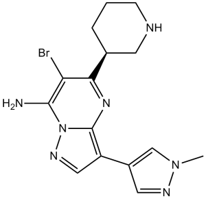
CHK1 抑制剂 MK-8776 是一种靶向细胞周期检查点激酶 1 (Chk1) 的药物,具有潜在的放射增敏和化疗增敏作用。Chk1 抑制剂 MK-8776 可特异性结合并抑制 Chk1,这可能导致肿瘤细胞绕过 Chk1 依赖的 S 期和 G2/M 期细胞周期阻滞,在进入有丝分裂前进行 DNA 修复;因此,肿瘤细胞可能对电离辐射和烷化剂化疗药物的 DNA 损伤作用更加敏感。 Chk1 是一种 ATP 依赖性丝氨酸/苏氨酸激酶,它在 DNA 损伤后磷酸化 cdc25 磷酸酶,导致 CDK-细胞周期蛋白复合物的酪氨酸磷酸化,从而抑制细胞周期,最终导致细胞周期阻滞。 6-溴-3-(1-甲基-4-吡唑基)-5-(3-哌啶基)-7-吡唑并[1,5-a]嘧啶胺是一种吡唑并嘧啶类化合物。 另见:Mk-8776(注释已移至)。 检查点激酶 1 (CHK1) 是一种重要的丝氨酸/苏氨酸激酶,它响应 DNA 损伤和 DNA 复制停滞。CHK1 对于在 DNA 抗代谢物暴露期间维持复制叉的活性至关重要。在人类肿瘤细胞系中,在抗代谢物暴露期间 CHK1 功能的缺失会导致双链 DNA 断裂的积累和细胞死亡。在此,我们进一步拓展了这些观察结果,并证实 CHK2 的缺失不会导致这些表型,反而可能减弱这些表型。此外,同时抑制细胞周期蛋白依赖性激酶 (CDK) 的活性足以完全拮抗所需的 CHK1 缺失表型。基于这些机制的观察结果促使我们开发了一种高通量、基于细胞的 γ-H2AX 诱导筛选方法,γ-H2AX 是双链 DNA 断裂的替代标志物。我们利用这种基于机制的功能性方法来优化 CHK1 小分子抑制剂。具体而言,该检测方法用于从机制上确定具有不同 CHK1、CHK2 和 CDK 选择性的化合物的最佳细胞内活性谱。通过这种方法,我们鉴定出 SCH 900776 是一种高效且功能最优的 CHK1 抑制剂,其固有拮抗作用极小。 SCH 900776 暴露可模拟短干扰 RNA 介导的 CHK1 基因敲除,并在体外和体内与 DNA 抗代谢药物产生协同作用,选择性地诱导肿瘤细胞背景下的双链 DNA 断裂和细胞死亡。[1] 许多抗癌药物会损伤 DNA 并使细胞周期进程停滞在 S 期或 G2 期。先前使用拓扑异构酶 I 抑制剂 SN38 的研究表明,Chk1 抑制剂 UCN-01 可以有效克服这种停滞并诱导有丝分裂灾难。UCN-01 的临床试验受限于其不利的药代动力学特性。SCH900776 是一种新型且更具选择性的 Chk1 抑制剂,可有效抑制 Chk1 并消除 SN38 诱导的细胞周期停滞。与 UCN-01 类似,SN38 诱导的细胞周期阻滞的消除会增强细胞死亡速率,但不会增加总体细胞死亡数。相反,SCH900776 使羟基脲的生长抑制浓度降低了 20 至 70 倍。阿糖胞苷也观察到了类似的增敏作用。吉西他滨的增敏作用提高了 5 至 10 倍,但顺铂、5-氟尿嘧啶或 6-硫鸟嘌呤则没有增敏作用。在羟基脲浓度略微减缓 DNA 复制且未明显激活 Chk1 的情况下,即可观察到增敏作用,但这会导致对 Chk1 的依赖性随时间增加。例如,在给予羟基脲 18 小时后加入 SCH900776,可诱导 DNA 双链断裂,这与复制叉的快速崩解相一致。此外,一些细胞系对单独使用SCH900776高度敏感,这些细胞只需较低浓度的SCH900776即可对羟基脲敏感。我们得出结论,某些肿瘤可能对SCH900776和羟基脲的联合用药非常敏感。延迟给药SCH900776可能比同时给药更有效。SCH900776目前正在进行I期临床试验,这些结果为未来的临床试验提供了理论依据和时间安排。[2] 目的:既往研究表明,涉及共济失调毛细血管扩张症突变基因和Rad3相关基因(ATR)以及Chk1激酶的复制检查点,会导致细胞系对阿糖胞苷产生耐药性。本研究旨在探讨在体内阿糖胞苷输注期间,临床急性髓系白血病 (AML) 中该检查点是否被激活,并评估阿糖胞苷与近期报道的 Chk1 抑制剂 SCH 900776 联合用药在体外的影响。实验设计:采用免疫印迹法检测阿糖胞苷输注前后采集的 AML 骨髓穿刺液。用阿糖胞苷处理人 AML 细胞系(有或无 SCH 900776 存在),并通过免疫印迹法、DNA 核苷酸掺入法和流式细胞术检测检查点激活情况。采用克隆形成实验检测 AML 细胞系、临床 AML 分离株和正常髓系祖细胞的长期效应。结果:免疫印迹分析显示,在阿糖胞苷输注 48 小时后,超过一半的含 Chk1 的 AML 细胞中 Chk1 磷酸化水平升高,这是检查点激活的标志。在人急性髓系白血病(AML)细胞系中,SCH 900776 不仅能抑制阿糖胞苷诱导的 Chk1 激活和 S 期阻滞,还能显著增强阿糖胞苷诱导的细胞凋亡。克隆形成实验表明,SCH 900776 在 AML 细胞系和临床 AML 样本中增强了阿糖胞苷的抗增殖作用,而其浓度对正常髓系祖细胞的影响可忽略不计。结论:这些结果不仅为临床 AML 中阿糖胞苷诱导的 S 期检查点激活提供了证据,而且表明选择性 Chk1 抑制剂可以克服 S 期检查点并增强阿糖胞苷的细胞毒性。因此,有必要进一步研究阿糖胞苷/SCH 900776 联合用药在 AML 中的应用。[3] - MK-8776 (SCH900776) 是一种选择性 Chk1 抑制剂,可消除 G2/M DNA 损伤检查点,使癌细胞对 DNA 损伤剂(化疗/放疗)敏感 [1][2] - 它在 p53 缺陷型肿瘤中表现出优先活性,利用了合成致死性。临床试验评估了MK-8776(SCH 900776)联合吉西他滨或放疗治疗晚期实体瘤(例如结直肠癌、肺癌)的疗效[3]。MK-8776是一种强效且选择性的小分子Chk1抑制剂,Chk1是DNA损伤反应和细胞周期检查点的关键调节因子[1]。MK-8776的作用机制是阻断G2/M期和S期检查点,迫使DNA未修复的癌细胞进入有丝分裂,最终导致有丝分裂灾难和细胞凋亡[1][2]。MK-8776旨在增强DNA靶向化疗的疗效,尤其适用于依赖Chk1生存的p53缺陷型肿瘤[1][2]。MK-8776已进入治疗晚期实体瘤的I期临床试验。肿瘤和血液系统恶性肿瘤,初步数据显示与吉西他滨联合使用具有抗肿瘤活性[3] |
| 分子式 |
C15H18BRN7
|
|
|---|---|---|
| 分子量 |
376.25
|
|
| 精确质量 |
375.08
|
|
| 元素分析 |
C, 47.88; H, 4.82; Br, 21.24; N, 26.06
|
|
| CAS号 |
891494-63-6
|
|
| 相关CAS号 |
SCH900776 (S-isomer);891494-64-7
|
|
| PubChem CID |
46239015
|
|
| 外观&性状 |
white to off-white Solid powder
|
|
| 密度 |
1.8±0.1 g/cm3
|
|
| 折射率 |
1.819
|
|
| LogP |
0.76
|
|
| tPSA |
86.06
|
|
| 氢键供体(HBD)数目 |
2
|
|
| 氢键受体(HBA)数目 |
5
|
|
| 可旋转键数目(RBC) |
2
|
|
| 重原子数目 |
23
|
|
| 分子复杂度/Complexity |
425
|
|
| 定义原子立体中心数目 |
1
|
|
| SMILES |
NC1=C(Br)C([C@H]2CNCCC2)=NC2=C(C3=CN(C)N=C3)C=NN12
|
|
| InChi Key |
GMIZZEXBPRLVIV-SECBINFHSA-N
|
|
| InChi Code |
InChI=1S/C15H18BrN7/c1-22-8-10(6-19-22)11-7-20-23-14(17)12(16)13(21-15(11)23)9-3-2-4-18-5-9/h6-9,18H,2-5,17H2,1H3/t9-/m1/s1
|
|
| 化学名 |
6-bromo-3-(1-methylpyrazol-4-yl)-5-[(3R)-piperidin-3-yl]pyrazolo[1,5-a]pyrimidin-7-amine
|
|
| 别名 |
|
|
| HS Tariff Code |
2934.99.9001
|
|
| 存储方式 |
Powder -20°C 3 years 4°C 2 years In solvent -80°C 6 months -20°C 1 month |
|
| 运输条件 |
Room temperature (This product is stable at ambient temperature for a few days during ordinary shipping and time spent in Customs)
|
| 溶解度 (体外实验) |
|
|||
|---|---|---|---|---|
| 溶解度 (体内实验) |
配方 1 中的溶解度: ≥ 2.5 mg/mL (6.64 mM) (饱和度未知) in 10% DMSO + 40% PEG300 + 5% Tween80 + 45% Saline (这些助溶剂从左到右依次添加,逐一添加), 澄清溶液。
例如,若需制备1 mL的工作液,可将100 μL 25.0 mg/mL澄清DMSO储备液加入到400 μL PEG300中,混匀;然后向上述溶液中加入50 μL Tween-80,混匀;加入450 μL生理盐水定容至1 mL。 *生理盐水的制备:将 0.9 g 氯化钠溶解在 100 mL ddH₂O中,得到澄清溶液。 配方 2 中的溶解度: ≥ 2.5 mg/mL (6.64 mM) (饱和度未知) in 10% DMSO + 90% (20% SBE-β-CD in Saline) (这些助溶剂从左到右依次添加,逐一添加), 澄清溶液。 例如,若需制备1 mL的工作液,可将 100 μL 25.0 mg/mL澄清DMSO储备液加入900 μL 20% SBE-β-CD生理盐水溶液中,混匀。 *20% SBE-β-CD 生理盐水溶液的制备(4°C,1 周):将 2 g SBE-β-CD 溶解于 10 mL 生理盐水中,得到澄清溶液。 View More
配方 3 中的溶解度: ≥ 2.5 mg/mL (6.64 mM) (饱和度未知) in 10% DMSO + 90% Corn Oil (这些助溶剂从左到右依次添加,逐一添加), 澄清溶液。 配方 4 中的溶解度: 4% DMSO +30% Propylene glycol : 5 mg/mL 1、请先配制澄清的储备液(如:用DMSO配置50 或 100 mg/mL母液(储备液)); 2、取适量母液,按从左到右的顺序依次添加助溶剂,澄清后再加入下一助溶剂。以 下列配方为例说明 (注意此配方只用于说明,并不一定代表此产品 的实际溶解配方): 10% DMSO → 40% PEG300 → 5% Tween-80 → 45% ddH2O (或 saline); 假设最终工作液的体积为 1 mL, 浓度为5 mg/mL: 取 100 μL 50 mg/mL 的澄清 DMSO 储备液加到 400 μL PEG300 中,混合均匀/澄清;向上述体系中加入50 μL Tween-80,混合均匀/澄清;然后继续加入450 μL ddH2O (或 saline)定容至 1 mL; 3、溶剂前显示的百分比是指该溶剂在最终溶液/工作液中的体积所占比例; 4、 如产品在配制过程中出现沉淀/析出,可通过加热(≤50℃)或超声的方式助溶; 5、为保证最佳实验结果,工作液请现配现用! 6、如不确定怎么将母液配置成体内动物实验的工作液,请查看说明书或联系我们; 7、 以上所有助溶剂都可在 Invivochem.cn网站购买。 |
| 制备储备液 | 1 mg | 5 mg | 10 mg | |
| 1 mM | 2.6578 mL | 13.2890 mL | 26.5781 mL | |
| 5 mM | 0.5316 mL | 2.6578 mL | 5.3156 mL | |
| 10 mM | 0.2658 mL | 1.3289 mL | 2.6578 mL |
1、根据实验需要选择合适的溶剂配制储备液 (母液):对于大多数产品,InvivoChem推荐用DMSO配置母液 (比如:5、10、20mM或者10、20、50 mg/mL浓度),个别水溶性高的产品可直接溶于水。产品在DMSO 、水或其他溶剂中的具体溶解度详见上”溶解度 (体外)”部分;
2、如果您找不到您想要的溶解度信息,或者很难将产品溶解在溶液中,请联系我们;
3、建议使用下列计算器进行相关计算(摩尔浓度计算器、稀释计算器、分子量计算器、重组计算器等);
4、母液配好之后,将其分装到常规用量,并储存在-20°C或-80°C,尽量减少反复冻融循环。
计算结果:
工作液浓度: mg/mL;
DMSO母液配制方法: mg 药物溶于 μL DMSO溶液(母液浓度 mg/mL)。如该浓度超过该批次药物DMSO溶解度,请首先与我们联系。
体内配方配制方法:取 μL DMSO母液,加入 μL PEG300,混匀澄清后加入μL Tween 80,混匀澄清后加入 μL ddH2O,混匀澄清。
(1) 请确保溶液澄清之后,再加入下一种溶剂 (助溶剂) 。可利用涡旋、超声或水浴加热等方法助溶;
(2) 一定要按顺序加入溶剂 (助溶剂) 。
| NCT Number | Recruitment | interventions | Conditions | Sponsor/Collaborators | Start Date | Phases |
| NCT00779584 | Completed | Drug: MK-8776 Drug: Gemcitabine |
Hodgkin Disease Neoplasms |
Merck Sharp & Dohme LLC | October 17, 2008 | Phase 1 |
| NCT01870596 | Completed | Drug: CHK1 Inhibitor SCH 900776 Drug: Cytarabine |
Adult Acute Monocytic Leukemia Adult Erythroleukemia |
National Cancer Institute (NCI) |
May 2013 | Phase 2 |
| NCT00907517 | Completed | Drug: MK-8776 Drug: Cytarabine |
Myelogenous Leukemia, Acute Leukemia, Lymphocytic, Acute |
Merck Sharp & Dohme LLC | July 29, 2009 | Phase 1 |
") Comparative efficacy of UCN-01 and SCH900776 at inhibiting Chk1 and abrogating SN38-induced cell cycle arrest.Mol Cancer Ther.2012 Feb;11(2):427-38. |
|---|
") The impact of checkpoint inhibitors on sensitivity of cells to SN38, hydroxyurea and cisplatin.Mol Cancer Ther.2012 Feb;11(2):427-38. |
") Analysis of cell cycle perturbation induced by hydroxyurea.Mol Cancer Ther.2012 Feb;11(2):427-38. |
") Impact of concentration and schedule of hydroxyurea and SCH900776 on DNA damage and cytotoxicityA.Mol Cancer Ther.2012 Feb;11(2):427-38. |
|---|
") Comparative efficacy of UCN-01 and SCH900776 at abrogating SN38-induced cell cycle arrest in cells suppressed for Chk1.A.MDA-MB-231ΔChk1 cells were incubated with 10 ng/mL SN38 for 24 h.Mol Cancer Ther.2012 Feb;11(2):427-38. |
 CHK1-IN-14
 MA203
MA203
 XL-844
 Prexasertib-d4
InvivoChem的所有产品仅用于作科学研究,不面向患者销售
Copyright 2020 InvivoChem LLC | All Rights Reserved 粤ICP备20063088号-1


 COA
COA
 463611831
