| 规格 | 价格 | 库存 | 数量 |
|---|---|---|---|
| 10 mM * 1 mL in DMSO |
|
||
| 1mg |
|
||
| 5mg |
|
||
| 50mg |
|
||
| 100mg |
|
||
| 250mg |
|
||
| 500mg |
|
||
| 1g |
|
||
| 2g |
|
||
| Other Sizes |
|

| 靶点 |
VEGFR1 (IC50 = 34 nM); VEGFR2 (IC50 = 13 nM); VEGFR3 (IC50 = 13 nM); FGFR1 (IC50 = 69 nM); FGFR2 (IC50 = 37 nM); FGFR3 (IC50 = 108 nM); PDGFRα (IC50 = 59 nM); PDGFRβ (IC50 = 65 nM)
|
|---|---|
| 体外研究 (In Vitro) |
体外活性:BIBF1120 抑制 PDGFR α 和 PDGFR β 型的 PDGFR 激酶活性,IC50 值分别为 59 nM 和 65 nM。此外,BIBF1120 抑制 FGFR 亚型,FGFR1、FGFR2 和 FGFR3 的 IC50 分别为 60 nM、37 nM 和 108 nM。 BIBF1120 与激酶结构域氨基端叶和羧基端叶之间缝隙中的 ATP 结合位点结合。吲哚酮支架与铰链区的 Cys919 的主链氮和 Glu917 的主链羰基氧形成两个氢键。 BIBF 1120 抑制 PDGF-BB 刺激的 BRP 增殖,在细胞测定中 EC50 为 79 nM。用 5% 血清加 PDGF-BB 刺激后,浓度低至 100 nM 的 BIBF1120 可阻断 MAPK 的激活。在人血管平滑肌细胞 (HUASMC) 培养物中,BIBF1120 可防止 PDGF-BB 刺激的增殖,EC50 为 69 nM。激酶测定:将 VEGFR2 的细胞质酪氨酸激酶结构域(根据数据库 SWISS-PROT P35968 中保存的序列,残基 797-1355)克隆到与 GST 融合的 pFastBac 中并提取。在存在或不存在 BIBF1120 在 25% DMSO 中进行系列稀释的情况下测定酶活性。每个微量滴定板都包含内部对照,例如空白、最大反应和历史参考化合物。所有孵育均在室温下在旋转摇床上进行。将 10 μL 每种 BIBF1120 稀释液添加到 10 μL 稀释激酶(0.8 μg/mL VEGFR2、10 mM Tris pH 7.5、2 mM EDTA 和 2 mg/mL BSA)中,并预孵育 1 小时。通过添加 30 μL 底物混合物开始反应,其中包含 62.4 mM Tris pH 7.5、2.7 mM DTT、5.3 mM MnCl2、13.3 mM 乙酸镁、0.42 mM ATP、0.83 mg/mL Poly-Glu-Tyr(4:1 )和 1.7 μg/mL Poly-Glu-Tyr(4:1)-生物素并孵育 1 小时。添加 50 μL 250 mM EDTA、20 mM HEPES、pH 7.4 终止反应。将 90 μL 反应混合物转移至链霉亲和素板并孵育 1-2 小时。用 PBS 洗涤 3 次后,添加 EU 标记抗体 PY20(建议在 DELFIA 测定缓冲液中按 1:2000 稀释 0.5 mg/mL 标记抗体)。 DELFIA 洗涤缓冲液洗涤 3 次即可去除过量的检测抗体。然后,在多标签阅读器上测量前 10 分钟,将每个孔与 100 μL DELFIA 增强溶液一起孵育。 [1] 细胞测定:细胞系(HUVEC、HUASMC 和 BRP)用于测定。在添加配体前两小时将 BIBF1120 添加到培养物中。产生细胞裂解物。蛋白质印迹使用标准 SDS-PAGE 方法进行,每泳道上样 50 至 75 μg 蛋白质。增强的化学发光有助于检测。使用单克隆抗体 M3807 和 M8159 分析总丝裂原激活蛋白激酶 (MAPK) 和磷酸化丝裂原激活蛋白激酶 (MAPK)。使用相应的多克隆抗体检测总 Akt,并使用其单克隆抗体分析磷酸化 Akt (Ser473)。单克隆抗体也用于检测裂解的 caspase-3,而 KDR (VEGFR2) 蛋白则使用相应的抗体进行检测。 [1]
激酶选择性特征。[1] 广泛的生化测试揭示了一种独特的、窄范围的激酶,这些激酶在药理学相关浓度下被Nintedanib/BIBF 1120抑制。靶向激酶包括所有三种VEGFR亚型(IC50,13-34 nmol/L)、PDGFRα和PDGFRβ(IC50,59和65 nmool/L)以及FGFR 1、2和3型(IC50分别为69、37和108 nmol/L;表1)。相应的人类和啮齿动物激酶也有类似的抑制作用。此外,BIBF 1120抑制FLT3(抑制急性髓系白血病细胞增殖已在先前显示;参考文献29)以及Src家族成员(Src、Lyn和Lck)。相比之下,受体酪氨酸激酶,如EGFR和HER2、InsR、IGF-IR或细胞周期激酶CDK1、CDK2和CDK4(表1)在低于1000nmol/L的浓度下没有受到抑制。 内皮细胞的信号通路、增殖和存活。用NintedanibBIBF 1120处理来源于脐静脉(HUVEC)和皮肤微血管(HSMEC)的VEGF刺激的内皮细胞,可抑制细胞增殖和凋亡(EC50,<10 nmol/L;表2),并在此之前抑制MAPK和Akt磷酸化(图2A)。抑制bFGF刺激的HUVEC增殖需要更高的药物浓度(EC50,290 nmol/L),尽管在低至100 nmol/L的浓度下,MAPK和Akt的激活至少部分受到抑制。在VEGF刺激和bFGF刺激的HUVEC中,凋亡标志物切割的caspase-3以浓度依赖的方式上调,在50 nmol/L BIBF 1120存在的情况下,TUNEL染色测量的凋亡HUVEC细胞的比例从对照细胞的2%增加到28%(补充图S1A)。 对周细胞和平滑肌细胞的影响。[1] 众所周知,周细胞对血管成熟和稳定很重要,可以表达PDGFR(30)Nintedanib/BIBF 1120抑制PDGF-BB刺激的BRPs增殖,EC50为79 nmol/L(表2),这与生化激酶抑制数据基本一致。信号通路分析表明,浓度低至100nmol/L的BIBF 1120可以阻断5%血清加PDGF-BB刺激后MAPK的激活。用5%血清加bFGF刺激BRP可以阻断MAPK磷酸化,但不是浓度依赖性的(图2B)。在用PDGF-BB或bFGF刺激至100nmol/L的浓度后,BIBF 1120明显抑制了Akt的激活;有趣的是,这种途径的抑制并没有导致切割的半胱氨酸天冬氨酸蛋白酶-3的增加。 在人血管平滑肌细胞(HUASMC)的培养物中,Nintedanib/BIBF 1120抑制了PDGF-BB刺激的增殖,EC50为69 nmol/L(表2),MAPK激活在低至100 nmol/L的浓度下受到抑制。用bFGF刺激的HUASMC的细胞裂解物在300 nmol/L以上的浓度下显示出对MAPK激活的抑制。Akt的磷酸化在bFGF中完全被阻断,而PDGF-BB在BIBF 1120低至100 mmol/L的浓度下被阻断。此外,在用BIBF 1120处理的bFGF刺激的HUASMC中,凋亡标志物切割的caspase-3上调(图2C)。 持续阻断VEGFR。[1] 为了确定Nintedanib/BIBF 1120对VEGFR-2的抑制持续时间,对VEGFR-2-转染的NIH3T3细胞进行了脉冲追逐实验(31)。将细胞暴露于50 nmol/L BIBF 1120 1小时,用PBS彻底清洗,在培养基中孵育8、24或32小时,然后用VEGF刺激10分钟。免疫沉淀后细胞裂解物的蛋白质印迹分析显示,在去除BIBF 1120后,受体磷酸化的抑制持续了至少32小时(补充图S1B)。 三氟尿苷和Nintedanib对体外培养的结直肠癌癌症细胞系的联合作用[2] 基于DLD-1、HT-29和HCT116细胞(图1A-C)单独使用三氟尿苷或尼达尼布的72小时生长抑制曲线,使用三条等效曲线(模式I、模式IIa和模式IIb)绘制等深线图。根据可用的剂量反应曲线,我们分析了两种药物在IC50点的联合作用。三氟尿苷在DLD-1、HT-29和HCT116细胞中的IC50值分别为4.3×10⁻⁶、3.8×10⁻⁶和1.8×10⁻² M,而尼达尼布的相应IC50值分别是3.4×10⁻³、1.4×10⁻⁴和2.5×10⁻⁵ M。在DLD-1和HT-29细胞中,暴露于联合处理72小时会产生相加效应(图1A和B)。在HCT116细胞中,上述组合处理产生了亚加性效应(图1C)。 |
| 体内研究 (In Vivo) |
在迄今为止测试的所有肿瘤模型中,包括在裸鼠中生长的人类肿瘤异种移植物和同基因大鼠肿瘤模型,BIBF1120在耐受良好的剂量(每天口服25-100 mg/kg)下具有高度活性。这在 3 天后肿瘤灌注的磁共振成像、5 天后血管密度和血管完整性降低以及深度生长抑制中显而易见。 [2]
BIBF 1120/尼达尼布影响肿瘤血管密度和周细胞。[1] 为了确认BIBF 1120影响肿瘤血管系统,用载体对照或BIBF 1120以100mg/kg的剂量连续五天治疗已建立FaDu异种移植物的小鼠。最后一次施用后,使用Meca 32和PDGFRβ特异性抗体对内皮细胞和周细胞进行免疫组化解剖和分析(图3B)。与对照肿瘤相比,用BIBF 1120治疗的小鼠异种移植物中的血管密度降低了76%(图3C;P<0.001)。用BIBF 1120治疗5天后,PDGFRβ阳性壁细胞的定量显示减少了64%(图3C;P<0.001)。对照组和BIBF 1120治疗小鼠的肿瘤切片中Meca 32和PDGFRβ的双重免疫荧光染色显示,对照组小鼠的Meca 32阳性内皮细胞和PDGFRα阳性周细胞明显相关(图3D,上图),而在BIBF 1120处理的小鼠中,与分隔肿瘤结节的瘤周肿瘤基质相比,Meca 32阴性和PDGFRγ阳性细胞主要在瘤内室中明显减少(图3D:右下两条虚线之间的区域)。在高倍镜下,在对照小鼠的肿瘤样本中可以看到Meca 32阳性和PDGFRβ阳性细胞之间的紧密关联,但在BIBF 1120治疗的肿瘤样本(图3D,左上角和左下角的箭头)中则没有。这些数据不仅显示BIBF 1120治疗后Meca 32阳性和PDGFRβ阳性细胞减少,而且在治疗5天后发现的大多数肿瘤血管中,这两种细胞类型之间的紧密联系也消失了。 体内抗肿瘤活性与小鼠独特的药代动力学特征和良好的耐受性有关。[1] 用50或100mg/kg的已建立的FaDu肿瘤异种移植物对小鼠进行连续的每日一次口服治疗,可显著抑制肿瘤生长,治疗组与对照组的T/C值分别为27%和11%(图4A)。BIBF 1120/尼达尼布即使在高剂量组中也具有良好的耐受性,在治疗期间没有明显的体重减轻。如补充表S1所述,在人肾细胞癌(图4B;Caki-1)、结直肠(HT-29)、卵巢(SKOV-3)、非小细胞肺(Calu-6)和前列腺癌(PAC-120)的异种移植物模型中也观察到明显的肿瘤生长抑制。此外,在同基因大鼠胶质母细胞瘤模型(细胞系GS-9L)中,观察到50、25和10mg/kg的疗效,T/C值分别为30%、45%和74%(补充表S1)。小鼠口服给药后的药代动力学研究(图4C)显示,给药后1小时的最大血浆浓度约为1000 nmol/L,给药24小时的谷血浆水平低于8 nmol/L。这种独特的药代动力学特征可以通过甲酯裂解BIBF 1120的快速代谢来解释,导致产生含有游离酸残基的主要代谢产物BIBF 1202(数据未显示)。 TFTD/Nintedanib联合治疗体内抗肿瘤疗效[2] 评估了TFTD单药治疗、Nintedanib单药治疗以及TFTD和Nintedanib联合治疗在人类结直肠癌癌症异种移植物模型中的体内疗效。 携带DLD-1肿瘤的裸鼠连续14天接受150mg/kg TFTD、40mg/kg<strong>尼达尼布</strong>或TFTD和尼达尼布联合治疗。在第15天,TFTD单药治疗和尼达尼布单药治疗导致体内肿瘤生长显著减少(P<0.01)(图2A)。此外,联合疗法比两种单一疗法表现出更大的抗肿瘤活性。 在荷肿瘤的裸鼠中评估上述治疗的疗效,所述肿瘤来源于耐5-FU的人结直肠癌癌症细胞DLD-1/5-FU(图2C)。TFTD单药治疗和尼达尼布单药治疗导致体内肿瘤生长显著减少(P<0.01)。两种单一疗法的抗肿瘤效果在5-FU耐药DLD-1细胞和亲本DLD-1细胞之间相似。这表明DLD-1/5-FU和任何一种单一疗法之间都没有发生交叉耐药性。与两种单一疗法相比,TFTD/尼达尼布联合疗法对DLD-1/5-FU表现出更大的抗肿瘤活性。因此,联合治疗对DLD-1/5-FU(肿瘤生长抑制率72.8%)和DLD-1(肿瘤生长抑制剂率61.5%)肿瘤显示出类似的抗肿瘤作用(数据未显示)。 |
| 酶活实验 |
体外激酶活性测定。[1]
将VEGFR-2的细胞质酪氨酸激酶结构域(根据数据库SWISS-PROT P35968中的序列,残基797-1355)克隆到与GST融合的pFastBac中,并按照补充方法进行提取。在标准条件下,使用无规聚合物(Glu/Tyr 4:1)并在100μmol/L ATP的存在下测定酶活性(详见补充方法)。对于所有其他激酶测定,将受体的整个细胞质结构域(从跨膜末端到COOH末端)克隆到含有GST的pFastBac载体中,并在标准条件下进行测定。 体外VEGFR-2激酶测定[3] 将VEGFR-2的细胞质激酶结构域(根据数据库SWISS-PROT P35968中的序列,残基797至1335)克隆到与谷胱甘肽-S-转移酶(GST)融合的pFastBac中。GST融合蛋白在SF-9昆虫细胞中表达,并用HEPEX(20 mM HEPES pH 7.4,100 mM NaCl,10 mM ss甘油磷酸,10 mM对硝基苯基磷酸,30 mM NaF,5 mM EDTA,5%甘油,1%Triton X-100,1 mM Na3VO4,0.1%SDS,0.5μg/mL pepstatin A,2.5μg/mL 3,4-二氯异香豆素,2.5μg/mL反式环氧琥珀酰-l-亮酰基-l-酰胺丁烷,抑肽酶20 KIU/mL,亮肽2μg/mL,苯甲脒1 mM和0.002%PMSF)提取。在25%DMSO中进行的抑制剂系列稀释存在或不存在的情况下测定酶活性。每个微量滴定板都包含内部对照,如空白、最大反应和历史参考化合物。所有培养均在室温下在旋转振荡器上进行。将10μL每种抑制剂稀释液加入10μL稀释的激酶(0.8μg/mL VEGFR-2、10 mM Tris pH 7.5、2 mM EDTA、2 mg/mL BSA)中,预孵育1小时。通过加入30μL底物混合物开始反应,该底物混合物含有62.4 mM Tris pH7.5、2.7 mM DTT、5.3 mM MnCl2、13.3 mM乙酸镁、0.42 mM ATP、0.83 mg/mL Poly-Glu-Tyr(4:1)和1.7μg/mL Poly-Glu-Ter(4:1。然后将90μL停止的溶液转移到链霉抗生物素蛋白板上,孵育1-2小时。用PBS洗涤三次EU标记的抗体后,加入PY20(建议稀释1:2000的0.5 mg/mL标记抗体在DELFIA测定缓冲液中)。通过三次洗涤DELFIA洗涤缓冲液去除过量的检测抗体。然后在多标记阅读器VICTOR上测量前10分钟,将每个孔与100μL DELFIA增强溶液一起孵育。IC50值通过使用具有可变斜率的非线性回归分析的S形曲线分析程序计算。 含有 VEGFR2 胞质酪氨酸激酶结构域(残基 797-1355,基于数据库 SWISS-PROT P35968 中存储的序列)的 pFastBac 克隆与 GST 融合并提取。酶活性测定在含有或不含连续稀释的 Nintedanib/BIBF1120 的 25% DMSO 中进行。每个微量滴定板上都有内部对照,包括空白、最大反应和历史参考化合物。在旋转摇床上,所有孵育均在室温下进行。将 10 μL 稀释激酶(0.8 μg/mL VEGFR2、10 mM Tris pH 7.5、2 mM EDTA 和 2 mg/mL BSA)与 10 μL 每种 BIBF1120 稀释液预孵育一小时。添加 30 μL 底物混合物,其中含有 13.3 mM 乙酸镁、6.2.4 mM Tris pH 7.5、2.7 mM DTT、5.3 mM MnCl2、0.42 mM ATP、0.83 mg/mL Poly-Glu- Tyr(4:1) 和 1.7 μg/mL Poly-Glu-Tyr(4:1)-生物素引发反应,然后孵育一小时。将 90 μL 反应混合物置于链霉亲和素板上并孵育 1 至 2 小时。添加 50 μL 250 mM EDTA、20 mM HEPES 和 pH 7.4 终止反应。用 EU 标记抗体用 PBS 洗涤 3 次后,添加 PY20(建议在 DELFIA 测定缓冲液中按 1:2000 稀释 0.5 mg/mL 标记抗体)。使用 3 次 DELFIA 洗涤缓冲液洗涤以除去多余的检测抗体。然后将 DELFIA 增强溶液 (100 μL) 在每个孔中孵育 10 分钟,然后在多标签阅读器上进行测量。 |
| 细胞实验 |
该测定使用细胞系 BRP、HUASMC 和 HUVEC。在添加配体之前两小时向培养物补充 BIBF1120。产生细胞裂解物。标准 SDS-PAGE 技术用于蛋白质印迹,每泳道上样 50–75 μg 蛋白质。改进的化学发光有助于检测。单克隆抗体 M3807 和 M8159 用于分析总丝裂原激活蛋白激酶 (MAPK) 和磷酸化丝裂原激活蛋白激酶 (MAPK)。使用磷酸化 Akt (Ser473) 的单克隆抗体对其进行分析,而相应的多克隆抗体则用于检测总 Akt。虽然相应的抗体用于检测 KDR (VEGFR2) 蛋白,但单克隆抗体也用于检测裂解的 caspase-3。
药物处理细胞中细胞信号级联的抑制。[1] 如上所述培养HUVEC、HUASMC和BRP。在加入配体前两小时,将Nintedanib/BIBF 1120加入培养物中。根据标准方案产生细胞裂解物。使用标准SDS-PAGE方法进行蛋白质印迹,每条泳道装载50至75μg蛋白质,通过增强化学发光检测。使用单克隆抗体分析总丝裂原活化蛋白激酶(MAPK)和磷酸化MAPK。 细胞毒性试验和体外联合作用评价[2] 用结晶紫法测定药物的细胞毒性。将细胞(2000-4000)在96孔微孔板中培养24小时,每孔含100µl培养基。将三氟啶和尼达尼布以10mM的浓度溶解在二甲亚砜中,并在无菌条件下使用培养基制备相应的溶液。将总共100µl的药物溶液(三氟尿苷:0.18–10µM;尼达尼布:0.18-10µM)加入培养基中。将平板温育72小时后,取出培养基,用4%戊二醛固定细胞30分钟。用0.1%结晶紫染色固定细胞2分钟,洗涤并溶解在0.05 M NaH2PO4/50%乙醇中。使用酶标仪在540nm波长下测量吸光度。 使用等深线图法分析了三氟尿苷和尼达尼布组合的细胞毒性作用。基于单独使用三氟尿苷和单独使用尼达尼布的生长抑制曲线,绘制了总共3条等效曲线(模式I、IIa和IIb)。三条曲线所包围的总面积代表了一个“可加性包络”。当实验观察到的IC50值包含在封套的左侧时,药物治疗的组合被认为显示出超加性(协同)相互作用,而当IC50值包含在内时,该组合被认为是加性的。当IC50值包含在封套的右侧并且在虚线方格内时,该组合被认为是亚加性的。最后,当IC50值超出平方时,该组合被认为具有保护作用。 |
| 动物实验 |
本实验采用体重在21至33克之间的无胸腺NMRI-nu/nu雌性小鼠,小鼠年龄为5至6周。小鼠适应环境后,在其右侧腹部皮下注射1至5×10⁶个(100 μL)SKOV-3、FaDu、Caki-1、H460、HT-29或PAC-120细胞。F344 Fischer大鼠适应环境后,在其右侧腹部皮下注射5×10⁶个(100 μL)GS-9L细胞。在预定的时间间隔内,从小鼠的眼眶后静脉丛抽取血液进行药代动力学分析,并使用高效液相色谱-质谱联用技术[1]检测血浆。
体内肿瘤模型。[1] 使用5周龄至6周龄的无胸腺NMRI-nu/nu雌性小鼠(21-31 g)。适应环境后,将1至5 × 10⁶个(100 μL)FaDu、Caki-1、SKOV-3、H460、HT-29或PAC-120细胞皮下注射到小鼠右侧腹部。将5 × 10⁶个(100 μL)GS-9L细胞皮下注射到F344 Fischer大鼠右侧腹部。为进行药代动力学分析,在指定时间点从小鼠眼眶后静脉丛采集血液,并使用高效液相色谱-质谱联用方法分析血浆。 [1] TFTD 的制备方法是将三氟尿苷和 TPI 按 1:0.5 的摩尔比混合于 0.5% 的羟丙甲纤维素 (HPMC) 溶液中。TFTD 的剂量以三氟尿苷的含量表示。TFTD 从第 1 天到第 14 天,每天两次,间隔 6 小时,以已报道的有效剂量 (150 mg/kg/天) 口服给药。尼达尼布从第 1 天到第 14 天,每天两次,间隔 6 小时,以已报道的有效剂量 (40 mg/kg/天) 口服给药 (14,24)。对照组小鼠按照与试验药物相同的给药方案,给予 10 ml/kg 的赋形剂(0.5% HPMC 溶液)[2]。 |
| 药代性质 (ADME/PK) |
小鼠口服给药后的药代动力学研究(图 4C)显示,给药后 1 小时血浆浓度峰值约为 1000 nmol/L,24 小时血浆谷浓度低于 8 nmol/L。这种独特的药代动力学特征可归因于尼达尼布/BIBF 1120 通过甲基酯裂解快速代谢,生成含有游离酸残基的主要代谢物 BIBF 1202(数据未显示)。[1]
DCE-MRI 检测到对肿瘤灌注和通透性的快速体内影响。 [1] 在裸鼠体内生长的人类FaDu(头颈部鳞状细胞癌)异种移植瘤,在每日口服100 mg/kg的尼达尼布/BIBF 1120治疗前和治疗72小时后,采用钆对比剂进行动态对比增强磁共振成像(DCE-MRI)分析。初始MRI扫描中肿瘤灌注和血管通透性清晰可见,治疗3天后明显降低(图3A);KTRANS值定量分析显示,与基线值和未治疗的对照组相比,尼达尼布/BIBF 1120治疗组肿瘤的KTRANS值显著降低(图3A)。 由于化合物2和3具有良好的细胞活性和选择性,因此选择它们进行体内试验。两种化合物在小鼠口服给药2小时后均达到良好的血浆浓度,并在给药24小时后几乎完全从血浆中清除(表3)。如先导化合物所示,在与HUVEC细胞测试浓度相似的浓度下,所有化合物均未抑制VEGF非依赖性细胞系的增殖(EC50 > 1 μM),尤其是HeLa、Calu-6和FaDu肿瘤细胞系。 与人体药代动力学相关的临床前药代动力学[4] 在多种动物中研究了尼达尼布(静脉输注或灌胃给药)的药代动力学和药物代谢。在50–2000 ng/mL的浓度范围内,尼达尼布在小鼠和大鼠中的平均血浆蛋白结合率> 97%,在猴子中为91–93%,在人中为98% [13, 17, 31]。白蛋白是主要的结合蛋白。大鼠服用[14C]放射性标记的尼达尼布后,放射性广泛分布于大多数组织(中枢神经系统[CNS]除外)。连续13天口服[14C]放射性标记的尼达尼布(30 mg/kg)后,部分组织中出现轻微蓄积,但血浆浓度未见类似蓄积。 临床药代动力学[4] 在健康受试者、肝功能损害志愿者以及特发性肺纤维化(IPF)或各种晚期癌症患者中研究了尼达尼布单药治疗的临床药代动力学。在健康志愿者中,仅进行了单次给药。表2列出了晚期癌症患者单次和稳态每日两次给药后尼达尼布的关键药代动力学参数。尼达尼布的药代动力学特征通过两项连续的群体药代动力学(PopPK)分析进一步表征。第一项分析基于非小细胞肺癌(NSCLC,n = 849)和特发性肺纤维化(IPF,n = 342)患者的合并药代动力学数据,第二项分析仅针对参与II期和III期临床试验的IPF患者(n = 933)。为便于比较,表3列出了基于PopPK分析的典型IPF或NSCLC患者多次给药后尼达尼布的关键药代动力学参数。结果表明,两组患者的关键药代动力学参数具有一致性。 |
| 毒性/毒理 (Toxicokinetics/TK) |
妊娠期和哺乳期影响
◉ 哺乳期用药概述 目前尚无关于尼达尼布在哺乳期临床应用的信息。由于尼达尼布与血浆蛋白的结合率超过97%,其在乳汁中的含量可能较低。然而,其半衰期约为9.7小时,可能会在婴儿体内蓄积。生产商建议在尼达尼布治疗期间停止母乳喂养。 ◉ 对母乳喂养婴儿的影响 截至修订日期,未找到相关的已发表信息。 ◉ 对哺乳和母乳的影响 截至修订日期,未找到相关的已发表信息。 体外药物相互作用的受害方和施害方特性[4] 已进行多项体外代谢、转运和药物相互作用研究,以定量评估尼达尼布的药物相互作用潜力。体外人肝细胞和/或人肝微粒体研究表明,尼达尼布是细胞色素P450 (CYP) 3A4同工酶的次要底物,并且(及其两种主要代谢物[BIBF 1202和BIBF 1202葡萄糖醛酸苷])抑制或诱导CYP同工酶的潜力极低,包括那些与人体药物代谢最相关或具有遗传多态性的同工酶(CYP1A2、CYP2B6、CYP2C8、CYP2C9、CYP2C19和CYP3A4)。在人肝微粒体中,尼达尼布主要通过酯酶水解(主要机制)和CYP3A4去甲基化(次要机制)生成代谢物BIBF 1202和BIBF 1053。 CYP依赖性代谢约占5%,而酯裂解约占25%。因此,尼达尼布作为CYP酶调节剂的靶标(例如与CYP抑制剂或诱导剂合用)发生药物相互作用的可能性极低。此外,尼达尼布作为CYP酶的激活剂(例如作为CYP酶抑制剂或诱导剂)发生药物相互作用的可能性也极低。进一步的体外实验数据表明,在临床相关浓度下,尼达尼布不会抑制人肝微粒体中尿苷二磷酸葡萄糖醛酸转移酶(UDP-葡萄糖醛酸转移酶,UGT)1A1(UGT1A1)的葡萄糖醛酸化作用。 UGT1A1负责在人肝微粒体中将代谢物BIBF 1202葡萄糖醛酸化为BIBF 1202葡萄糖醛酸苷。此外,BIBF 1202还可被多种肠道UGT(UGT1A7、UGT1A8和UGT1A10)葡萄糖醛酸化。由于所有半数抑制浓度(IC50)值均显著高于治疗血浆浓度,因此口服尼达尼布后基于UGT抑制的临床相关药物相互作用的可能性较小。使用转染MDCK细胞进行的体外实验表明,尼达尼布是外排转运蛋白P-gp的底物,并能弱抑制P-gp(表1)。利用表达不同药物转运蛋白的细胞系进行的研究表明,尼达尼布并非有机阴离子转运多肽(OATP)1B1、OATP1B3、OATP2B1、有机阳离子转运蛋白(OCT)2、多药耐药相关蛋白2(MRP-2)或外排乳腺癌耐药蛋白(BCRP)的底物,但却是OCT1的弱底物。在临床相关浓度下,尼达尼布不抑制OATP1B1、OATP1B3、OATP2B1、OCT1、OCT2、P-gp或BRCP介导的转运。 |
| 参考文献 |
|
| 其他信息 |
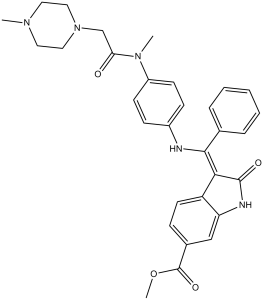
尼达尼布属于吲哚酮类化合物,是一种激酶抑制剂,以乙磺酸盐的形式用于治疗特发性肺纤维化和癌症。它具有抗肿瘤、酪氨酸激酶抑制、血管内皮生长因子受体拮抗、成纤维细胞生长因子受体拮抗和血管生成抑制等多种作用。它是一种芳香酯、甲基酯、吲哚酮类化合物、烯胺、芳香胺、芳香酰胺和N-烷基哌嗪。它是尼达尼布(1+)的共轭碱。
尼达尼布是一种激酶抑制剂。尼达尼布的作用机制是作为一种蛋白激酶抑制剂。 另见:尼达尼布(注释已移至)。 药物适应症 Ofev适用于成人特发性肺纤维化(IPF)的治疗。 通过阻断血管内皮生长因子(VEGF)信号通路抑制肿瘤血管生成是肿瘤学领域的一种新型治疗方法。临床前研究表明,阻断其他促血管生成受体酪氨酸激酶,例如血小板衍生生长因子受体(PDGFR)和成纤维细胞生长因子受体(FGFR),可能改善长期临床疗效。BIBF 1120是一种吲哚啉酮衍生物,在酶学测定中能有效阻断VEGF受体(VEGFR)、PDGFR和FGFR的激酶活性(IC50为20-100 nmol/L)。 BIBF 1120 可抑制三种参与血管生成的细胞类型(内皮细胞、周细胞和平滑肌细胞)中的丝裂原活化蛋白激酶和 Akt 信号通路,从而抑制细胞增殖(EC50 为 10-80 nmol/L)并诱导细胞凋亡。在迄今为止测试的所有肿瘤模型中,包括裸鼠体内的人源肿瘤异种移植模型和同源大鼠肿瘤模型,BIBF 1120 在耐受性良好的剂量(25-100 mg/kg,每日口服)下均表现出高度活性。通过 3 天后肿瘤灌注的磁共振成像评估,BIBF 1120 可在 5 天后降低血管密度和血管完整性,并显著抑制肿瘤生长。BIBF 1120 在细胞培养中的一个显著药效学特征是其通路抑制作用持续存在(给药 1 小时后可持续长达 32 小时),提示其受体解离动力学缓慢。尽管BIBF 1120在体内通过甲基酯裂解迅速代谢,导致平均停留时间较短,但每日一次口服给药在异种移植模型中仍具有完全疗效。这些独特的药代动力学和药效学特性可能有助于解释BIBF 1120的临床观察结果,该药物目前正处于III期临床开发阶段。[1] 三氟尿苷/替吡嘧啶(TFTD)是一种用于治疗转移性结直肠癌的复方药物,曾用名TAS-102。它由两种活性药物成分组成:三氟尿苷,一种抗肿瘤的胸苷类核苷类似物;以及替吡嘧啶,后者可提高三氟尿苷在体内的生物利用度。TFTD用于治疗对标准疗法耐药的不可切除的晚期或复发性结直肠癌患者。本研究探讨了三氟尿苷联合口服三重血管激酶抑制剂尼达尼布对人结直肠癌细胞系的抗癌作用。采用结晶紫染色法测定了其对DLD-1、HT-29和HCT116细胞系的细胞毒性。等效线图分析表明,三氟尿苷联合尼达尼布对DLD-1和HT-29细胞的生长抑制具有叠加效应,而对HCT116细胞的生长抑制则呈亚叠加效应。随后,将人结直肠癌细胞系皮下植入裸鼠体内,以评估三氟尿苷联合尼达尼布治疗在体内的肿瘤生长抑制效果。从第1天到第14天,每天两次口服给予小鼠TFTD(150 mg/kg/天)和/或尼达尼布(40 mg/kg/天)。联合治疗对DLD-1、DLD-1/5-FU、HT-29和HCT116异种移植瘤的肿瘤生长抑制率分别为61.5%、72.8%、67.6%和67.5%。该抑制率显著高于TFTD或尼达尼布单药治疗的效果(P<0.05)。这些结果表明TFTD和尼达尼布联合治疗结直肠癌异种移植瘤的有效性。采用液相色谱-串联质谱法测定了HT-29和HCT116肿瘤中DNA掺入的三氟尿苷浓度。连续14天接受TFTD和尼达尼布治疗后,肿瘤细胞的摄取水平高于单独使用TFTD治疗的水平。临床前研究结果表明,TFTD联合尼达尼布治疗是结直肠癌的一种有前景的治疗选择。[2] 通过阻断血管内皮生长因子(VEGF)信号通路抑制肿瘤血管生成是肿瘤治疗领域的一种新方法。临床前研究提示,阻断其他促血管生成激酶,例如成纤维细胞和血小板衍生生长因子受体(FGFR和PDGFR),可能提高药物治疗癌症的疗效。6位取代的吲哚啉酮被鉴定为VEGF、PDGF和FGF受体激酶的选择性抑制剂。特别是,6-甲氧羰基取代的吲哚啉酮表现出极佳的选择性。优化筛选出对VEGF相关内皮细胞增殖具有强效抑制作用的化合物,这些化合物对周细胞和平滑肌细胞也具有额外疗效。相反,未观察到对肿瘤细胞增殖的直接抑制作用。化合物2(BIBF 1000)和3(BIBF 1120)均为口服药物,在体内肿瘤模型中显示出令人鼓舞的疗效,且耐受性良好。三重血管激酶抑制剂3目前正在进行治疗非小细胞肺癌的III期临床试验。[3]尼达尼布是一种口服小分子酪氨酸激酶抑制剂,已获批用于治疗特发性肺纤维化和晚期非小细胞肺癌(腺癌组织学类型)。尼达尼布可与血管内皮生长因子(VEGF)、血小板衍生生长因子(PDGF)和成纤维细胞生长因子(FGF)的激酶结构域竞争性结合。在健康志愿者和晚期癌症患者的研究中,尼达尼布的药代动力学特征与时间无关。口服给药后约2-4小时达到血浆尼达尼布的峰值浓度,之后至少呈双指数下降。在所研究的剂量范围内(每日一次50-450 mg,每日两次150-300 mg),尼达尼布的暴露量与剂量成正比。尼达尼布通过酯水解代谢,生成游离酸部分,随后经葡萄糖醛酸化并经粪便排出。不到1%的药物相关放射性物质经尿液排出。尼达尼布的末端消除半衰期约为10-15小时。每日两次重复给药后,药物蓄积可忽略不计。性别和肾功能对尼达尼布的药代动力学无影响,而种族、低体重、高龄和吸烟的影响均在尼达尼布暴露量的患者间变异范围内,无需调整剂量。不建议中度或重度肝功能损害患者使用尼达尼布,轻度肝功能损害患者应密切监测并相应调整剂量。尼达尼布与其他药物发生相互作用的可能性较低,尤其是与经细胞色素P450酶代谢的药物。同时使用强效P-糖蛋白转运体抑制剂或诱导剂可能会影响尼达尼布的药代动力学。在每日两次、每次200 mg的剂量下,尼达尼布不具有致心律失常作用。[4] |
| 分子式 |
C31H33N5O4
|
|---|---|
| 分子量 |
539.62
|
| 精确质量 |
539.253
|
| 元素分析 |
C, 69.00; H, 6.16; N, 12.98; O, 11.86
|
| CAS号 |
656247-17-5
|
| 相关CAS号 |
Nintedanib esylate;656247-18-6;Nintedanib-13C,d3;Nintedanib-d3;1624587-84-3;Nintedanib-d8;1624587-87-6
|
| PubChem CID |
135423438
|
| 外观&性状 |
Yellow solid powder
|
| 密度 |
1.3±0.1 g/cm3
|
| 沸点 |
742.2±60.0 °C at 760 mmHg
|
| 闪点 |
402.7±32.9 °C
|
| 蒸汽压 |
0.0±2.5 mmHg at 25°C
|
| 折射率 |
1.658
|
| LogP |
2.59
|
| tPSA |
94.22
|
| 氢键供体(HBD)数目 |
2
|
| 氢键受体(HBA)数目 |
7
|
| 可旋转键数目(RBC) |
8
|
| 重原子数目 |
40
|
| 分子复杂度/Complexity |
892
|
| 定义原子立体中心数目 |
0
|
| SMILES |
O=C(C([H])([H])N1C([H])([H])C([H])([H])N(C([H])([H])[H])C([H])([H])C1([H])[H])N(C([H])([H])[H])C1C([H])=C([H])C(=C([H])C=1[H])/N=C(\C1C([H])=C([H])C([H])=C([H])C=1[H])/C1=C(N([H])C2C([H])=C(C(=O)OC([H])([H])[H])C([H])=C([H])C1=2)O[H]
|
| InChi Key |
CPMDPSXJELVGJG-UHFFFAOYSA-N
|
| InChi Code |
InChI=1S/C31H33N5O4/c1-34-15-17-36(18-16-34)20-27(37)35(2)24-12-10-23(11-13-24)32-29(21-7-5-4-6-8-21)28-25-14-9-22(31(39)40-3)19-26(25)33-30(28)38/h4-14,19,33,38H,15-18,20H2,1-3H3
|
| 化学名 |
methyl 2-hydroxy-3-[N-[4-[methyl-[2-(4-methylpiperazin-1-yl)acetyl]amino]phenyl]-C-phenylcarbonimidoyl]-1H-indole-6-carboxylate
|
| 别名 |
BIBF1120; Nintedanib; BIBF-1120; Intedanib; BIBF 1120; trade name: Vargatef
|
| HS Tariff Code |
2934.99.9001
|
| 存储方式 |
Powder -20°C 3 years 4°C 2 years In solvent -80°C 6 months -20°C 1 month |
| 运输条件 |
Room temperature (This product is stable at ambient temperature for a few days during ordinary shipping and time spent in Customs)
|
| 溶解度 (体外实验) |
|
|||
|---|---|---|---|---|
| 溶解度 (体内实验) |
配方 1 中的溶解度: 10 mg/mL (18.53 mM) in 50% PEG300 50% Saline (这些助溶剂从左到右依次添加,逐一添加), 悬浮液;超声助溶。
*生理盐水的制备:将 0.9 g 氯化钠溶解在 100 mL ddH₂O中,得到澄清溶液。 配方 2 中的溶解度: 10 mg/mL (18.53 mM) in 1% CMC 0.5% Tween-80 (这些助溶剂从左到右依次添加,逐一添加), 悬浊液; 超声助溶。 View More
配方 3 中的溶解度: 30% PEG400+0.5% Tween80+5% propylene glycol: 30mg/mL 1、请先配制澄清的储备液(如:用DMSO配置50 或 100 mg/mL母液(储备液)); 2、取适量母液,按从左到右的顺序依次添加助溶剂,澄清后再加入下一助溶剂。以 下列配方为例说明 (注意此配方只用于说明,并不一定代表此产品 的实际溶解配方): 10% DMSO → 40% PEG300 → 5% Tween-80 → 45% ddH2O (或 saline); 假设最终工作液的体积为 1 mL, 浓度为5 mg/mL: 取 100 μL 50 mg/mL 的澄清 DMSO 储备液加到 400 μL PEG300 中,混合均匀/澄清;向上述体系中加入50 μL Tween-80,混合均匀/澄清;然后继续加入450 μL ddH2O (或 saline)定容至 1 mL; 3、溶剂前显示的百分比是指该溶剂在最终溶液/工作液中的体积所占比例; 4、 如产品在配制过程中出现沉淀/析出,可通过加热(≤50℃)或超声的方式助溶; 5、为保证最佳实验结果,工作液请现配现用! 6、如不确定怎么将母液配置成体内动物实验的工作液,请查看说明书或联系我们; 7、 以上所有助溶剂都可在 Invivochem.cn网站购买。 |
| 制备储备液 | 1 mg | 5 mg | 10 mg | |
| 1 mM | 1.8532 mL | 9.2658 mL | 18.5316 mL | |
| 5 mM | 0.3706 mL | 1.8532 mL | 3.7063 mL | |
| 10 mM | 0.1853 mL | 0.9266 mL | 1.8532 mL |
1、根据实验需要选择合适的溶剂配制储备液 (母液):对于大多数产品,InvivoChem推荐用DMSO配置母液 (比如:5、10、20mM或者10、20、50 mg/mL浓度),个别水溶性高的产品可直接溶于水。产品在DMSO 、水或其他溶剂中的具体溶解度详见上”溶解度 (体外)”部分;
2、如果您找不到您想要的溶解度信息,或者很难将产品溶解在溶液中,请联系我们;
3、建议使用下列计算器进行相关计算(摩尔浓度计算器、稀释计算器、分子量计算器、重组计算器等);
4、母液配好之后,将其分装到常规用量,并储存在-20°C或-80°C,尽量减少反复冻融循环。
计算结果:
工作液浓度: mg/mL;
DMSO母液配制方法: mg 药物溶于 μL DMSO溶液(母液浓度 mg/mL)。如该浓度超过该批次药物DMSO溶解度,请首先与我们联系。
体内配方配制方法:取 μL DMSO母液,加入 μL PEG300,混匀澄清后加入μL Tween 80,混匀澄清后加入 μL ddH2O,混匀澄清。
(1) 请确保溶液澄清之后,再加入下一种溶剂 (助溶剂) 。可利用涡旋、超声或水浴加热等方法助溶;
(2) 一定要按顺序加入溶剂 (助溶剂) 。
Post-marketing Surveillance of Ofev Capsules in Chronic Fibrosing Interstitial Lung Diseases With a Progressive Phenotype in Japan
CTID: NCT04559581
Phase: Status: Active, not recruiting
Date: 2024-11-18
Cancer Res. 2008 Jun 15;68(12):4774-82. |
") |
Cancer Res. 2008 Jun 15;68(12):4774-82. |
 VEGFR2-IN-7
 SYHA1813
 VEGFR-2-IN-38
 BHEP
BHEP
InvivoChem的所有产品仅用于作科学研究,不面向患者销售
Copyright 2020 InvivoChem LLC | All Rights Reserved 粤ICP备20063088号-1


 COA
COA
")
")
 463611831
