| 规格 | 价格 | 库存 | 数量 |
|---|---|---|---|
| 10 mM * 1 mL in DMSO |
|
||
| 1mg |
|
||
| 5mg |
|
||
| 10mg |
|
||
| 50mg |
|
||
| 100mg |
|
||
| 250mg |
|
||
| 500mg |
|
||
| 1g |
|
||
| 2g |
|
||
| Other Sizes |
|
| 靶点 |
Broad spectrum anthelmintic
|
|---|---|
| 体外研究 (In Vitro) |
体外活性:硝唑尼特可减少细胞培养物中寄生虫的生长 90% 以上,几乎没有证据表明与药物相关的细胞毒性。 Nitazoxanide 是一种新型噻唑类抗寄生虫药,对多种原生动物和蠕虫表现出优异的体外活性。硝唑尼特及其代谢物替唑尼特在体外对肠杆菌、溶组织内阿米巴和阴道毛虫的活性比甲硝唑更强。 Nitazoxanide 可有效抑制 HBV 和 HCV 复制。在含有 HCV 复制子的细胞中,硝唑尼特可增强硝唑尼特加 IFN 的后续治疗效果,但不增强硝唑尼特加 2CmeC 的效果。 Nitazoxanide 可诱导 2.2.15 细胞产生的多种 HBV 蛋白(HBsAg、HBeAg、HBcAg)减少,但不影响 HBV RNA 转录。 Nitazoxanide 对溶组织内阿米巴的 IC50 和 IC90 值分别为 0.017 和 0.776 mg/mL,对肠杆菌的 IC50 和 IC90 值分别为 0.004 和 0.067 mg/mL,对阴道毛滴虫的 IC50 和 IC90 值分别为 0.034 和 2.046 mg/mL。硝唑尼特对溶组织内阿米巴的毒性比甲硝唑和阿苯达唑更强。
NTZ/硝唑尼特以剂量依赖的方式显著抑制JEV在培养细胞中的复制,50%有效浓度值为0.12±0.04μg/ml,这是培养细胞中无毒的浓度(50%细胞毒性浓度=18.59±0.31μg/ml)。计算出的化疗指数为154.92。与模拟处理的细胞相比,NTZ处理的细胞在感染后12、24、36和48小时的病毒产量显著降低。发现NTZ在病毒感染的早期中期发挥其抗JEV作用。[2] 含有溴而非硝基的NTZ/硝唑尼特衍生物(表1)对贾第鞭毛虫滋养体均无活性(IC50>50μM),但RM4820除外,其显示出中等抑制活性(IC50为18.8μM)低于RM4802和RM4805。MTZ的疗效低于NTZ和TIZ,但仍高于本研究中测试的任何其他药物。 为了观察由硝唑尼特诱导的G.lamblia滋养体的形态变化,并将其与MTZ诱导的形态变化进行比较,用50μM NTZ/Nitazoxanide或MTZ或DMSO作为对照处理融合的无菌滋养体培养物。光学显微镜观察显示,用NTZ处理3小时后,大约50%的滋养体是不动的;处理5小时后,95%以上的滋养体不动,形成大的多细胞聚集体;在NTZ处理24小时时未发现活动滋养体(数据未显示)。它们都表现出异常的液泡细胞质结构。透射电子显微镜的检查(图1)在很大程度上证实了这些发现,并表明在NTZ处理1小时后,相当多的滋养体在细胞质内已经表现出相对较小的异常细胞质内含物(图1A和B)。在处理3小时后,可以在大量寄生虫中观察到含有膜内含物或膜堆叠的较大液泡(图1C),在处理24小时后,这些寄生虫受到严重损伤,表现为细胞质组织的解离(图1D),或者在许多情况下,表现为含有膜残基的大液泡(图1E)。然而,除此之外,滋养体的细胞骨架元件,如与腹侧盘或鞭毛和基体相关的细丝,没有明显改变。在单独在DMSO中孵育的对照制剂中,未检测到寄生虫超微结构的明显变化(图1F)。[1] 在第一系列实验中,Caco2细胞在DMSO作为溶剂对照或30μM NTZ/Nitazoxanide硝唑尼特存在下与越来越多的滋养体(每孔103至106个寄生虫)一起孵育。然后通过实时PCR定量附着在Caco2细胞上的寄生虫(图4)。在没有任何药物的情况下,在每孔105个寄生虫的初始接种密度下,70%至90%的滋养体在24至48小时内保持附着在Caco2细胞上。在接种密度高出10倍(106个寄生虫/孔)时,该值降至近50%,表明与Caco2细胞表面的结合是可饱和的,并且取决于是否存在合适的结合位点和/或宿主细胞表面受体。在30μM NTZ的存在下,接种密度为105个滋养体,24小时后仍附着在Caco2细胞上的寄生虫数量降至对照值的20%以下。基于这些发现,我们研究了不同噻唑类药物与MTZ相比的效果。用新鲜的Caco2生长培养基补充汇合的Caco2细胞。向每个孔中加入滋养子(105),并加入许多噻唑类、MTZ和DMSO(图5)。24小时后收获细胞,通过实时PCR定量附着的滋养体。只有那些在无菌培养物中表现出强烈抑制作用的化合物(NTZ、TIZ、RM4802、RM4805和MTZ)会干扰Caco2共培养系统中的滋养体附着。与无菌培养相比,RM4802、RM4805和MTZ在Caco2共培养中的疗效与NTZ和TIZ相当(图5)。低于15μM的浓度对任何测试药物都没有显示出任何明显的抑制作用。[1] 对原生动物和厌氧细菌的研究表明,硝唑尼特/Nitazoxanide能够抑制丙酮酸铁氧还蛋白氧化还原酶(PFOR),这是一种对厌氧能量代谢至关重要的酶。然而,对PFOR酶依赖性电子转移反应的干扰可能不是硝唑尼特表现出抗原生动物活性的唯一途径,硝唑嗪对蠕虫的活性机制尚不清楚。 Nitazoxanide/硝唑尼特已被证明对细小隐球菌和肠杆菌具有体外活性。它已被证明可以单独抑制微小隐球菌子孢子的生长,并且还与阿奇霉素和利福平联合具有体外活性,分别抑制微小隐杆菌的生长83.9%和79.8%,而单独使用时为56.1%。同样,硝唑尼特及其衍生物替唑奈德的体外研究表明,其对肠道G.的疗效优于甲硝唑。具体而言,替唑硝胺对肠支原体甲硝唑敏感株的活性是甲硝唑的8倍,对耐药株的活性则是甲硝唑的两倍。 硝唑尼特还显示出对许多其他寄生虫和微生物病原体的广泛体外活性,包括肠杆菌、角膜弧菌、溶组织杆菌、阴道毛滴虫、人型棘球蚴、细粒棘球蚴和肝片吸虫。Nitazoxanide/硝唑尼特和替唑奈德的抗菌性能已对241种厌氧菌进行了测试,其中大部分在体外受到抑制,MIC90在0.06 mg/L至4 mg/L之间。硝唑嗪还对艰难梭菌以及甲硝唑敏感和甲硝唑耐药的幽门螺杆菌菌株显示出体外和体内抗菌活性[4]。 |
| 体内研究 (In Vivo) |
硝唑尼特以 250 mg/kg/天的剂量口服给药 11 天,可部分有效减少限生仔猪腹泻模型中的寄生虫负担,但 125 mg/kg/天的剂量则无效。硝唑尼特会引起仔猪药物相关性腹泻,这可能会影响其治疗效果。
NTZ/硝唑尼特可降低致死剂量乙脑攻击小鼠的死亡率[2] 为了评估NTZ对受致死剂量乙脑攻击的小鼠的保护作用,从感染后1天开始,每天以指定剂量灌胃给药NTZ,持续25天。感染乙脑并接受安慰剂(DMSO)治疗的小鼠(乙脑+DMSO组)从感染后5天开始出现乙脑的临床症状,包括肢体瘫痪、运动受限、竖毛、身体僵硬和全身震颤,所有小鼠(10/10只小鼠)在感染后9天内死亡。相比之下,感染JEV并接受NTZ治疗的小鼠(JEV+NTZ组,100mg/kg/天)从感染后11天开始出现JE的临床症状,在这10只小鼠中,1只在感染后12天内死亡,9只在实验期(25天)内存活(图6A)。NTZ介导的保护似乎是剂量依赖性的,因为接受50mg/kg/天、75mg/kg/天和100mg/kg/天NTZ的感染小鼠分别导致30%、70%和90%的小鼠存活(图6A)。这些数据表明,NTZ治疗降低了JEV感染小鼠的死亡率,并保护小鼠免受JEV致死剂量的攻击。模拟感染JEV并接受NTZ治疗的小鼠(mock+NTZ组)没有显示出可检测到的异常行为迹象,与模拟感染并接受DMSO治疗的小鼠相似(mock+DMSO组)(图6A)。对实验小鼠脑样本中JEV滴度的分析表明,与JEV+DMSO组相比,NTZ治疗显著降低了JEV+NTZ组脑中的病毒载量(图6B)。通过免疫组织化学检查实验小鼠的脑样本中是否存在病毒NS3蛋白。病毒NS3蛋白在神经元细胞的细胞质中被染色为棕色沉积物(附加文件1,箭头细胞)。JEV+DMSO组小鼠的脑切片显示,NS3染色阳性细胞的数量明显高于JEV+NTZ组。在Mock+NTZ或Mock+DMSO组小鼠的切片中未检测到NS3染色的阳性细胞。 对小鼠的研究。[3] 在急性隐孢子虫病的抗IFN-γ条件SCID小鼠模型中测试了NTZ/Nitazoxanide硝唑尼特单独或与PRM联合使用的疗效。虽然一项初步研究表明,Nitazoxanide/NTZ可以部分减少卵囊脱落(数据未显示),但我们无法在随后的几项实验中复制这一结果。以下是两项独立试验的合并结果,这些试验未能在该模型中显示疗效。单独使用NTZ治疗的任何组与安慰剂对照组之间的卵囊脱落对数没有差异(图1)。相比之下,所有接受PRM治疗的小鼠排出的卵囊水平明显低于单独接受NTZ或安慰剂治疗的小鼠(P<0.001)。NTZ和PRM的联合给药在降低卵囊脱落水平方面并不比单独使用PRM更有效。一般来说,在任何一组小鼠之间都没有观察到平均体重的显著差异(数据未显示)。 Student Newman-Keuls方差分析显示,单独使用NTZ或安慰剂治疗的小鼠的粘膜感染程度明显大于使用PRM治疗的动物(图2;P<0.001)。与单独接受PRM的小鼠相比,NTZ和PRM的联合给药没有显著改变粘膜感染的程度。 仔猪研究。[3] 仔猪模型提供了一个额外的优势,因为仔猪会因感染而腹泻。除了在治疗开始前对仔猪实施安乐死外,25只感染细小隐球菌的动物中有4只因与腹泻相关的健康状况不佳而实施安乐死,其中包括1只来自安慰剂组的仔猪(在感染后第5天),2只来自接受250mg硝唑尼特Nitazoxanide/NTZ/kg的组(在感染后第8天和第9天),1只来自PRM治疗组(在接种后第5天)。在攻击后第8天,对另外3头仔猪(1头安慰剂处理的仔猪、1头PRM处理的仔猪和1头未感染的对照仔猪)实施安乐死,以比较粘膜感染程度。对卵囊脱落水平和粘膜感染程度的分析表明,250mg/kg/天的NTZ显著降低了这些仔猪的粘膜感染程度,但不如500mg/kg/天的PRM有效(图3和图4)。 除PRM治疗组外,所有感染仔猪在攻击后56小时内均出现不同程度的腹泻。腹泻持续到实验结束,13天后(治疗开始后11天)。表2提供了每个治疗组观察到的腹泻天数的累积分析。对这些数据的卡方分析显示,治疗组之间存在非常显著的差异(总体皮尔逊卡方值为88.096,5 df;P<0.001)。以125mg/kg/天的剂量给予硝唑尼特/NTZ的未感染仔猪没有腹泻(24天观察中为0天)。相比之下,在36个观察日中的22个观察日,以250mg/kg/天的剂量给予NTZ的未感染仔猪出现了明显的药物性腹泻(Pearson卡方检验和Fisher精确检验[双尾]的P≤0.001)。在感染组中,只有PRM治疗组的腹泻频率低于感染安慰剂对照组。PRM治疗组仔猪出现腹泻的观察天数百分比明显低于其他任何感染组(卡方P<0.001,Fisher精确双尾检验P<0.001,与感染的安慰剂对照组、接受125 mg/kg/天NTZ的组和接受250 mg/kg/天NT Z的组进行比较)。 |
| 细胞实验 |
细胞系:贾第鞭毛虫滋养体在人结肠癌 Caco2 细胞中培养,数量不断增加(每孔 103–106 个寄生虫)。
浓度:30 μM 孵育时间:24 小时 结果:当硝唑尼特未添加时存在并且初始接种密度为每孔 105 个寄生虫,70-90% 的滋养体在 24-48 小时内粘附在 Caco2 细胞上。在 30 μM 硝唑尼特存在且接种密度为 105 个滋养体的情况下, 24小时后仍附着在Caco2细胞上的寄生虫数量下降至对照值的20%以下。 病毒、细胞和硝唑尼特Nitazoxanide/NTZ给药[2] JEV毒株(SH-JEV01)在3天大的BALB/c小鼠中生长,并使用BHK-21细胞通过空斑试验进行滴定,如下所述。BHK-21 细胞在37°C的Dulbecco改良Eagle培养基(DMEM)中,在含有5%CO2的气氛中,补充了10%胎牛血清(FBS)。 将NTZ/硝唑尼特(纯度≥98%)以50μg/μl的浓度溶解在培养级DMSO 中。除非另有说明,否则在1小时的吸附期后立即加入NTZ溶液,并在实验期间将其保持在培养基中。对照组接受等量的DMSO(终浓度≤0.06%),其不影响细胞活力或病毒复制。 细胞毒性试验[2] BHK-21细胞以每孔5×10³个细胞的密度接种在96孔板中。孵育24小时后,在37°C下用0.1至32μg/ml的不同浓度的NTZ处理细胞48小时。单独用DMSO处理的细胞用作对照。采用MTT法评估NTZ的细胞毒性。细胞存活率以DMSO处理的对照细胞总数的百分比计算。CC50定义为抑制指数生长细胞增殖50%的浓度,按所述计算。 NTZ/硝唑尼特在BHK-21细胞中的抗病毒作用分析[2] 六孔板中的BHK-21细胞以0.001的MOI感染JEV。在1小时的吸附期后,用浓度为0.01至10μg/ml的NTZ处理细胞,并在37°C下孵育48小时或指定时间。通过空斑试验和qRT-PCR确定病毒产量。病毒滴度的降低计算如下:病毒滴度降低%=[1-(PFUJEV+NTZ/PFUJEV+DMSO)]×100。EC50定义为在细胞中抑制病毒产量50%的浓度,如所述计算。 NTZ/硝唑尼特在细胞培养中的活性。[3] 将选择性克隆的MDBK细胞接种在96孔微升平板上,以检测其对细小隐球菌感染的敏感性。为了确定NTZ的剂量反应,72小时后,当细胞融合时,将3.0×10⁴ C.parvum卵囊与或不与药物一起添加到每个孔中。硫酸巴诺霉素(PRM)用作阳性对照药物。所有药物稀释液均在添加了5%胎牛血清、500 U青霉素、500μg链霉素/ml、1 mM丙酮酸钠、2 mM l-谷氨酰胺和0.2%二甲亚砜(DMSO)(培养基)的Dulbecco最低必需培养基中制备。将培养基添加到含有感染微小隐球菌卵囊的MDBK细胞的孔中作为阴性对照。所有药物浓度和对照品均进行了四次测试。孵育48小时(37°C,8%CO2)后,单层被甲醇固定,并在间接免疫荧光测定中反应,以确定感染强度。固定孔用含有1%正常山羊血清(NGS)的磷酸盐缓冲盐水(PBS)复水15分钟。复水后,将寄生虫反应性兔抗血清在含有1%NGS的PBS中稀释1:1000,加入孔中,在室温下孵育1小时。所有孔用PBS洗涤三次,用异硫氰酸荧光素偶联的山羊抗兔免疫球蛋白G抗体在含有1%NG的PBS中以1:100稀释检测结合抗体。在室温下温育1小时后,用PBS洗涤孔三次并干燥。在紫外光显微镜下,用专门为此目的设计的微型计算机视频成像设备定量微小念珠菌感染的程度。感染抑制百分比计算如下:1−(药物孔中的平均寄生虫数/对照孔中的寄生虫平均数)×100。所有结果的抑制分数分别为0、1、2、3和4,抑制率分别为0至30%、31至55%、56至70%、71至90%和91至100%。 细胞毒性试验。[3] 通过CellTiter 96 AQueous非放射性细胞增殖试验测定NTZ/硝唑尼特和PRM对MDBK细胞的细胞毒性。每种细胞毒性试验的对照包括(i)在培养基中孵育的未感染细胞,(ii)在培养基中孵育了感染细胞,以及(iii)暴露于培养基中含有3.0×104卵囊当量的冻融裂解物的细胞。根据光密度(OD)计算细胞毒性百分比,如下所示:[(未感染细胞的平均OD-感染细胞的均值OD)/未感染细胞平均OD]×100。所有结果的细胞毒性评分分别为0、1、2、3和4,细胞毒性百分比分别为0至5%、6至25%、26至50%、51至75%和76至100%。细胞毒性评分为0、1和2分别表示无毒性、轻度毒性和中度毒性,评分为3和4表示MDBK细胞的重度毒性。当感染细胞的OD大于未感染细胞的OD时,会产生负百分比毒性。 |
| 动物实验 |
动物模型:将日本脑炎病毒(JEV)腹腔注射到3周龄、体重12~14克的雌性昆明小鼠体内。[2]
剂量:50、75或100 mg/kg/天 给药途径:灌胃给药 结果:50 mg/kg/天、75 mg/kg/天和100 mg/kg/天的小鼠存活率分别为30%、70%和90%。 硝唑尼特(NTZ)/硝唑尼特在重症联合免疫缺陷(SCID)小鼠中的活性。[3] 抗γ干扰素(IFN-γ)预处理的SCID小鼠模型已在先前文献中描述。简而言之,断奶后(3至4周龄)的雄性近交系CB-17 SCID小鼠被饲养在经IACUC批准的微隔离笼中。在开始药物试验之前,将动物随机分为7组,每组7只小鼠。每只小鼠均腹腔注射1 mg XMG1.2(一种IFN-γ中和单克隆抗体)进行免疫。两小时后,7组中的6组小鼠每只均口服接种10⁷个卵囊。药物治疗于感染后第6天开始,此时卵囊开始从粪便中排出。治疗方案如下:第1组,200 mg/kg体重/天;第2组,100 mg/kg体重/天;第3组小鼠每日接受200 mg/kg NTZ联合2500 mg/kg PRM治疗;第4组小鼠每日接受100 mg/kg NTZ联合2500 mg/kg PRM治疗;第5组小鼠每日接受2500 mg/kg PRM治疗。NTZ溶于100% DMSO,每日分两次口服,每次30 μl。PRM溶于饮用水中,配制成10 mg/ml(16.2 mM)的浓度,根据每日饮水量计算,剂量为2500 mg/kg/天。第6组为未感染小鼠,每日接受200 mg/kg NTZ联合2500 mg/kg PRM治疗(药物毒性对照组)。第7组由7只小鼠组成,每天两次口服30 μl DMSO(安慰剂对照组)。所有小鼠均接受为期10天的治疗,并在治疗结束后继续观察5天。在整个研究期间,每周三次通过显微镜观察每只感染动物改良抗酸染色粪便涂片的30个高倍视野来确定卵囊排出量。结果以每组排出卵囊的平均对数±95%置信区间表示。在整个研究期间,每周一次测定小鼠体重。结果以每组平均体重±95%置信区间表示。在尸检时,取胃幽门区、小肠中段、回肠、盲肠和近端结肠的组织切片进行组织学分析,以确定黏膜感染程度。根据感染程度,每个部位被赋予一个评分,如下所示:0,无感染; 1. 极难发现的寄生虫形态;2. 稀少但易于发现的寄生虫形态;3. 大量但呈局灶性分布的寄生虫形态;4. 寄生虫形态广泛存在,覆盖大部分黏膜表面;5. 寄生虫形态广泛存在,覆盖整个黏膜表面。数据以五个部位的平均总分±95%置信区间表示。 硝唑尼特(NTZ)在仔猪腹泻模型中的活性。[3] 从四窝仔猪中剖腹产获得的31头无菌仔猪,按照先前描述的方法,在实验期间饲养于无菌隔离器中。31头仔猪中的26头在产仔24小时后接种卵囊。由于这些实验并非同时进行,因此根据体外卵囊脱囊率(脱囊率)计算了5 × 10⁶个脱囊卵囊的感染剂量。体外脱囊率的测定方法为:将卵囊在0.75%牛磺胆酸溶液中于37℃孵育45分钟。确定体外脱囊率后,相应调整接种量,使13头仔猪接种2 × 10⁷个卵囊,另13头仔猪接种7 × 10⁶个卵囊。每天观察仔猪两到三次,注意腹泻、精神萎靡、厌食等症状以及整体外观。腹泻的定义为:与未感染的对照仔猪相比,感染仔猪的排便次数、粪便量和含水量增加两倍。每日测量仔猪体重并采集粪便样本。攻毒后3天内,根据仔猪的体重、卵囊排出时间和腹泻情况进行分组。随后,仔猪开始接受每日治疗,治疗方案分别为:250 mg/kg 硝唑尼特/NTZ(5头仔猪)、125 mg/kg NTZ(6头仔猪)、500 mg/kg PRM(5头仔猪)或安慰剂(牛奶;9头仔猪)。5头未感染的对照动物作为药物毒性对照;其中3头仔猪接受250 mg/kg/天的NTZ治疗,2头仔猪接受125 mg/kg/天的NTZ治疗。26头感染仔猪中有1头因病情严重而被实施安乐死,并被排除在研究之外。所有药物均通过牛奶饲喂,每日分两次给药,持续11天。每天对每头仔猪进行改良抗酸染色粪便涂片检查,并测定其中卵囊的数量。由于仔猪感染小隐孢子虫后会出现腹泻,因此在测定排出卵囊数量时必须考虑粪便性状的任何变化。特别是,水样腹泻会因液体含量增加而有效稀释粪便,从而影响粪便涂片中检测到的卵囊数量。因此,我们设计了一个评分系统,该系统同时考虑了粪便的性状和检测到的卵囊数量。该系统的评分标准如下:0,未检测到卵囊;1,≤10个卵囊;2,≤25个卵囊;3,≤50个卵囊;4,≤100个卵囊;以及 5,>100 个卵囊。结果以各治疗组的平均卵囊排出评分±平均值的标准误差表示。存活的仔猪在治疗开始后 11 天被安乐死,并取六个肠道部位(胃幽门区、三个等距分布的小肠部位、盲肠和结肠)进行组织学分析,以评估黏膜感染程度。根据上述抗 IFN-γ 预处理的 SCID 小鼠的感染程度,对每个部位进行评分。结果以每头仔猪六个部位的总评分表示。 NTZ/硝唑尼特在小鼠模型中的抗病毒作用分析[2] 将三周龄雌性中国昆明小鼠(体重 12–14 g)随机分为六组(每组 10 只小鼠)。 JEV + NTZ 组感染日本脑炎病毒 (JEV) 后接受 NTZ 治疗(50、75 或 100 mg/kg/天)。JEV + DMSO 组感染 JEV 后接受安慰剂(DMSO)治疗。Mock + NTZ 组进行 JEV 假感染,并接受 NTZ 治疗。Mock + DMSO 组进行 JEV 假感染,并接受安慰剂(DMSO)治疗。感染时,小鼠腹腔注射 6×10⁴ PFU 的 JEV(相当于 50 × LD₅₀ 的 JEV)。NTZ 治疗时,将 NTZ 溶解于 DMSO 中,通过灌胃法给药,即将灌胃针插入食道,将 NTZ 直接输送到胃内。 NTZ 的总剂量分别为 50、75 或 100 mg/kg/天,从感染后第 1 天开始,每天连续给药,最多持续 25 天。每天监测小鼠的发病率和死亡率。 |
| 药代性质 (ADME/PK) |
吸收、分布和排泄
与片剂相比,混悬液的相对生物利用度为70%。与食物同服时,片剂的AUC和Cmax分别增加2倍和50%,而口服混悬液的AUC和Cmax分别增加45%至50%和≤10%。 替唑尼特经尿液、胆汁和粪便排泄,替唑尼特葡萄糖醛酸苷经尿液和胆汁排泄。口服硝唑尼特约2/3的剂量经粪便排泄,1/3经尿液排泄。 硝唑尼特经尿液和粪便清除。其代谢物替唑尼特也存在于尿液、血浆和母乳中。该药物在尿液中未检测到原形。 代谢/代谢物 该药物的活性代谢物是替唑尼特(去乙酰硝唑尼特)。硝唑尼特代谢途径的初始反应是水解生成替唑尼特,随后进行结合反应,主要是葡萄糖醛酸化,生成替唑尼特葡萄糖醛酸苷。该药物口服混悬液的生物利用度与口服片剂不同。与片剂相比,混悬液的生物利用度为70%。与空腹服用相比,与食物同服时,血浆中替唑尼特和替唑尼特葡萄糖醛酸苷的AUCt增加近两倍,最大浓度增加近50%。当口服混悬液与食物同服时,替唑尼及其葡萄糖醛酸苷的AUC增加约50%,而Cmax增加不到10%。 生物半衰期 7.3小时 |
| 毒性/毒理 (Toxicokinetics/TK) |
肝毒性
硝唑尼特治疗与血清转氨酶水平升高或临床上明显的急性肝损伤无关。然而,关于硝唑尼特长期治疗的研究很少,而且该药物的大多数对照试验采用的是短期疗程,未进行血清转氨酶监测。硝唑尼特已用作慢性丙型肝炎的辅助治疗,通常与聚乙二醇干扰素联合使用,可联合或不联合利巴韦林;在这些研究中,大多数患者的血清转氨酶水平有所改善,未报告急性肝炎加重或黄疸病例。 可能性评分:E(不太可能是临床上明显的肝损伤的原因)。 妊娠和哺乳期影响 ◉ 哺乳期用药概述 有限的信息表明,母亲服用 500 mg 硝唑尼特后,母乳中活性代谢物替唑尼特的浓度较低,预计不会对母乳喂养的婴儿造成任何不良影响,尤其是在婴儿超过 2 个月大的情况下。但在获得更多数据之前,尤其是在哺乳新生儿或早产儿期间,可能更倾向于选择其他药物。 ◉ 对母乳喂养婴儿的影响 截至修订日期,未找到相关的已发表信息。 ◉ 对泌乳和母乳的影响 截至修订日期,未找到相关的已发表信息。 蛋白结合 蛋白结合率极高(大于 99%),与血浆蛋白结合。 药物相互作用 [4] 目前,尚未进行尼他唑尼的体内药物相互作用研究。由于尼他唑尼与血浆蛋白的结合率超过 99%,因此在同时服用尼他唑尼和其他血浆蛋白结合率高且治疗指数窄的药物时应谨慎。建议同时服用华法林和尼他唑尼的患者监测凝血酶原时间。 副作用[4] 硝唑尼特通常耐受性良好,在人体试验中未观察到显著不良事件。不良事件通常较轻且短暂,主要与胃肠道相关,例如腹痛、腹泻和恶心。在参与临床试验的2000多名未感染HIV的患者中,发生率低于1%的不良事件包括厌食、胀气、食欲增加、唾液腺肿大、发热、感染、不适、肌酐水平升高、血清丙氨酸氨基转移酶水平升高、瘙痒、出汗、巩膜苍白发黄、鼻炎、头晕和尿色异常。此外,接受硝唑尼特治疗的患者,其心电图、生命体征、血液学、临床化学或尿液分析参数均未出现显著变化。尼他唑尼在空腹或随餐服用时,最大剂量为 4 克时耐受性良好,但胃肠道副作用的发生频率会随着剂量水平的增加而显著增加。 |
| 参考文献 | |
| 其他信息 |
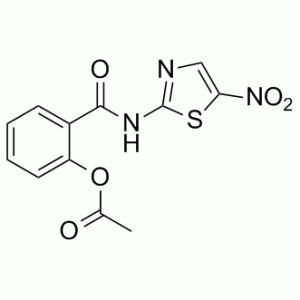
药效学
本药的总体作用是通过干扰微生物生存和增殖所需的重要能量途径来抑制其活性。硝唑尼特通过干扰丙酮酸铁氧还蛋白/黄素蛋白氧化还原酶依赖的电子传递反应发挥抗原生动物活性,该反应是多种微生物厌氧能量代谢所必需的。因此,小隐孢子虫的子孢子和蓝氏贾第鞭毛虫的滋养体受到抑制,从而缓解腹泻症状。干扰PFOR酶依赖的电子传递反应可能只是硝唑尼特发挥抗原生动物活性的众多途径之一。 乙酸[2-[[(5-硝基-2-噻唑基)氨基]-氧甲基]苯基]酯是一种羧酸酯,属于苯甲酰胺类化合物。它在功能上与水杨酰胺类药物相关。 硝唑尼特属于噻唑类药物。硝唑尼特 (NTZ) 是一种广谱抗感染药物,可显著调节多种细胞外和细胞内原生动物、蠕虫、厌氧菌和微需氧菌以及病毒的存活、生长和增殖。该药可有效治疗健康人群的胃肠道感染,包括小隐孢子虫或蓝氏贾第鞭毛虫感染。它通常耐受性良好。硝唑尼特是治疗健康(非免疫抑制)成人和儿童由小隐孢子虫或蓝氏贾第鞭毛虫感染引起的疾病的一线标准治疗药物,也可用于治疗其他原生动物或蠕虫引起的疾病。近年来,由于该药物能够抑制多种RNA和DNA病毒的复制,因此被研究作为一种广谱抗病毒药物。 硝唑尼特是一种抗原生动物药。 硝唑尼特是一种抗菌药物,对多种寄生虫和原生动物有效,主要在美国用于治疗贾第虫病和隐孢子虫病。据报道,硝唑尼特治疗期间不会引起血清转氨酶升高或临床上明显的肝损伤。 硝唑尼特是一种具有抗原生动物活性的合成苯甲酰胺类药物。硝唑尼特通过干扰丙酮酸铁氧还蛋白/黄素蛋白氧化还原酶依赖的电子传递反应发挥其抗原生动物活性,该反应对厌氧能量代谢至关重要。PFOR酶会还原硝唑尼特,从而损害能量代谢。然而,干扰PFOR酶依赖的电子转移反应可能并非硝唑尼特发挥抗原生动物活性的唯一途径。硝唑尼特对蓝氏贾第鞭毛虫和微小隐孢子虫均有效。 硝唑尼特是一种小分子药物,其临床试验阶段最高为IV期(涵盖所有适应症),于2002年首次获批,用于治疗阿米巴病和腹泻,并有26项在研适应症。 噻唑类药物是一类新型抗感染药物,其母体化合物为硝基噻唑类药物硝唑尼特[2-乙酰氧基-N-(5-硝基-2-噻唑基)苯甲酰胺] (NTZ)。NTZ对多种感染动物和人类的蠕虫、原生动物和肠道细菌具有广谱活性。在体内,硝唑尼特(NTZ)可迅速脱乙酰化为替唑尼特(TIZ),后者具有相似的活性。我们在此比较研究了NTZ、TIZ、多种其他修饰的噻唑类化合物以及甲硝唑(MTZ)对在无菌培养条件下以及与人结肠癌细胞系Caco2共培养条件下生长的蓝氏贾第鞭毛虫滋养体的体外作用。噻唑类化合物的修饰一方面包括用溴取代噻唑环上的硝基,另一方面包括改变苯环上甲基的位置。在七种用溴取代硝基的化合物中,只有一种化合物RM4820在无菌培养条件下对蓝氏贾第鞭毛虫的增殖表现出中等程度的抑制作用,但在与Caco2细胞共培养条件下则没有抑制作用,其半数抑制浓度(IC50)为18.8 μM;相比之下,硝基噻唑(NTZ)和替唑尼的IC50值为2.4 μM,甲硝唑(MTZ)的IC50值为7.8 μM。此外,苯环3位甲基化或羧化导致活性显著降低,而5位甲基化则完全消除了硝基噻唑类化合物的抗寄生虫作用。用硝基噻唑处理的滋养体在治疗后2至3小时内,腹盘上即出现明显的病变,而用甲硝唑处理则在较晚的时间点导致背膜严重受损。[1] 背景:日本脑炎病毒(JEV)对公共卫生造成重大影响。据估计,高危地区仍有30亿人未接种疫苗,且某些亚洲国家的未接种疫苗人数还在不断增加。因此,迫切需要开发针对日本脑炎的新型治疗药物。硝唑尼特 (NTZ) 是一种噻唑类抗感染药物,获准用于治疗寄生虫性胃肠炎。近期研究表明,NTZ 具有抗病毒特性。本研究在培养细胞和小鼠模型中评估了 NTZ 对日本脑炎病毒 (JEV) 的抗病毒活性。方法:用不同浓度的 NTZ 处理 JEV 感染的细胞。通过病毒滴度测定检测 JEV 在对照细胞和 NTZ 处理细胞中的复制情况。在 JEV 感染的不同时间点给予 NTZ,以确定 NTZ 影响 JEV 复制的阶段。小鼠感染致死剂量的 JEV,并在感染后第 1 天开始灌胃给予 NTZ。本研究评估了NTZ对感染日本脑炎病毒(JEV)小鼠的保护作用。结果显示:NTZ以剂量依赖的方式显著抑制培养细胞中JEV的复制,其50%有效浓度(EC50)为0.12 ± 0.04 μg/ml,该浓度对培养细胞无毒(50%细胞毒性浓度为18.59 ± 0.31 μg/ml)。计算得到的化疗指数为154.92。与对照组相比,NTZ处理组在感染后12、24、36和48小时的病毒产量均显著降低。NTZ在病毒感染的早期至中期发挥其抗JEV作用。 NTZ的抗日本脑炎病毒(JEV)作用也在体内得到证实,每日灌胃给予100 mg/kg/天NTZ的小鼠中,90%的小鼠免受致死剂量JEV的攻击。结论:体外和体内数据均表明NTZ具有抗JEV活性,提示NTZ可能用于治疗日本脑炎。[2] 硝唑尼特(NTZ)是一种目前正在进行人体临床试验以评估其治疗慢性隐孢子虫病疗效的药物,本研究在细胞培养和两种动物模型中评估了NTZ的活性。NTZ的抑制活性与巴龙霉素(PRM)进行了比较,巴龙霉素是一种对小隐孢子虫部分有效的药物。浓度为 10 μg/ml (32 μM) 的 NTZ 可使细胞培养中的寄生虫生长持续减少 90% 以上,且几乎没有药物相关的细胞毒性,而 PRM 在 2000 μg/ml (3.2 mM) 的浓度下仅能减少 80%。与体外疗效相反,在感染了小隐孢子虫且经抗 γ-干扰素处理的 SCID 小鼠中,无论 NTZ 的剂量为 100 或 200 mg/kg 体重/天,连续给药 10 天,均未能有效降低寄生虫负荷。NTZ 与 PRM 联合治疗的效果并不优于单独使用 PRM。最后,在无菌仔猪腹泻模型中,口服 NTZ 250 mg/kg/天,连续给药 11 天,可部分降低寄生虫负荷,但 125 mg/kg/天的剂量则无效。然而,较高剂量的硝唑尼特(NTZ)在仔猪中诱发了药物相关性腹泻,这可能影响了其治疗效果。正如我们之前报道的,PRM在500 mg/kg/天的剂量下能显著降低仔猪体内的寄生虫负荷。我们的结果表明,在所有测试模型中,仔猪腹泻模型最能模拟慢性隐孢子虫病患者对硝唑尼特治疗的部分反应。[3]硝唑尼特是一种新型噻唑类抗寄生虫药物,体外对多种原生动物和蠕虫均表现出优异的活性。它通过口服给药,生物利用度良好,耐受性良好,主要表现为轻微的胃肠道副作用。目前尚未发现药物相互作用。硝唑尼特已获准用于治疗1岁及以上患者的贾第鞭毛虫引起的腹泻和1-11岁儿童的隐孢子虫引起的腹泻。目前,它正在申请获准用于治疗成人隐孢子虫感染以及用于治疗免疫功能低下患者。它是抗寄生虫药物库的重要补充。[4] |
| 分子式 |
C12H9N3O5S
|
|---|---|
| 分子量 |
307.28
|
| 精确质量 |
307.026
|
| 元素分析 |
C, 46.90; H, 2.95; N, 13.67; O, 26.03; S, 10.44
|
| CAS号 |
55981-09-4
|
| 相关CAS号 |
Nitazoxanide-d4;1246819-17-9
|
| PubChem CID |
41684
|
| 外观&性状 |
Light yellow to yellow solid powder
|
| 密度 |
1.5±0.1 g/cm3
|
| 熔点 |
202ºC
|
| 折射率 |
1.673
|
| LogP |
1.79
|
| tPSA |
142.35
|
| 氢键供体(HBD)数目 |
1
|
| 氢键受体(HBA)数目 |
7
|
| 可旋转键数目(RBC) |
4
|
| 重原子数目 |
21
|
| 分子复杂度/Complexity |
428
|
| 定义原子立体中心数目 |
0
|
| SMILES |
S1C(=C([H])N=C1N([H])C(C1=C([H])C([H])=C([H])C([H])=C1OC(C([H])([H])[H])=O)=O)[N+](=O)[O-]
|
| InChi Key |
YQNQNVDNTFHQSW-UHFFFAOYSA-N
|
| InChi Code |
InChI=1S/C12H9N3O5S/c1-7(16)20-9-5-3-2-4-8(9)11(17)14-12-13-6-10(21-12)15(18)19/h2-6H,1H3,(H,13,14,17)
|
| 化学名 |
[2-[(5-nitro-1,3-thiazol-2-yl)carbamoyl]phenyl] acetate
|
| 别名 |
NSC-697855; NTZ; NSC 697855;NSC697855; NITAZOXANIDE; 55981-09-4; Alinia; Nitazoxanida; 2-((5-nitrothiazol-2-yl)carbamoyl)phenyl acetate; Nitazoxamide; Nitazoxanidum; Daxon; Alinia, Colufase, Daxon, Nitazoxamide
|
| HS Tariff Code |
2934.99.9001
|
| 存储方式 |
Powder -20°C 3 years 4°C 2 years In solvent -80°C 6 months -20°C 1 month |
| 运输条件 |
Room temperature (This product is stable at ambient temperature for a few days during ordinary shipping and time spent in Customs)
|
| 溶解度 (体外实验) |
DMSO : 61~100 mg/mL ( 198.51~325.44 mM )
|
|---|---|
| 溶解度 (体内实验) |
配方 1 中的溶解度: ≥ 3.25 mg/mL (10.58 mM) (饱和度未知) in 10% DMSO + 40% PEG300 + 5% Tween80 + 45% Saline (这些助溶剂从左到右依次添加,逐一添加), 澄清溶液。
例如,若需制备1 mL的工作液,可将100 μL 32.5 mg/mL澄清DMSO储备液加入到400 μL PEG300中,混匀;然后向上述溶液中加入50 μL Tween-80,混匀;加入450 μL生理盐水定容至1 mL。 *生理盐水的制备:将 0.9 g 氯化钠溶解在 100 mL ddH₂O中,得到澄清溶液。 配方 2 中的溶解度: 10% DMSO+40% PEG300+5% Tween-80+45% Saline: ≥ 3.25 mg/mL (10.58 mM) 请根据您的实验动物和给药方式选择适当的溶解配方/方案: 1、请先配制澄清的储备液(如:用DMSO配置50 或 100 mg/mL母液(储备液)); 2、取适量母液,按从左到右的顺序依次添加助溶剂,澄清后再加入下一助溶剂。以 下列配方为例说明 (注意此配方只用于说明,并不一定代表此产品 的实际溶解配方): 10% DMSO → 40% PEG300 → 5% Tween-80 → 45% ddH2O (或 saline); 假设最终工作液的体积为 1 mL, 浓度为5 mg/mL: 取 100 μL 50 mg/mL 的澄清 DMSO 储备液加到 400 μL PEG300 中,混合均匀/澄清;向上述体系中加入50 μL Tween-80,混合均匀/澄清;然后继续加入450 μL ddH2O (或 saline)定容至 1 mL; 3、溶剂前显示的百分比是指该溶剂在最终溶液/工作液中的体积所占比例; 4、 如产品在配制过程中出现沉淀/析出,可通过加热(≤50℃)或超声的方式助溶; 5、为保证最佳实验结果,工作液请现配现用! 6、如不确定怎么将母液配置成体内动物实验的工作液,请查看说明书或联系我们; 7、 以上所有助溶剂都可在 Invivochem.cn网站购买。 |
| 制备储备液 | 1 mg | 5 mg | 10 mg | |
| 1 mM | 3.2544 mL | 16.2718 mL | 32.5436 mL | |
| 5 mM | 0.6509 mL | 3.2544 mL | 6.5087 mL | |
| 10 mM | 0.3254 mL | 1.6272 mL | 3.2544 mL |
1、根据实验需要选择合适的溶剂配制储备液 (母液):对于大多数产品,InvivoChem推荐用DMSO配置母液 (比如:5、10、20mM或者10、20、50 mg/mL浓度),个别水溶性高的产品可直接溶于水。产品在DMSO 、水或其他溶剂中的具体溶解度详见上”溶解度 (体外)”部分;
2、如果您找不到您想要的溶解度信息,或者很难将产品溶解在溶液中,请联系我们;
3、建议使用下列计算器进行相关计算(摩尔浓度计算器、稀释计算器、分子量计算器、重组计算器等);
4、母液配好之后,将其分装到常规用量,并储存在-20°C或-80°C,尽量减少反复冻融循环。
计算结果:
工作液浓度: mg/mL;
DMSO母液配制方法: mg 药物溶于 μL DMSO溶液(母液浓度 mg/mL)。如该浓度超过该批次药物DMSO溶解度,请首先与我们联系。
体内配方配制方法:取 μL DMSO母液,加入 μL PEG300,混匀澄清后加入μL Tween 80,混匀澄清后加入 μL ddH2O,混匀澄清。
(1) 请确保溶液澄清之后,再加入下一种溶剂 (助溶剂) 。可利用涡旋、超声或水浴加热等方法助溶;
(2) 一定要按顺序加入溶剂 (助溶剂) 。
 Forward genetic screen for identification of NTZ resistant worm mutants.Mol Biochem Parasitol.2014 Jan;193(1):1-8. |
|---|
Dose response curves ofC. elegansmutants resistant to other classes of drugs to NTZ in a six day lethality assay.Mol Biochem Parasitol.2014 Jan;193(1):1-8. |
Combination of NTZ with albendazole (ALB) and pyrantel (PYR).Mol Biochem Parasitol.2014 Jan;193(1):1-8. |
 Ivermectin (IVM) susceptibility of the two NTZ resistant mutants identified in forward genetic screens at 44–45 hours.Mol Biochem Parasitol.2014 Jan;193(1):1-8. |
|---|
Effect of NTZ onC. elegansN2 wild-type nematodes.Mol Biochem Parasitol.2014 Jan;193(1):1-8. |
 Ipflufenoquin
 HDAC6-IN-27
 Antimalarial agent 34
 DNDI-6174
InvivoChem的所有产品仅用于作科学研究,不面向患者销售
Copyright 2020 InvivoChem LLC | All Rights Reserved 粤ICP备20063088号-1


 COA
COA


 463611831
