| 规格 | 价格 | 库存 | 数量 |
|---|---|---|---|
| 25mg |
|
||
| 50mg |
|
||
| 100mg |
|
||
| 250mg |
|
||
| 500mg |
|
||
| 1g |
|
||
| 2g |
|
||
| Other Sizes |
|

| 靶点 |
Natural alkaloid; iNOS; TGF-β/Smad; anti-inflammatory; antifibrosis; antitumor; antiviral; bocavirus minute virus of canines (MVC)
Prostate cancer-related targets (Akt, Cyclin D1, Bcl-2, Bax) (IC50: ~40 μM for PC-3 cells; ~45 μM for DU145 cells at 72 hours) [2] - TGFβ-Smad signaling pathway (TGFβ1, Smad2, Smad3, Smad7) [3] - Bocavirus MVC replication-related targets (viral capsid protein VP1/VP2, non-structural protein NS1) (EC50 = 32.6 μM for inhibiting MVC replication) [4] - NF-κB, STAT3, MAPK signaling pathways [1] |
|---|---|
| 体外研究 (In Vitro) |
一种名为氧化苦参碱的生物碱提取自苦参根,已被证明具有抗炎、抗纤维化、抗肿瘤和保护心脏的特性。氧化苦参碱的潜在信号通路包括:δ-阿片受体-Bcl-2、CD40、核因子红细胞2相关因子2/血红素加氧酶-1信号通路、二甲基精氨酸二甲氨基水解酶/不对称二甲基精氨酸代谢通路、Janus激酶/信号转录转导子和激活子、Toll 样受体 9/TRAF6、δ-阿片受体激活的 B 细胞核因子 kappa 轻链增强子和 δ-阿片受体-Bcl-2 [1]。氧化苦参碱以时间和剂量依赖性方式显着抑制 PC-3 和 DU145 细胞系的生长。另一方面,氧化苦参碱治疗并没有抑制 PNT1B 健康人前列腺细胞的增殖 [2]。
氧化苦参碱是一种生物碱,来源于中药苦参。氧化苦参碱已被证明具有抗炎、抗病毒和抗癌特性。本研究旨在探讨氧化苦参碱对人前列腺癌症细胞的抗癌作用及其潜在的分子机制。MTT分析表明,氧化苦参碱以时间和剂量依赖的方式显著抑制前列腺癌症细胞的增殖。此外,流式细胞术和末端脱氧核苷酸转移酶介导的dUTP-生物素缺口末端标记分析表明,氧化苦参碱治疗可能以剂量依赖性方式诱导前列腺癌症细胞凋亡。此外,蛋白质印迹分析显示,在癌症衰竭细胞中,p53和bax的表达显著增加,Bcl-2的表达显著降低,且呈剂量依赖性。[2] 氧化苦参碱(OMT)作为苦参的主要活性成分,具有抗氧化、抗炎、抗肿瘤和抗病毒等多种药理活性,目前广泛用于治疗病毒性肝炎;然而,其对细小病毒感染的影响尚未有报道。在本研究中,我们研究了OMT对感染犬微小病毒(MVC)的Walter Reed犬细胞/3873D的细胞存活率、病毒DNA复制、病毒基因表达、细胞周期和凋亡的影响。发现OMT浓度低于4 mmol/L(无细胞毒性)时,可以抑制MVC DNA复制,并在mRNA和蛋白质水平上降低病毒基因表达,这与MVC感染早期细胞周期S期阻滞的抑制有关。此外,OMT显著提高了细胞存活率,减少了MVC感染的细胞凋亡,并降低了活化半胱氨酸天冬氨酸蛋白酶3的表达。我们的研究结果表明,OMT在对抗细小病毒感染方面具有潜在的应用[4]。 氧化苦参碱(Oxymatrine, Matrine N-oxide)以剂量和时间依赖性方式抑制人前列腺癌细胞(PC-3、DU145)增殖,72小时IC50分别为~40 μM(PC-3)和~45 μM(DU145)。它诱导G0/G1期细胞周期阻滞,下调Akt磷酸化和Cyclin D1表达,通过降低Bcl-2、升高Bax水平促进凋亡[2] - 在TGFβ1刺激的大鼠肝星状细胞(HSC-T6)中,氧化苦参碱(Oxymatrine, Matrine N-oxide)(50-200 μM)抑制细胞活化和胶原合成。它下调TGFβ1、Smad2、Smad3表达,上调抑制性Smad7水平,阻断TGFβ-Smad信号传导[3] - 在人胚肾HEK293T细胞中,氧化苦参碱(Oxymatrine, Matrine N-oxide)抑制博卡病毒MVC复制,EC50为32.6 μM。50 μM时,分别减少病毒VP1/VP2和NS1基因表达~65%和~70%,缓解MVC诱导的凋亡(凋亡率从~42%降至~18%)[4] - 它调控多条信号通路:在LPS刺激的RAW264.7巨噬细胞中,抑制NF-κB和STAT3激活,阻断MAPK(ERK1/2、p38)磷酸化,减少促炎细胞因子(TNF-α、IL-6)产生[1] - 浓度高达100 μM时,对正常人前列腺上皮细胞(PrEC)和肝细胞(LO2)无明显细胞毒性[2][3] |
| 体内研究 (In Vivo) |
小鼠肿瘤的体积和重量呈剂量依赖性显着减少。氧化苦参碱在体内诱导细胞凋亡,从而减缓前列腺癌细胞的增殖[2]。氧化苦参碱显着减少实验大鼠肝组织中胶原蛋白的形成和沉积。当CCl4诱导SD大鼠肝纤维化时,氧化苦参碱可通过上调Smad 7的表达、下调Smad 3和CBP的表达来调节TGFβ-Smad通路的纤维化信号转导[3]。
氧化苦参碱在体内减少前列腺癌症细胞增殖[2] 为了研究氧化苦参碱对体内肿瘤生长的影响,使用PC-3皮下异种移植物将三种浓度的氧化苦参碱腹腔注射到裸鼠体内。结果表明,小鼠肿瘤的体积(图4A)和重量(图4B)以剂量依赖的方式显著降低。TUNEL检测表明,凋亡细胞的数量以剂量依赖的方式显著增加(图4C)。根据体外分析,凋亡相关蛋白p53和bcl-2的表达以剂量依赖的方式降低,bax的表达增加(图4D)。因此,氧化苦参碱可能通过促进体内细胞凋亡来减少前列腺癌症细胞的生长。 氧化苦参碱治疗的大鼠肝组织中胶原沉积和实质重排显著减少。与肝硬化模型大鼠相比,这些大鼠的半定量组织学评分(2.43+/-0.47 microm2 vs 3.76+/-0.68 microm2,P<0.05)和胶原蛋白的平均面积显著降低(94.41+/-37.26 microm2 vs 290.86+/-89.37 microm2,P<0.05)。治疗动物Smad 3 mRNA的基因表达显著降低。治疗组大鼠Smad 3 mRNA的A值低于模型组大鼠(0.034+/-0.090 vs 0.167+/-0.092,P<0.05)。相反,受试动物Smad 7 mRNA的A值显著增加(0.175+/-0.065 vs 0.074+/-0.012,P<0.05)。与模型大鼠相比,治疗组大鼠CBP mRNA的表达明显降低,其a值降低(0.065+/-0.049 vs 0.235+/-0.025,P<0.001)。 结论:氧化苦参碱能有效减少实验大鼠肝组织胶原的产生和沉积。氧化苦参碱可以促进SD大鼠CCl4诱导的肝纤维化中Smad 7的表达,抑制Smad 3和CBP的表达,可以调节TGF-β-Smad通路的纤维化信号转导[3]。 在PC-3前列腺癌异种移植裸鼠模型中,腹腔注射氧化苦参碱(Oxymatrine, Matrine N-oxide)(50 mg/kg、100 mg/kg,每日1次,持续21天)抑制肿瘤生长,肿瘤体积缩小率分别为~52%(50 mg/kg)和~70%(100 mg/kg),肿瘤重量抑制率分别为~48%和~65%。它下调肿瘤组织中p-Akt、Cyclin D1、Bcl-2表达,上调Bax水平[2] - 在四氯化碳(CCl4)诱导的肝纤维化大鼠模型中,腹腔注射氧化苦参碱(Oxymatrine, Matrine N-oxide)(40 mg/kg、80 mg/kg,每日1次,持续8周)缓解肝纤维化。它减少肝组织胶原沉积(羟脯氨酸含量分别降低~35%和~55%),下调肝组织中TGFβ1、Smad2/3、α-SMA表达,上调Smad7水平[3] |
| 酶活实验 |
oxymatrine/氧化苦参碱是一种从槐属植物根中提取的生物碱成分,已被证明具有抗炎、抗纤维化和抗肿瘤作用,并具有保护心肌损伤等能力。氧化苦参碱临床应用中涉及的潜在信号通路可能包括TGF-β/Smad、toll样受体4/活化B细胞核因子κ轻链增强子、toll状受体9/TRAF6、Janus激酶/信号转导和转录激活因子、磷脂酰肌醇-3激酶/Akt、δ阿片受体-arrestin-Bcl-2、CD40、表皮生长因子受体、核因子红细胞介素-2相关因子2/血红素加氧酶-1信号通路和二甲基精氨酸二甲基氨基水解酶/不对称二甲基精胺代谢通路。在这项工作中,研究人员总结了最近对氧化苦参碱相关信号通路的研究,为进一步研究其临床应用提供了线索和参考[1]。
低分子量(Hirt)DNA的分离和Southern Blot[4] 将WRD细胞(每孔1×10^6个)接种并培养在60mm培养皿中,然后用MVC(MOI)感染 = 10) 在37°C下培养1小时。细胞用PBS洗涤,分别在含有4 mmol/L oxymatrine/OMT(无毒浓度)的新鲜培养基中生长24、36和48小时。用PBS洗涤细胞两次,在2%十二烷基硫酸钠(SDS)中裂解,然后用蛋白酶K(0.5mg/mL)处理。收获Hirt DNA,用1%琼脂糖凝胶分离,并转移到Hybond N+膜上。根据制造商的方案,使用DIG High Prime DNA标记和检测起始试剂盒II将印迹与pI MVC基因组探针杂交,探针范围从nt 1到nt 5402。在ChemiDoc™MP成像系统(BIO-RAD)中检测到信号。Hirt DNA检测是在关的实验室进行的,所使用的方法在之前已有描述(Zhang等人,2017)[4]。 Akt激酶活性实验:将重组Akt激酶与ATP、特异性肽底物及0-100 μM 氧化苦参碱(Oxymatrine, Matrine N-oxide)在37°C下孵育45分钟,ELISA法检测磷酸化底物,计算激酶抑制率[2] - TGFβ-Smad通路活性实验:提取HSC-T6细胞核蛋白,与生物素标记的Smad特异性DNA探针及50-200 μM 氧化苦参碱(Oxymatrine, Matrine N-oxide)孵育,链霉亲和素偶联试剂检测DNA-蛋白复合物,定量Smad转录活性[3] - 病毒复制相关酶实验:博卡病毒MVC感染的HEK293T细胞用10-80 μM 氧化苦参碱(Oxymatrine, Matrine N-oxide)处理48小时,提取病毒RNA,实时PCR检测VP1/VP2和NS1基因表达水平[4] |
| 细胞实验 |
细胞增殖试验[2]
将DU145、PC-3和PNT1B细胞系接种到96孔板中,孵育过夜,并用oxymatrine/氧化苦参碱(0、2、4、6和8mg/ml)处理。使用MTT法测定细胞存活率。将细胞(3×104个细胞/孔)接种到96孔板中,在37°C的5%CO2中孵育过夜。随后,用不同浓度的氧化苦参碱(0、2、4、6和8mg/ml)孵育细胞。加入MTT(10 ml;5 mg/ml),将混合物在37°C的黑暗中孵育2小时。使用酶标仪在490 nm的波长下测量吸光度。 流式细胞术分析[2] 用不同浓度的oxymatrine/氧化苦参碱(0、4和8mg/ml)处理人前列腺癌症细胞系。用氧化苦参碱处理48小时后,将细胞胰蛋白酶消化并以1000 x g离心,用PBS洗涤沉淀物两次。重新悬浮细胞并用PBS洗涤三次。根据制造商的说明,使用膜联蛋白V-异硫氰酸荧光素/碘化丙啶(膜联蛋白V-FITC/IP)细胞凋亡检测试剂盒检测凋亡细胞。 蛋白质印迹分析[2] 经过oxymatrine/氧化苦参碱处理后,使用十二烷基硫酸钠聚丙烯酰胺电泳凝胶提取和分离蛋白质。然后将蛋白质转移到聚偏二氟乙烯膜上。将膜封闭并与以下一抗一起孵育:小鼠抗人p53单克隆抗体(1:1000稀释)、小鼠抗人bcl-2单克隆抗体(1:1 000稀释)、小鼠反人bax单克隆抗体(1∶1000稀释)和小鼠抗人GAPDH单克隆抗体(1:5000稀释),在4°C下过夜。用Tris缓冲盐水和吐温洗涤后,将膜与辣根过氧化物酶偶联的山羊抗小鼠二抗(1:10000稀释)一起孵育,并使用增强的化学发光检测试剂进行可视化。 前列腺癌细胞实验:PC-3/DU145细胞接种于96孔板,用0-100 μM 氧化苦参碱(Oxymatrine, Matrine N-oxide)处理24-72小时,MTT法检测细胞活力;碘化丙啶染色后流式细胞术分析细胞周期;Annexin V-FITC/PI双染色检测凋亡;Western blot分析Akt、p-Akt、Cyclin D1、Bcl-2、Bax表达[2] - 肝星状细胞实验:HSC-T6细胞经TGFβ1刺激后,用50-200 μM 氧化苦参碱(Oxymatrine, Matrine N-oxide)处理48小时,天狼星红染色检测胶原合成;Western blot和PCR分析TGFβ1、Smad2、Smad3、Smad7、α-SMA表达[3] - 病毒感染细胞实验:HEK293T细胞用博卡病毒MVC(感染复数MOI=0.1)感染后,用10-80 μM 氧化苦参碱(Oxymatrine, Matrine N-oxide)处理24-72小时,空斑实验测定病毒滴度;流式细胞术检测凋亡细胞;Western blot分析病毒蛋白表达[4] - 巨噬细胞炎症实验:RAW264.7巨噬细胞用20-100 μM 氧化苦参碱(Oxymatrine, Matrine N-oxide)预处理2小时后,加入LPS刺激,ELISA法检测细胞因子(TNF-α、IL-6)水平;Western blot分析NF-κB、STAT3、MAPK磷酸化水平[1] |
| 动物实验 |
小鼠:50 mg/kg 和 100 mg/kg;腹腔注射
小鼠:本研究采用 BALB/c 纯合裸鼠 (nu/nu)。24 只荷瘤小鼠随机分为三组:对照组用 PBS 处理,另外两组分别用不同浓度的苦参碱(50 mg/kg 和 100 mg/kg 体重)处理。苦参碱每日腹腔注射给药。 大鼠:本研究采用 100 只健康雄性 SD 大鼠(体重 140-160 g)。所有 100 只大鼠随机分为三组:对照组 (n=20)、治疗组 (n=40) 和模型组 (n=40)。模型组大鼠皮下注射溶于液体石蜡的300 g/L四氯化碳溶液,剂量为3 mL/kg,每周两次。治疗组大鼠除注射四氯化碳外,还每周两次腹腔注射10 mg/kg苦参碱。体内异种移植[2] BALB/c纯合子(nu/nu)裸鼠(6-8周龄;体重18-20 g),由本实验室自行繁殖,饲养于特定病原体清除(SPF)环境中。将PC-3细胞(3×10⁶)悬浮于100 μl PBS中,皮下注射至受体小鼠左侧腋窝。第五天,将24只荷瘤小鼠随机分为三组:对照组用PBS处理,另外两组分别用不同浓度的苦参碱(50 mg/kg和100 mg/kg体重)处理。苦参碱每日腹腔注射给药。肿瘤体积采用公式A × B² × π/6计算,其中A为肿瘤最长边的长度,B为垂直于A方向的肿瘤长度。治疗五周后,采用颈椎脱臼法处死小鼠,并测量肿瘤重量。 将100只健康雄性SD大鼠随机分为三组:正常组(n = 20)、苦参碱治疗组(n = 40)和四氯化碳诱导纤维化组(n = 40)。通过皮下注射四氯化碳(CCl4,溶于浓度为300 g/L的液体石蜡中,注射剂量为3 mL/kg,每周两次,持续8周)诱导实验性肝纤维化。同时,治疗组大鼠经腹腔注射给予苦参碱,剂量为10 mg/kg,每周两次。采用苏木精-伊红(H&E)染色和马松染色观察胶原沉积情况。采用酶联免疫吸附试验(ELISA)检测血清TGF-β1浓度。分别采用原位杂交(ISH)和免疫组织化学(IH)检测Smads和CBP(CREB结合蛋白)的基因表达。所有实验图像均使用专门的图像分析软件进行扫描和分析。[3] 前列腺癌异种移植模型:将PC-3细胞皮下接种于裸鼠。当肿瘤体积达到约 100 mm³ 时,将小鼠随机分为对照组和氧化苦参碱(苦参碱 N-氧化物)治疗组。将药物溶于生理盐水中,每日一次腹腔注射 50 mg/kg 或 100 mg/kg,连续 21 天。每 3 天测量一次肿瘤体积;处死小鼠并收集肿瘤组织进行蛋白质印迹和免疫组织化学分析[2] - 肝纤维化模型:每周两次腹腔注射 CCl4,持续 8 周,以诱导大鼠肝纤维化。从第 1 周到第 8 周,每日一次腹腔注射氧化苦参碱(苦参碱 N-氧化物)(40 mg/kg、80 mg/kg)。治疗结束后,处死大鼠;收集肝组织进行组织学(HE 染色、天狼星红染色)和分子生物学分析;检测血清肝功能指标[3] |
| 药代性质 (ADME/PK) |
在大鼠中,口服氧化苦参碱(苦参碱N-氧化物)(50 mg/kg)的口服生物利用度约为28%[1]
- 血浆消除半衰期(t1/2)为5.6小时,给药后2小时达到血浆峰浓度(Cmax)1.8 μg/mL[1] - 它广泛分布于各种组织中,在肝脏、肾脏和脾脏中的浓度较高[1] |
| 毒性/毒理 (Toxicokinetics/TK) |
相互作用
当归(Radix Angelicae sinensis (Oliv.) Diels)和苦参(Radix Sophora flavescens Ait.)的组合在中医中被广泛用于治疗痤疮、心脏病和肝炎等炎症性疾病。阿魏酸钠(SF)和苦参碱(OMT)分别是当归和苦参的有效成分。本研究探讨了SF和OMT组合的协同抗炎作用及其对RAW 264.7细胞中炎症相关介质的调节作用。体内实验中,采用二甲苯诱导的小鼠耳肿胀模型和角叉菜胶诱导的大鼠足爪肿胀模型评估了SF和OMT组合的抗炎作用。体外实验中,采用实时荧光定量PCR(RT-PCR)芯片分析检测脂多糖(LPS)激活的RAW 264.7细胞中趋化因子和细胞因子的mRNA表达水平。采用酶联免疫吸附试验(ELISA)检测LPS刺激的RAW 264.7细胞上清液中白细胞介素-11(IL-11)、C反应蛋白(CRP)和干扰素-γ(INF-γ)的水平。SF和OMT联合用药可显著抑制二甲苯诱导的小鼠耳廓水肿和角叉菜胶诱导的大鼠足爪水肿,而单独使用上述剂量的SF或OMT均未观察到疗效。与单独使用SF或OMT相比,SF和OMT联合用药在下调LPS刺激的RAW 264.7细胞中炎症相关介质的mRNA表达方面效果更佳。 ELISA结果显示,SF和OMT联合用药以剂量依赖的方式协同抑制LPS诱导的IL-11、CRP和INF-γ的产生。SF和OMT联合用药表现出协同抗炎作用,其活性可能与其对炎症相关介质(尤其是IL-11、CRP和INF-γ)的调节有关。 阿魏酸钠(SF)和苦参碱(OMT)是从中药中提取的化合物,在中国分别用于治疗心脏和肝脏疾病多年。本研究旨在探讨SF和OMT联合用药的镇痛效果及其机制。采用醋酸扭体试验和福尔马林试验建立动物疼痛模型,结果表明SF和OMT联合用药具有显著的剂量依赖性镇痛作用。体外实验表明,联合治疗抑制了辣椒素诱导的背根神经节神经元内钙离子浓度升高。重要的是,全细胞膜片钳实验证实了SF和OMT对辣椒素诱导电流的协同抑制作用。我们的结果提示,SF和OMT具有显著的镇痛作用,这可能与瞬时受体电位香草酸受体1(TRPV1)的协同抑制有关。本研究旨在探讨苦参碱-黄芩苷(OB)组合对2.2.15细胞中乙型肝炎病毒(HBV)复制以及HSC-T6细胞中α平滑肌肌动蛋白(αSMA)表达、I型胶原合成的影响。分别培养和处理2.2.15细胞和HSC-T6细胞。采用ELISA法检测培养上清液中的HBsAg和HBeAg,并采用荧光定量PCR法测定HBV DNA水平。从HSC-T6细胞中提取总RNA,并反转录为cDNA。cDNA经PCR扩增,其含量以β-肌动蛋白为内参进行校正。从HSC-T6细胞中提取的总细胞蛋白经电泳分离。分离后的蛋白电泳转移至硝酸纤维素膜。显色后,蛋白条带的含量以β-肌动蛋白进行校正。在2.2.15细胞培养体系中,OB组对HBsAg和HBeAg分泌的抑制率显著高于苦参碱组(HBsAg,P = 0.043;HBeAg,P = 0.026);OB组的HBV DNA水平显著低于苦参碱组(P = 0.041)。在HSC-T6细胞中,与苦参碱组相比,OB组的α-SMA mRNA和蛋白表达水平显著降低(mRNA,P = 0.013;蛋白,P = 0.042);与苦参碱组相比,OB组的I型胶原蛋白mRNA和蛋白表达水平也显著降低(mRNA,P < 0.01;蛋白,P < 0.01)。作者得出结论:OB组合对2.2.15细胞中的HBV复制具有更好的抑制作用,并且在体外对HSC-T6细胞中α-SMA表达和I型胶原蛋白合成的抑制作用也优于苦参碱。苦参碱已被证实可通过抗炎和抗凋亡作用保护肝脏、肠道和心脏免受缺血再灌注损伤。为了探究这种保护作用是否适用于脑缺血损伤,作者研究了苦参碱潜在的神经保护作用及其机制。雄性Sprague-Dawley大鼠被随机分为四组:永久性大脑中动脉闭塞(pMCAO)组、高剂量组(pMCAO+苦参碱120 mg/kg)、低剂量组(pMCAO+苦参碱60 mg/kg)和假手术组。作者采用永久性大脑中动脉闭塞模型,在脑缺血后立即腹腔注射苦参碱,并在随后的几天内每日一次。MCAO后24小时,采用改良的六分制量表评估神经功能缺损;测量脑含水量;并通过免疫组织化学、Western blotting和RT-PCR检测NF-κB的表达。 72小时后,采用2,3,5-氯化三苯基四氮唑(TTC)染色分析梗死体积。与pMCAO组相比,高剂量组的神经功能缺损改善(P < 0.05),梗死体积减少(P < 0.001),脑水肿减轻(P < 0.05)。与这些指标一致,免疫组化、Western blot和RT-PCR分析表明,高剂量组中NF-κB表达显著降低。低剂量苦参碱对pMCAO大鼠的NF-κB表达无影响。氧化苦参碱可减少pMCAO诱导的梗死体积,这种作用可能是通过降低NF-κB表达实现的。 体外实验表明,浓度高达100 μM的氧化苦参碱(苦参碱N-氧化物)对正常前列腺上皮细胞(PrEC)和肝细胞(LO2)无显著细胞毒性[2][3]。 体内实验表明,给予异种移植小鼠100 mg/kg剂量的氧化苦参碱(苦参碱N-氧化物)21天,或给予肝纤维化大鼠80 mg/kg剂量的氧化苦参碱8周,均未引起体重、器官指数或血清ALT/AST/肌酐水平的显著变化[2][3]。 小鼠急性腹腔注射氧化苦参碱(苦参碱N-氧化物)的LD50为[此处应填写具体数值]。约350毫克/千克[1] |
| 参考文献 |
|
| 其他信息 |
治疗用途
抗心律失常药;抗病毒药 本研究旨在评估苦参碱胶囊治疗慢性乙型肝炎的疗效和安全性。本研究为一项随机、双盲、安慰剂对照的多中心试验。注射用苦参碱作为阳性对照药物。共有216例慢性乙型肝炎患者入组,随访24周,其中108例服用苦参碱胶囊,36例服用苦参碱注射剂,72例服用安慰剂。治疗前后观察患者的临床症状、肝功能、血清乙型肝炎病毒标志物及不良反应。216例患者中,6例脱落,11例因不符合标准而被排除。因此,本研究分析了苦参碱在患者中的疗效和安全性。在胶囊治疗组中,76.47%的患者ALT水平恢复正常,38.61%和31.91%的患者HBV DNA和HBeAg均转阴。在注射治疗组中,83.33%的患者ALT水平恢复正常,43.33%和39.29%的患者HBV DNA和HBeAg均转阴。在安慰剂治疗组中,40.00%的患者ALT水平恢复正常,7.46%和6.45%的患者HBV DNA和HBeAg均转阴。胶囊治疗组的完全缓解率和部分缓解率分别为24.51%和57.84%,注射治疗组分别为33.33%和50.00%,安慰剂组分别为2.99%和41.79%。两组患者之间无显著差异,但均显著高于安慰剂组。胶囊、注射剂和安慰剂的不良反应发生率分别为7.77%、6.67%和8.82%,彼此之间无统计学差异。/结论是/苦参碱是治疗慢性乙型肝炎的一种有效且安全的药物。 苦参碱(苦参碱N-氧化物)是从苦参(Sophora flavescens Ait.)的根中分离得到的喹诺里西啶生物碱。苦参(Sophora tonkinensis Gapnep)[1][2][3][4] - 其抗肿瘤机制包括抑制Akt信号通路、诱导细胞周期阻滞和凋亡以及调节NF-κB/STAT3/MAPK通路[1][2] - 它通过阻断TGFβ-Smad信号通路、抑制肝星状细胞活化和胶原合成来减轻肝纤维化[3] - 其抗博卡病毒MVC病毒作用是通过抑制病毒基因表达和复制以及减少病毒诱导的细胞凋亡来实现的[4] - 它在治疗前列腺癌、肝纤维化和病毒感染方面具有潜在的临床应用价值,且全身毒性低,口服生物利用度中等[1][2][3][4] |
| 分子式 |
C15H24N2O2
|
|
|---|---|---|
| 分子量 |
264.36
|
|
| 精确质量 |
264.183
|
|
| 元素分析 |
C, 68.15; H, 9.15; N, 10.60; O, 12.10
|
|
| CAS号 |
16837-52-8
|
|
| 相关CAS号 |
|
|
| PubChem CID |
114850
|
|
| 外观&性状 |
White to off-white solid powder
|
|
| 熔点 |
208 °C
|
|
| 蒸汽压 |
0mmHg at 25°C
|
|
| 折射率 |
1.637
|
|
| LogP |
-0.35
|
|
| tPSA |
49.74
|
|
| 氢键供体(HBD)数目 |
0
|
|
| 氢键受体(HBA)数目 |
2
|
|
| 可旋转键数目(RBC) |
0
|
|
| 重原子数目 |
19
|
|
| 分子复杂度/Complexity |
400
|
|
| 定义原子立体中心数目 |
4
|
|
| SMILES |
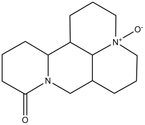
C1C[C@@H]2[C@H]3CCC[N+]4([C@H]3[C@@H](CCC4)CN2C(=O)C1)[O-]
|
|
| InChi Key |
XVPBINOPNYFXID-VNSSVHEPSA-N
|
|
| InChi Code |
InChI=1S/C15H24N2O2/c18-14-7-1-6-13-12-5-3-9-17(19)8-2-4-11(15(12)17)10-16(13)14/h11-13,15H,1-10H2/t11-,12+,13+,15+,17+/m0/s1
|
|
| 化学名 |
(4R,41R,7aS,13aR,13bR)-10-oxododecahydro-1H,5H-dipyrido[2,1-f:3,2,1-ij][1,6]naphthyridine 4(41H)-oxide
|
|
| 别名 |
|
|
| HS Tariff Code |
2934.99.9001
|
|
| 存储方式 |
Powder -20°C 3 years 4°C 2 years In solvent -80°C 6 months -20°C 1 month |
|
| 运输条件 |
Room temperature (This product is stable at ambient temperature for a few days during ordinary shipping and time spent in Customs)
|
| 溶解度 (体外实验) |
|
|||
|---|---|---|---|---|
| 溶解度 (体内实验) |
配方 1 中的溶解度: ≥ 2.08 mg/mL (7.87 mM) (饱和度未知) in 10% DMSO + 40% PEG300 + 5% Tween80 + 45% Saline (这些助溶剂从左到右依次添加,逐一添加), 澄清溶液。
例如,若需制备1 mL的工作液,可将100 μL 20.8 mg/mL澄清DMSO储备液加入400 μL PEG300中,混匀;然后向上述溶液中加入50 μL Tween-80,混匀;加入450 μL生理盐水定容至1 mL。 *生理盐水的制备:将 0.9 g 氯化钠溶解在 100 mL ddH₂O中,得到澄清溶液。 配方 2 中的溶解度: ≥ 2.08 mg/mL (7.87 mM) (饱和度未知) in 10% DMSO + 90% (20% SBE-β-CD in Saline) (这些助溶剂从左到右依次添加,逐一添加), 澄清溶液。 例如,若需制备1 mL的工作液,可将 100 μL 20.8 mg/mL澄清DMSO储备液加入900 μL 20% SBE-β-CD生理盐水溶液中,混匀。 *20% SBE-β-CD 生理盐水溶液的制备(4°C,1 周):将 2 g SBE-β-CD 溶解于 10 mL 生理盐水中,得到澄清溶液。 View More
配方 3 中的溶解度: ≥ 2.08 mg/mL (7.87 mM) (饱和度未知) in 10% DMSO + 90% Corn Oil (这些助溶剂从左到右依次添加,逐一添加), 澄清溶液。 配方 4 中的溶解度: 100 mg/mL (378.27 mM) in PBS (这些助溶剂从左到右依次添加,逐一添加), 澄清溶液; 超声助溶. 1、请先配制澄清的储备液(如:用DMSO配置50 或 100 mg/mL母液(储备液)); 2、取适量母液,按从左到右的顺序依次添加助溶剂,澄清后再加入下一助溶剂。以 下列配方为例说明 (注意此配方只用于说明,并不一定代表此产品 的实际溶解配方): 10% DMSO → 40% PEG300 → 5% Tween-80 → 45% ddH2O (或 saline); 假设最终工作液的体积为 1 mL, 浓度为5 mg/mL: 取 100 μL 50 mg/mL 的澄清 DMSO 储备液加到 400 μL PEG300 中,混合均匀/澄清;向上述体系中加入50 μL Tween-80,混合均匀/澄清;然后继续加入450 μL ddH2O (或 saline)定容至 1 mL; 3、溶剂前显示的百分比是指该溶剂在最终溶液/工作液中的体积所占比例; 4、 如产品在配制过程中出现沉淀/析出,可通过加热(≤50℃)或超声的方式助溶; 5、为保证最佳实验结果,工作液请现配现用! 6、如不确定怎么将母液配置成体内动物实验的工作液,请查看说明书或联系我们; 7、 以上所有助溶剂都可在 Invivochem.cn网站购买。 |
| 制备储备液 | 1 mg | 5 mg | 10 mg | |
| 1 mM | 3.7827 mL | 18.9136 mL | 37.8272 mL | |
| 5 mM | 0.7565 mL | 3.7827 mL | 7.5654 mL | |
| 10 mM | 0.3783 mL | 1.8914 mL | 3.7827 mL |
1、根据实验需要选择合适的溶剂配制储备液 (母液):对于大多数产品,InvivoChem推荐用DMSO配置母液 (比如:5、10、20mM或者10、20、50 mg/mL浓度),个别水溶性高的产品可直接溶于水。产品在DMSO 、水或其他溶剂中的具体溶解度详见上”溶解度 (体外)”部分;
2、如果您找不到您想要的溶解度信息,或者很难将产品溶解在溶液中,请联系我们;
3、建议使用下列计算器进行相关计算(摩尔浓度计算器、稀释计算器、分子量计算器、重组计算器等);
4、母液配好之后,将其分装到常规用量,并储存在-20°C或-80°C,尽量减少反复冻融循环。
计算结果:
工作液浓度: mg/mL;
DMSO母液配制方法: mg 药物溶于 μL DMSO溶液(母液浓度 mg/mL)。如该浓度超过该批次药物DMSO溶解度,请首先与我们联系。
体内配方配制方法:取 μL DMSO母液,加入 μL PEG300,混匀澄清后加入μL Tween 80,混匀澄清后加入 μL ddH2O,混匀澄清。
(1) 请确保溶液澄清之后,再加入下一种溶剂 (助溶剂) 。可利用涡旋、超声或水浴加热等方法助溶;
(2) 一定要按顺序加入溶剂 (助溶剂) 。
InvivoChem的所有产品仅用于作科学研究,不面向患者销售
Copyright 2020 InvivoChem LLC | All Rights Reserved 粤ICP备20063088号-1


 COA
COA
 463611831
