| 规格 | 价格 | 库存 | 数量 |
|---|---|---|---|
| 10 mM * 1 mL in DMSO |
|
||
| 1mg |
|
||
| 5mg |
|
||
| 10mg |
|
||
| 25mg |
|
||
| 50mg |
|
||
| 100mg |
|
||
| 250mg |
|
||
| 500mg |
|
||
| 1g |
|
||
| Other Sizes |
|
| 靶点 |
FGFR1 (IC50 = 10 nM); cFMS (IC50 = 20 nM); FLT3 (IC50 = 160 nM); KDR (IC50 = 350 nM); LCK (IC50 = 860 nM); FLT1 (IC50 = 880 nM); NTRK3 (IC50 = 890 nM)
|
|---|---|
| 体外研究 (In Vitro) |
体外活性:在M-NFS-60、Bac1.2F5和M-07e细胞中,Pexidartinib抑制CSF1依赖性增殖,IC50分别为0.44 μM、0.22 μM和0.1 μM。激酶检测:Pexidartinib (PLX-3397) 是一种有效、选择性和 ATP 竞争性 CSF1R (cFMS) 和 c-Kit 抑制剂,对 c-Kit 和 CSF1R 的选择性比其他相关激酶(例如 FLT3)高 10 至 100 倍、KDR (VEGFR2)、LCK、FLT1 (VEGFR1) 和 NTRK3 (TRKC),IC50 分别为 160、350、860、880 和 890 nM。
CSF1R活性在体外促进T细胞淋巴瘤生长 [5] 在明确T细胞淋巴瘤(TCL)中CSF1R的表达与激活后,我们采用功能缺失策略,通过分子与药理学互补方法探究其潜在致癌作用。首先使用临床可用且合理设计的CSF1R选择性酪氨酸激酶抑制剂(培西达替尼,PLX3397)(25,49)。为验证培西达替尼对CSF1R的抑制效果,用培西达替尼处理具有CSF1R自分泌激活的TCL细胞。结果显示培西达替尼治疗后CSF1R磷酸化显著降低(图2A,补充图4A)。重要的是,培西达替尼对部分TCL细胞中表达的致癌激酶NPM-ALK的磷酸化水平无影响(补充图4B)。此外,培西达替尼暴露导致细胞增殖呈剂量依赖性下降(图2B及补充图4D-E),但在不表达CSF1R的TCL细胞中未观察到此效应,证实了该FDA批准药物的相对选择性(补充图4C)。与此一致,培西达替尼治疗增加TCL细胞凋亡,表现为磷脂酰丝氨酸外翻(图2C-E)、PARP裂解及Caspase 3裂解(图2F与补充图4F)。为深入探究CSF1R在T细胞淋巴瘤生长中的作用并排除TKI脱靶效应可能,成功在T细胞淋巴瘤衍生细胞系中建立多西环素诱导的CSF1R靶向shRNA稳定表达体系。 CSF1R激活与不同信号通路磷酸化相关 [5] CSF1R的生理性结合与激活引发下游磷酸化依赖性信号传导,调控髓系细胞存活与分化(33,34)。然而,CSF1R激活在多数非髓系细胞中调控的下游信号尚不明确。为表征CSF1R激活的信号通路,在T细胞淋巴瘤系中用培西达替尼抑制CSF1R活性后,采用无偏倚磷酸化蛋白质组学分析。筛选选用Karpas 299 T细胞淋巴瘤系,因其兼具CSF1配体分泌与强表面CSF1R表达。该筛选共鉴定1936个独立磷酸酪氨酸肽段(对应1123种蛋白质),同时识别8045个独立磷酸丝氨酸/磷酸苏氨酸肽段组合(对应3136种蛋白质)(补充表1-2)。培西达替尼处理细胞中CSF1R自身磷酸化肽段显著减少,进一步验证了该方法(表1,补充图5A及补充表3)。对三个技术重复进行层次聚类分析,显示培西达替尼处理细胞形成独立聚类(图4A与补充图5B),共涵盖551种磷酸化肽段显著修饰的独特蛋白。其中451个为丝氨酸残基修饰,122个为酪氨酸残基修饰,113个为苏氨酸残基修饰(补充表3)。通过京都基因与基因组百科全书(KEGG在线软件)对CSF1R抑制后的信号通路分析显示,这些变化与PI3K/AKT调控信号通路的差异性磷酸化一致(补充表1,图4B)。此外,还鉴定到参与细胞过程(包括代谢、细胞周期进程和肌动蛋白-细胞骨架动力学)的蛋白质修饰(图4B,补充表4及补充图5C)。为深入探索CSF1R信号下游激活的通路,在培西达替尼抑制CSF1R后进行无偏倚基因表达谱芯片分析。经培西达替尼抑制CSF1R后,217个基因表达显著改变(n=3, p<0.01;图4C)。重要的是,CSF1R抑制与细胞因子(JAK/STAT)信号相关基因表达变化相关(图4D)。类似地,磷酸化蛋白质组学筛选显示JAK/STAT信号通路相关蛋白(包括STAT1、STAT3、STAT5和SOS2)存在差异性磷酸化(补充表2)。因筛选所用T细胞系携带致癌性NPM-ALK融合,故纳入ALK抑制剂克唑替尼处理细胞作为对照。与ALK抑制相比,培西达替尼处理细胞中46%的基因表达变化(n=154个基因)具有特异性(图4E),63个基因的表达受培西达替尼或克唑替尼共同调控(图4E)。 |
| 体内研究 (In Vivo) |
在 MMTV-PyMT 小鼠中,Pexidartinib(40 mg/kg,口服)通过 CD45+CD11b+Ly6C−Ly6G−F4/80+ 显着抑制稳态和 PTX 诱导的肿瘤浸润。 Pexidartinib/PTX 治疗还导致乳腺肿瘤内 CD31+ 血管密度显着降低,同时诱导细胞凋亡和坏死。在携带 GL261 肿瘤的 C57 小鼠中,Pexidartinib (po) 抑制胶质母细胞瘤侵袭。在 cmo 小鼠中,PLX3397 通过减少尾部和爪子的侵蚀性骨病变以及循环 MIP-1α 的水平,显着减轻自身炎症性疾病。在患有 B16F10 黑色素瘤的小鼠中,Pexidartinib(45 mg/kg,口服)可增强 CD8 介导的黑色素瘤免疫治疗。[1]
PLX3397治疗显著降低了喂食食物和高脂肪饮食的小鼠脂肪组织中的巨噬细胞数量,而不影响总髓细胞水平。尽管如此,PLX3397并没有显著改变葡萄糖稳态,也没有影响高脂肪饮食诱导的内脏脂肪细胞因子表达(Il-6和Tnfa)的增加,对应激激酶JNK和ERK的磷酸化以及巨噬细胞极化的影响有限。 化疗诱导的TAM募集的阻断 [1] 为探究肿瘤浸润性TAM是否调控乳腺上皮细胞(MEC)对细胞毒疗法的敏感性,我们通过免疫学和药理学手段在体内阻断TAM浸润(补充图S4),并评估治疗小鼠肿瘤的髓系细胞浸润情况(补充图S5)。荷原位乳腺肿瘤的小鼠接受中和性单克隆抗体(mAb)CSF1(克隆5S1)或CD11b(克隆M1/70)治疗,或采用对CSF1/cKIT受体酪氨酸激酶具有高效(nM级)选择性的ATP竞争性抑制剂(培西达替尼),实施单药或与紫杉醇(PTX)联用治疗。CD11b是表达于粒细胞、巨噬细胞、单核细胞、树突状细胞(DC)及自然杀伤细胞的整合素细胞黏附分子,部分调控细胞向组织和肿瘤实质的跨内皮迁移。PLX3397对cKIT和CSF1R的选择性较其他相关激酶(如KDR)高10-100倍(见补充图S6A与方法;参考文献20)。 对浸润乳腺肿瘤的主要髓系亚型进行荧光激活细胞分选分析显示:无论单药或联用PTX,经αCSF1单抗或培西达替尼治疗后,CD45+CD11b+Ly6C−Ly6G−F4/80+ TAM的募集均显著减少,但对CD45+CD11b+LY6Ghigh 未成熟髓系细胞(iMC)或CD45+CD11blow/−Ly6C−CD22−Ly6G−CD11chighMHCIIhigh DC的浸润无影响(图3A、B;补充图S5A、B)。αCD11b单抗治疗则同时降低TAM和iMC浸润(图3A)。分析经αCSF1或PLX3397治疗后残存于乳腺肿瘤组织的TAM成熟分化状态,发现CD11b、CD11c、F4/80、CD45及MHCII表达均无显著变化(补充图S5B)。但乳腺肿瘤切片检查显示仍存在一群血管周非CSF1依赖的F4/80+ TAM(图3C)。TAM募集的阻断是CSF1/CSF1R抑制的直接效应:体外CSF1R抑制有效阻断了CD11b+单核细胞对对照组或PTX处理的pMEC条件培养基的趋化反应,而对CD3+ T淋巴细胞趋化无影响(图3D;补充图S3G)。该结果在体内得到印证:晚期MMTV-PyMT小鼠经PLX3397治疗显著抑制了CD45+CD11b+Ly6C−Ly6G−F4/80+ TAM的基础状态及PTX诱导的肿瘤浸润(图3E;补充图S5),且未改变TAM成熟/分化状态(补充图S6B)。 随后,我们以αCSF1、αCD11b或培西达替尼(对比对照组)治疗80日龄MMTV-PyMT小鼠或荷同基因原位PyMT来源肿瘤(约1.0 cm)的小鼠5天,继以4周期PTX(10 mg/kg,静脉注射;补充图S4)。研究终点(原发瘤达2.0 cm或小鼠100日龄)时,αCSF1/PTX、αCD11b/PTX或PLX3397/PTX联合治疗组小鼠的原发瘤负荷较单药治疗组显著降低(图4A、B;补充图S7A)。在接受PLX3397/卡铂(CBDCA)联合治疗的同基因荷PyMT来源乳腺肿瘤小鼠中也观察到类似结果(图4B)。 MMTV-PyMT小鼠乳腺肿瘤的进展遵循明确的癌症发展阶段,与女性乳腺癌进程相似,包括旺炽型导管增生组织、伴早期间质浸润的导管原位癌及低分化浸润性导管癌(15, 21)。基于此分期标准,我们发现:与同龄单用PTX或培西达替尼治疗的小鼠相比,接受培西达替尼/PTX联合治疗的小鼠乳腺肿瘤晚期癌变进展减少(图4C;补充图S7B)。此外,PLX3397/PTX治疗组小鼠中出现的晚期癌灶存在大面积坏死(补充图S7C),其特征为凋亡细胞增多(通过cleaved caspase 3阳性检测;图4D),而上皮增殖无伴随改变(补充图S7D)。 血管密度降低伴随化疗敏感性提升 [1] 已知TAM为发育中乳腺肿瘤提供VEGF,从而调控组织血管生成程序(22–24)。MMTV-PyMT小鼠对顺铂(CDDP)的化疗敏感性部分受髓系来源VEGF调控(25);因此,我们探究TAM清除是否改变PTX治疗的MMTV-PyMT小鼠的VEGF表达和/或CD31+血管密度。虽然培西达替尼显著降低总VEGF mRNA表达(图4E),但70%的降幅与血管密度变化无关(图4F)。相比之下,PLX3397/PTX联合治疗导致乳腺肿瘤内CD31+血管密度显著降低,且与凋亡及坏死诱导同步(图4F)。 CSF1信号阻断增强化疗诱导的抗肿瘤免疫及CTL浸润 [1] 因人类乳腺癌组织分析显示基质TAM密度与CD8+ T细胞浸润呈负相关(补充表S1),我们推测清除TAM将促进CD8+ CTL浸润,从而营造抗肿瘤免疫微环境。通过流式细胞术或免疫组化(IHC)分析αCSF1/PTX或培西达替尼/PTX治疗小鼠的肿瘤浸润T淋巴细胞,发现乳腺肿瘤中CD4+和CD8+ T细胞显著增加(图5A、B;补充图S8A)。与此一致,PLX3397/PTX治疗的MMTV-PyMT小鼠乳腺组织细胞因子mRNA表达显示:细胞毒性效应分子(如IFN-γ、颗粒酶A、颗粒酶B、穿孔素-1)及1型DC效应分子(IL12p35、IFN-α)的mRNA表达升高(图5C)。相反,免疫抑制分子精氨酸酶-1的表达因PLX3397/PTX治疗下降(图5C)。此免疫微环境重编程伴随CD45+CD11blow/−CD19−Ly6G−Ly6ClowCD11chighMHCIIhigh DC的肿瘤浸润增加(图5D),表明PLX3397/PTX联合治疗通过表达高水平细胞毒性效应分子的T淋巴细胞激发抗肿瘤免疫应答。 巨噬细胞清除以CD8∙ CTL依赖方式增强化疗反应 [1] 为探究培西达替尼/PTX治疗小鼠乳腺肿瘤化疗敏感性增强是否依赖CD8+ T细胞应答,我们在接受PTX、PLX3397或两者联用的晚期MMTV-PyMT小鼠中清除CD8+ T细胞。研究证实:PLX3397/PTX联合治疗提升的化疗敏感性及疗效改善确为CD8+ T细胞依赖性反应(图6A、B;补充图S8B)。我们发现CD8清除还导致接受PLX3397/PTX联合治疗的小鼠肿瘤分级升高且cleaved caspase-3阳性细胞减少(图6C、D)。综上,这些数据表明CSF1R信号阻断引发的细胞毒性反应增强具有CD8+ T细胞依赖性。 巨噬细胞清除联合化疗以CD8依赖方式阻断转移 [1] 乳腺癌患者长期生存常因原发瘤手术切除后播散性转移受限。人类乳腺癌白细胞谱分析显示总生存期(OS)及转移扩散受肿瘤浸润T淋巴细胞和巨噬细胞谱系调控。在MMTV-PyMT小鼠中,尽管单一CSF1R信号阻断或PTX治疗均未抑制肺转移发生,但接受培西达替尼/PTX联合治疗的小鼠肺转移减少>85%,且部分依赖CD8+ T细胞(图6E)。 培西达替尼治疗新生小鼠减少小胶质细胞及BrdU阳性增殖细胞 [2] PLX3397(一种集落刺激因子1受体(CSF1R)抑制剂)常用于清除小胶质细胞。本研究中,新生小鼠经腹腔注射PLX3397(图3A)。从出生后第0天(P0)至P7每日两次给药显著减少小胶质细胞数量(p=0.015,图3B,C)。PLX3397还降低视网膜距视神经500 μm处BrdU阳性增殖细胞数量(p=0.021,图3D,E)。为验证增殖细胞为视网膜前体细胞,我们共标BrdU与视网膜前体细胞标志物Pax6、Chx10(亦称视觉系统同源框2(Vsx2))。PLX3397显著减少BrdU/Chx10双阳性细胞,且呈减少BrdU/Pax6双阳性细胞趋势(p=0.038,图3F,G),但未改变cleaved caspase-3阳性细胞数量(图3H)。 培西达替尼/PLX3397治疗显著降低普通和高脂饮食小鼠脂肪组织巨噬细胞数量,但不影响总髓系细胞水平。尽管如此,PLX3397未明显改变葡萄糖稳态,未影响高脂饮食诱导的内脏脂肪细胞因子(Il-6和Tnfa)表达升高,且对应激激酶JNK和ERK磷酸化及巨噬细胞极化影响有限。 结论:高脂饮食诱导的脂肪组织巨噬细胞浸润可能并非机体葡萄糖稳态受损的触发因素,抗CSF1疗法或不适于治疗胰岛素抵抗 [4]。 在正常饮食小鼠中,PLX3397(50 mg/kg,隔日口服,连续2周)使附睾和皮下脂肪组织中的巨噬细胞数量减少40–55%,但不影响血液或脂肪组织中的髓样细胞总数、白细胞及嗜酸性粒细胞水平,也未改变空腹/进食血糖、血浆胰岛素、葡萄糖耐量或胰岛素耐量。在高脂饮食诱导的肥胖小鼠中,PLX3397同样显著降低脂肪组织巨噬细胞数量,但未改善空腹/进食血糖、血浆胰岛素、葡萄糖耐量或胰岛素敏感性,也未降低高脂饮食诱导的内脏脂肪中TNFα和IL-6的mRNA表达,且未抑制JNK和ERK的磷酸化。此外,PLX3397不影响肥胖小鼠的体重、摄食量、脂肪量、血浆瘦素水平及血浆ALT和AST活性(提示无明显肝毒性)。[4] 激活CSF1R促进PTCL体内生长[5] 为了进一步研究这些发现的治疗相关性,我们通过 培西达替尼抑制CSF1R来评估t细胞淋巴瘤异种移植物的生长。小鼠CSF1不结合人CSF1R(48,59);因此,我们在NSG小鼠产生的Karpas 299异种移植物中评估了自分泌依赖性CSF1R的激活。荷瘤小鼠用含假或培西达替尼的鼠粮治疗,没有发现与治疗相关的毒性。培西达替尼治疗小鼠肿瘤生长减少约50% (n=24, p<0.05;图6A和B),并且在培西达替尼处理的肿瘤蛋白提取物中观察到细胞凋亡增加(图6C)。使用这些异种移植物的蛋白质提取物检测CSF1R (Y699)和p70S6K (T389)的磷酸化作为药效学生物标志物,在培西达替尼处理的小鼠中观察到磷酸化显著降低(图6D)。在类似设计的实验中,使用supp - m2细胞(需要外源性CSF1),并在转基因表达人CSF1(48)或非CSF1表达对照的免疫缺陷小鼠中产生异种移植物。与对照小鼠相比,CSF1生成小鼠的肿瘤体积增加了约3倍(n=32, p<0.001;图6E和F)。重要的是,培西达替尼治疗的CSF1转基因小鼠的肿瘤生长受到抑制(n=15, p<0.001;图6E和F)。然而,在培西达替尼治疗的对照组小鼠中,肿瘤生长没有明显变化(图6E和F)。总的来说,这些发现表明,CSF1R的激活,无论是自分泌依赖还是旁分泌依赖,都能促进t细胞淋巴瘤的生长,并进一步支持CSF1R作为这些淋巴瘤的合理治疗靶点(补充图7)。 |
| 酶活实验 |
Pexidartinib (PLX3397)的生化选择性和效力:[1]
Pexidartinib(PLX3397)选择性抑制c-Fms和c-Kit受体酪氨酸激酶,生化IC50值分别为0.02µM和0.01µM(图S6A)Pexidartinib(PLX3397)通过使用基于支架和X射线结构的发现方法被鉴定为一种有效的CSF-1R和c-KIT激酶抑制剂。在226种不同激酶的全面筛选中,包括所有蛋白激酶亚家族和几种脂质激酶的代表,0.03µM和1.0µM的Pexidartinib (PLX3397)仅显著抑制了其他五种激酶Pexidartinib(PLX3397)是基于抑制小鼠粒细胞白血病细胞系M-NFS-60的CSF1依赖性增殖而选择的,IC50为0.44µM,小鼠巨噬细胞系Bac1.2F5的IC50为0.22µM。依赖添加SCF生长的人急性巨核细胞白血病细胞系M-07e被Pexidartinib (PLX3397)抑制,IC50为0.1µM。这些亚微摩尔效力证实,培昔达替尼(PLX3397)可以进入细胞并抑制Fms驱动的细胞生长。[1] Pexidartinib (PLX-3397) 是一种 ATP 竞争性、强效、选择性的 CSF1R (cFMS) 和 c-Kit 抑制剂,与其他相关激酶、FLT3、KDR (VEGFR2)、LCK、FLT1 相比,对 c-Kit 和 CSF1R 具有选择性(VEGFR1) 和 NTRK3 (TRKC),IC50 值分别为 160、350、860、880 和 890 nM。 |
| 细胞实验 |
CSF1R活性促进T细胞淋巴瘤体外生长[1]
在确定CSF1R在TCL中的表达和激活后,我们采用了功能丧失策略,通过互补的分子和药理学方法来解决其在这些TCL中潜在的致癌作用。我们首先使用了一种临床上可用且设计合理的酪氨酸激酶抑制剂,该抑制剂对CSF1R具有选择性(Pexidartinib,PLX3397)。为了证实pexidartinib处理对CSF1R的抑制作用,用pexidarinib处理具有CSF1R自分泌激活的TCL细胞。用pexidartinib治疗后,观察到CSF1R磷酸化显著降低(图2A,补充图4A)。重要的是,pexidartinib对在部分评估的TCL细胞中表达的致癌激酶NPM-ALK的磷酸化水平没有显示出任何影响(补充图4B)。此外,暴露于pexidartinib后,观察到增殖呈剂量依赖性下降(图2B和补充图4D-E),但在不表达CSF1R的TCL细胞中没有观察到这些影响,这支持了这种FDA批准的药物的相对选择性(补充图4C)。与这些发现一致,pexidartinib治疗与TCL细胞凋亡增加有关,如磷脂酰丝氨酸暴露(图2C-E)、PARP切割和Caspase 3切割所示。[1] 通过应用基于支架和 X 射线结构的发现方法,发现 PLX3397 是 CSF-1R 和 c-KIT 激酶的强抑制剂。 SelectScreenTM 分析服务提供了 IC50 数据。 白细胞趋化性实验:为了细胞迁移,从心脏穿刺后的FVB/n小鼠外周血中收集pbl,并将其(105个细胞/ 100µl含0.1% BSA的DMEM)播种到transwell过滤器的顶部腔(3-µm)。将过滤器放置在24孔板中,该板含有从车辆或PTX (20 nM)预处理的mmtv - pymt衍生mec分离的条件培养基。在某些条件下,上腔中加入Pexidartinib/PLX3397 (50 nM)。孵育6小时后,分离下室培养基,流式细胞术分析CD11b、CD3和7AAD。每个实验组取3份样品,重复实验2次。[1] |
| 动物实验 |
使用C57BL/6小鼠(葡萄糖稳态实验)和MacGreen小鼠(流式细胞术分析)。Pexidartinib/PLX3397以50 mg/kg剂量溶于5% DMSO + 25% PEG300的去离子水中,通过口服灌胃隔日给药,治疗周期为2周。代谢测定包括:禁食5小时后口服葡萄糖(2 mg/g)行葡萄糖耐量试验,或腹腔注射胰岛素(0.6 mU/g)行胰岛素耐量试验,尾静脉采血测血糖;同时测定空腹/进食血糖及血浆胰岛素(AlphaLISA法)。实验结束后取附睾和皮下脂肪组织,经胶原酶消化分离基质血管部分,用流式细胞术检测CD45+CD11b+CSF1R+髓样细胞、CD64+F4/80+巨噬细胞及其M1(CD11c+CD206-)和M2(CD11c-CD206+)亚型;同时提取内脏脂肪RNA行qPCR(CD68、MCP-1、IL-1β、TNFα、IL-6等),并用蛋白免疫印迹检测p-JNK、p-ERK水平[1].
我们使用了两种乳腺肿瘤小鼠模型来分析化疗反应(补充图S3)。第一种模型使用MMTV-PyMT小鼠(补充图S3A)。将80日龄的MMTV-PyMT雌性同窝小鼠按初始肿瘤体积随机分组,分别喂食溶于小鼠饲料的PLX3397或对照饲料。Pexidartinib/PLX3397溶于小鼠饲料中,使每只动物每日平均剂量为40 mg/kg。当PLX3397治疗的MMTV-PyMT小鼠达到85日龄时,每5天通过眼眶后静脉丛静脉注射给予紫杉醇(PTX)。 PTX以10 mg/kg动物/次注射的剂量给药,用PBS稀释。在PLX3397治疗开始后,每5天用游标卡尺测量一次肿瘤负荷。在收集组织前,用PBS对小鼠进行心脏灌注以清除外周血。在第二次注射PTX后2天,对PBS灌注的MMTV-PyMT小鼠的乳腺肿瘤组织进行流式细胞术和qRT-PCR分析,并确定转移负荷和肿瘤分级。原发肿瘤负荷通过对活体镇静小鼠进行游标卡尺测量来确定。转移负荷通过对福尔马林固定、石蜡包埋的肺组织进行连续切片来评估,将整个肺组织切片,并在H&E染色后,每隔100 µm取6张切片,确定转移灶(>5个细胞)的数量。对每组超过 10 只小鼠的肺组织进行了分析[1]。10 周龄小鼠分别喂食普通饲料或高脂饲料 10 周,然后每隔一天通过灌胃给予 Pexidartinib/PLX3397(50 mg/kg)治疗,持续 3 周,随后监测葡萄糖耐量、胰岛素敏感性并评估脂肪组织免疫细胞。PLX3397 治疗显著降低了普通饲料组和高脂饲料组小鼠脂肪组织中的巨噬细胞数量,而未影响总髓系细胞水平。尽管如此,PLX3397并未显著改变葡萄糖稳态,也未影响高脂饮食诱导的内脏脂肪细胞因子(IL-6和TnFA)表达增加,并且对应激激酶JNK和ERK的磷酸化以及巨噬细胞极化的影响有限。[1] 临床前小鼠模型和动物饲养[1] 本研究使用了FVB/n品系中在MMTV启动子控制下携带PyMT转基因的小鼠。我们使用了两种乳腺肿瘤发生的小鼠模型来分析化疗反应(补充图S3)。第一种模型使用了MMTV-PyMT小鼠(补充图S3A)。 80日龄的MMTV-PyMT雌性同窝小鼠按初始肿瘤体积随机分组,分别饲喂含Pexidartinib/PLX3397的鼠粮或对照鼠粮。PLX3397配制于鼠粮中,使每只动物每日平均剂量为40 mg/kg。当PLX3397治疗的MMTV-PyMT小鼠达到85日龄时,每5天通过眶后静脉丛静脉注射给予紫杉醇(PTX)。每次注射剂量为10 mg/kg,用PBS稀释。在PLX3397治疗开始后,每5天用游标卡尺测量肿瘤负荷。在收集组织前,用PBS对小鼠进行心脏灌注以清除外周血。在第二次注射紫杉醇(PTX)后2天,对PBS灌注的MMTV-PyMT小鼠的乳腺肿瘤组织进行流式细胞术和qRT-PCR分析,以确定转移负荷和肿瘤分级。原发肿瘤负荷通过对活体镇静小鼠进行游标卡尺测量来确定。转移负荷通过对福尔马林固定、石蜡包埋的肺组织进行连续切片来评估,将整个肺组织切片,并在H&E染色后,每隔100 µm取6张切片,确定转移灶(>5个细胞)的数量。每组分析超过10只小鼠的肺组织。 为了评估肿瘤分级,分期特征分析技术通过量化每个阶段转化腺体所占的面积,将肿瘤组织分为3个组织学进展阶段。肿瘤进展遵循以下过程:首先是“癌前阶段”,其特征为癌前增生和腺瘤/小鼠肠道上皮,但保留了一些正常的乳腺导管和腺泡形态;然后是上皮细胞密度更高的“早期癌”,伴有部分间质浸润;最后是侵袭性强、有丝分裂指数高的“晚期癌”。 免疫组化分析是在对100日龄MMTV-PyMT小鼠(详见补充图S4A)进行研究结束后,对组织切片进行分析。载体对照组小鼠仅注射PBS。我们还使用了同源原位可植入肿瘤模型(在所有图中均称为PyMT-植入模型,详见补充图S4B)。在本模型中,首先从3或4只100日龄MMTV-PyMT小鼠的乳腺肿瘤中分离出肿瘤细胞,然后经胶原酶A消化(参见前文流式细胞术分析的讨论)制备单细胞悬液。将100万个肿瘤细胞悬液稀释于培养基和基底膜提取物中,并原位注射到10周龄未交配的FVB/n雌性小鼠的乳腺脂肪垫(第四乳腺)中。植入后,待肿瘤生长至平均直径1.0 cm后,将小鼠纳入研究。根据肿瘤大小,将小鼠随机分组,并按上述方法分别接受Pexidartinib/PLX3397和紫杉醇(PTX)治疗。在某些研究中,使用卡铂(CBDCA),每次注射剂量为10 mg/kg小鼠,给药方式与紫杉醇类似(见上文)。对于植入肿瘤的小鼠,在开始PLX3397治疗后,每2至3天使用游标卡尺测量肿瘤负荷,并在研究结束时通过流式细胞术、免疫组化和qRT-PCR分析乳腺组织(补充图S3B)。免疫耗竭小鼠在第1天腹腔注射1.0 mg抗CD8免疫球蛋白G(YTS169.4)或同型对照大鼠免疫球蛋白,之后每5天腹腔注射500 µg。 PLX3397/Pexidartinib治疗[2] PLX3397(Pexidartinib)以0.25和1 mg/kg的剂量(腹腔注射,每日两次)治疗P0至P7的新生小鼠。将PLX3397配制成250 mg/ml的DMSO(100%二甲基亚砜)溶液作为储备液。用PBS稀释储备液,配制成注射液(0.25 mg/ml PBS溶液,含0.1% DMSO)。对照组注射等量的0.1% DMSO PBS溶液。BrdU的注射方法与上述相同。在出生后第7天(P7),处死小鼠并摘除眼球。 小鼠饲养于无特定病原体(SPF)条件下。将SUPM2或Karpas 299细胞(2 × 10⁶)皮下注射到免疫缺陷的NSG小鼠或转基因表达人源化CSF1配体的Rag2/Il2rg免疫缺陷小鼠体内(48)。注射后,小鼠分别饲喂含pexidartinib(275mg PLX3397/kg)的营养全面的饲料或对照饲料,均由Plexxikon公司根据MTA协议提供。通过每日监测临床状况(外观、活动和体况)来评估药物给药引起的毒性。在pexidartinib或对照治疗后约14天,测量肿瘤大小并对小鼠实施安乐死。 使用免疫缺陷NSG小鼠或Rag2/Il2rg背景的转基因小鼠(后者可转基因表达人源化CSF1配体)。将2×10⁶个Karpas 299或SUP-M2细胞皮下注射至小鼠胁腹部。成瘤后,小鼠被随机分配饲喂含 PLX3397/Pexidartinib的饲料(275 mg PLX3397/kg营养完全饲料)或对照饲料。每日监测小鼠的临床状态(外观、活动度、体况)以评估药物相关毒性。治疗约14天后(具体时间见结果描述,Figure 6中为10天治疗),测量肿瘤体积,然后人道处死小鼠。取部分肿瘤组织提取蛋白,通过免疫印迹检测CSF1R磷酸化(Y699)、p70S6K磷酸化(T389)、PARP剪切体和caspase-3剪切体等药效和凋亡标志物。[5] |
| 药代性质 (ADME/PK) |
吸收、分布和排泄
健康受试者单次给药和患者多次给药后,平均Cmax为8625 ng/mL,平均AUC为77465 ng·h/mL。中位Tmax为2.5小时,达到稳态血药浓度所需时间约为7天。与高脂餐同服pexidartinib可使药物Cmax和AUC增加100%,Tmax延迟2.5小时。 pexidartinib主要经粪便排泄,粪便排泄占pexidartinib总清除量的65%。通过此途径,粪便中约44%的化合物以原药形式回收。肾脏排泄占培西达替尼排泄总量的27%,其中超过10%的化合物以N-葡萄糖醛酸苷代谢物的形式存在。 培西达替尼的表观分布容积约为187升。在大鼠中,培西达替尼已被证实能够渗透到中枢神经系统。 表观清除率约为5.1升/小时。 代谢/代谢物 培西达替尼主要通过肝脏CYP3A4介导的氧化和UGT1A4介导的葡萄糖醛酸化进行代谢。UGT1A4介导的葡萄糖醛酸化后,会形成一种主要的无活性N-葡萄糖醛酸苷代谢物,单次给药培西达替尼后,该代谢物的暴露量比母体药物高约10%。根据体外研究结果,CYP1A2 和 CYP2C9 也可能在药物代谢中发挥次要作用。 生物半衰期 消除半衰期约为 26.6 小时。 |
| 毒性/毒理 (Toxicokinetics/TK) |
肝毒性
在pexidartinib治疗期间,血清转氨酶水平升高很常见,发生率在50%至90%之间,其中12%至20%的患者转氨酶水平超过正常值上限的5倍。此外,高达20%的治疗患者会出现碱性磷酸酶水平升高。在注册试验中,5%的患者出现了临床上明显的肝损伤,伴有黄疸。肝损伤的发生时间通常在2至6周之间,肝酶升高的模式为混合型或胆汁淤积型。自身免疫和免疫过敏特征并不明显。肝活检显示胆管损伤和缺失,至少有3例非TGCT疾病研究中的患者出现了胆管减少和胆管消失综合征的特征,其中1例最终接受了肝移植。培西达替尼的临床应用有限,其引起急性肝损伤的频率和类型尚未明确。 可能性评分:B(可能导致临床上明显的肝损伤)。 妊娠和哺乳期影响 ◉ 哺乳期用药概述 目前尚无培西达替尼在哺乳期临床应用的信息。由于培西达替尼与血浆蛋白的结合率超过99%,因此其在乳汁中的含量可能很低。然而,制造商建议在接受pexidartinib治疗期间以及末次给药后1周内停止母乳喂养。 ◉ 对母乳喂养婴儿的影响 截至修订日期,未找到相关的已发表信息。 ◉ 对泌乳和母乳的影响 截至修订日期,未找到相关的已发表信息。 蛋白质结合 根据体外血浆蛋白结合研究的结果,pexidartinib与血清蛋白的结合率约为99%,其中与人血清白蛋白的结合率为99.9%,与α1-酸性糖蛋白的结合率为89.9%。 肝毒性概述 Pexidartinib是一种口服小分子多激酶抑制剂,用作抗肿瘤药物,用于治疗腱鞘巨细胞瘤。佩西达替尼治疗期间会导致血清转氨酶和碱性磷酸酶升高,并且与多起临床上明显的肝损伤病例有关,这些病例表现为进行性肝内胆管损伤,其中一些病例最终导致肝移植或死亡。 |
| 参考文献 |
|
| 其他信息 |
药效学
Pexidartinib 的作用机制是抑制腱鞘巨细胞瘤的生长。在包含有症状腱鞘巨细胞瘤患者的临床试验中,与安慰剂相比,pexidartinib 具有更高的总体缓解率,表现为患者症状和功能预后得到改善。Pexidartinib 的作用机制是抑制肿瘤细胞因子和受体酪氨酸激酶的激活和信号传导,这些因子和激酶在肿瘤细胞增殖和存活中起着核心作用。与高脂肪食物同服pexidartinib可能会增加不良反应的发生率和严重程度,包括肝毒性。 Pexidartinib是一种吡咯并吡啶类化合物,其化学名称为5-氯-1H-吡咯并[2,3-b]吡啶,在3位被[6-({[6-(三氟甲基)吡啶-3-基]甲基}氨基)吡啶-3-基]甲基取代。它是一种强效的多靶点受体酪氨酸激酶抑制剂,可抑制CSF-1R、KIT和FLT3(IC50分别为20 nM、10 nM和160 nM)。已获FDA批准用于治疗有症状的腱鞘巨细胞瘤(TGCT)成人患者。它是一种EC 2.7.10.1(受体蛋白酪氨酸激酶)抑制剂和抗肿瘤药物。它是一种吡咯并吡啶类化合物、有机氯化合物、氨基吡啶类化合物、有机氟化合物和仲氨基化合物。 Pexidartinib是一种选择性酪氨酸激酶抑制剂,其作用机制是通过抑制集落刺激因子(CSF1)/CSF1受体通路。Pexidartinib最初由第一三共株式会社研发,并于2019年8月获得美国食品药品监督管理局(FDA)批准,成为首个用于治疗成人症状性腱鞘巨细胞瘤的全身性疗法。腱鞘巨细胞瘤是一种罕见的非恶性肿瘤,会导致滑膜和腱鞘增厚和过度生长,进而损伤周围关节组织。腱鞘巨细胞瘤常伴有严重的症状,并可能导致患者功能显著受限,生活质量下降。虽然手术切除是目前腱鞘巨细胞瘤的标准治疗方法,但对于某些类型的肿瘤,手术被认为临床疗效不佳,且终生复发风险较高。Pexidartinib 的作用机制是阻断腱鞘巨细胞瘤中激活的免疫反应。临床试验表明,pexidartinib 可改善腱鞘巨细胞瘤患者的症状和功能预后。Pexidartinib 为口服制剂,常用商品名为 Turalio。 Pexidartinib 是一种激酶抑制剂。 pexidartinib 的作用机制是作为激酶抑制剂、酪氨酸激酶抑制剂、集落刺激因子受体 1 型 (CSF-1R) 抑制剂、细胞色素 P450 3A 诱导剂、细胞色素 P450 2B6 抑制剂和 UGT1A1 抑制剂。 Pexidartinib 是一种口服小分子多激酶抑制剂,用于治疗腱鞘巨细胞瘤,是一种抗肿瘤药物。佩西达替尼治疗期间血清转氨酶和碱性磷酸酶升高发生率较高,并与多例临床表现明显的肝损伤病例有关,这些病例以进行性肝内胆管损伤为特征,其中一些病例最终导致肝移植或死亡。 佩西达替尼是一种小分子受体酪氨酸激酶 (RTK) 抑制剂,可抑制原癌基因受体酪氨酸激酶 (KIT)、集落刺激因子-1 受体 (CSF1R) 和 FMS 样酪氨酸激酶 3 (FLT3),具有抗肿瘤活性。口服后,佩西达替尼靶向并结合携带内部串联重复 (ITD) 突变的 KIT、CSF1R 和 FLT3,抑制其磷酸化。这导致肿瘤细胞增殖受到抑制。 FLT3、CSF1R 和 FLT3 在多种癌细胞类型中过度表达或发生突变,并在肿瘤细胞增殖和转移中发挥重要作用。 PEXIDARTINIB 是一种小分子药物,目前处于 IV 期临床试验阶段(涵盖所有适应症),于 2019 年首次获批,用于治疗肿瘤和腱鞘巨细胞瘤,并有 16 项在研适应症。该药物已被 FDA 列入黑框警告。 免疫调节通路影响癌症发展的多个方面。本文证明,乳腺癌中巨噬细胞和 T 细胞的丰度均可作为无复发生存期和总生存期的预后指标。我们提供的证据表明,化疗反应部分受这些白细胞的调控;细胞毒性疗法可诱导乳腺上皮细胞产生单核细胞/巨噬细胞募集因子,包括集落刺激因子1 (CSF1) 和白细胞介素-34,这些因子共同增强CSF1受体 (CSF1R) 依赖的巨噬细胞浸润。使用CSF1R信号通路拮抗剂阻断巨噬细胞募集,并联合紫杉醇治疗,可通过减缓原发肿瘤发展和减少肺转移,提高荷瘤小鼠的生存率。这些乳腺癌发生过程的改善伴随着血管密度降低和抗肿瘤免疫程序的出现,从而以CD8+ T细胞依赖的方式抑制肿瘤生长。这些数据为靶向巨噬细胞募集/反应通路(尤其是 CSF1R)联合细胞毒性疗法提供了理论依据,并确定了可能从这种新型治疗方法中获益的乳腺癌患者群体。 意义:这些发现表明,化疗反应部分受肿瘤免疫微环境调控,常用的细胞毒性药物可诱导肿瘤细胞产生单核细胞/巨噬细胞募集因子,进而增强巨噬细胞向乳腺腺癌的浸润。阻断介导巨噬细胞募集的通路,联合化疗,可显著降低原发肿瘤进展,减少转移,并通过 CD8+ T 细胞依赖性机制提高生存率,这表明肿瘤的免疫微环境可以被重新编程,从而促进抗肿瘤免疫并提高对细胞毒性疗法的反应。[1] 目的:在小鼠中,视网膜发育在出生后阶段持续进行,并伴随着视网膜前体细胞的增殖。既往研究表明,在出生后阶段,小胶质细胞数量从出生后第0天(P0)到第7天(P7)逐渐增加。然而,小胶质细胞如何参与视网膜发育仍不清楚。 方法:本研究采用两种方法探讨小胶质细胞在视网膜发育中的作用,即小胶质细胞的激活和丢失,分别使用脂多糖(LPS)和PLX3397(Pexidartinib)。 结果:在新生小鼠腹腔注射(ip)1 mg/kg LPS可增加P7时视网膜小胶质细胞的数量。与对照组相比,LPS处理组5-溴-2'-脱氧尿苷(BrdU)阳性增殖细胞数量增加。这些增殖细胞主要与视网膜前体细胞标志物配对盒6(Pax6)共定位。然而,PLX3397 治疗导致的小胶质细胞耗竭减少了 BrdU 阳性增殖细胞的数量。此外,前粒蛋白缺乏也减少了小胶质细胞和视网膜前体细胞的数量。 结论:这些发现表明小胶质细胞调节未成熟视网膜细胞的增殖。[2] 背景:Pexidartinib 是一种新型口服小分子酪氨酸激酶抑制剂,对集落刺激因子 1 受体具有很强的选择性。这项 I 期、非随机、开放标签、多剂量研究评估了 pexidartinib 在亚洲有症状的晚期实体瘤患者中的安全性和有效性。材料和方法:患者接受 pexidartinib 治疗:第 1 组,600 mg/d;第 2 组,1000 mg/d,持续 2 周,然后 800 mg/d。主要目标是评估pexidartinib的安全性和耐受性,并确定II期推荐剂量;次要目标是评估疗效和药代动力学特征。结果:所有11例患者(6例男性,5例女性;中位年龄64岁,范围23-82岁;队列1 n=3;队列2 n=8)均出现至少1例治疗期间出现的不良事件;其中5例出现至少1例≥3级不良事件,最常见的不良事件(18%)包括:天冬氨酸氨基转移酶升高、血碱性磷酸酶升高、γ-谷氨酰转移酶升高和贫血。II期推荐剂量为:1000 mg/d,持续2周,之后800 mg/d。 Pexidartinib暴露量、血浆浓度-时间曲线下面积(AUC0-8h)和最大血浆浓度(Cmax)在第1天和第15天随着pexidartinib剂量的增加而增加,而达到Cmax的时间(Tmax)在所有剂量组间均保持一致。多次每日给药后,pexidartinib暴露量以及血浆中脂联素和集落刺激因子1的水平升高。一名(13%)腱鞘巨细胞瘤患者出现客观肿瘤缓解。结论:这是首项评估pexidartinib在亚洲晚期实体瘤患者中疗效的研究。在先前为西方患者确定的推荐 2 期剂量下,Pexidartinib 在本研究人群中安全且耐受性良好(由 Daiichi Sankyo 资助;clinicaltrials.gov 编号:NCT02734433)。[3] 背景和目标:肥胖中脂肪组织巨噬细胞过度积累与介导炎症反应有关,这些炎症反应会损害葡萄糖稳态并促进胰岛素抵抗。集落刺激因子 1 (CSF1) 控制巨噬细胞分化,本研究旨在探讨 CSF1 受体抑制剂 Pexidartinib/PLX3397 对小鼠脂肪组织巨噬细胞水平的影响,并了解其对葡萄糖稳态的影响。 方法:将 10 周龄小鼠分别喂食普通饲料或高脂饲料 10 周,然后每隔一天通过灌胃给予 PLX3397 (50 mg/kg) 治疗,持续 3 周,随后监测葡萄糖耐量、胰岛素敏感性并评估脂肪组织免疫细胞。 结果:PLX3397 治疗显著降低了普通饲料组和高脂饲料组小鼠脂肪组织中的巨噬细胞数量,但未影响总髓系细胞水平。尽管如此,PLX3397并未显著改变葡萄糖稳态,也未影响高脂饮食诱导的内脏脂肪细胞因子(IL-6和TnFA)表达增加,并且对应激激酶JNK和ERK的磷酸化以及巨噬细胞极化的影响有限。 结论:我们的结果表明,高脂饮食诱导的脂肪组织巨噬细胞浸润可能并非全身葡萄糖稳态受损的触发因素,抗CSF1疗法不太可能用于治疗胰岛素抵抗。[4] 目的:外周T细胞淋巴瘤临床表现侵袭性强,通常预后不良,因为目前可用的疗法很少能实现完全或持久的缓解。近期证据表明,淋巴瘤相关巨噬细胞在T细胞淋巴瘤进展过程中发挥着关键作用,但恶性T细胞与其微环境之间相互作用的具体信号通路仍知之甚少。集落刺激因子1受体(CSF1R,CD115)是组织驻留巨噬细胞维持稳态所必需的。有趣的是,已有报道称其在部分肿瘤中存在异常表达。本文评估了CSF1R在T细胞淋巴瘤中的表达及其致癌作用。 实验设计:我们在多种体外和体内模型中开展了功能缺失研究,包括使用临床可用的酪氨酸激酶抑制剂Pexidartinib进行药理学抑制。此外,我们还进行了蛋白质组学和基因组学筛选,以发现CSF1R信号下游激活的信号通路。 结果:我们观察到CSF1R在许多T细胞淋巴瘤中异常表达,包括相当数量的外周和皮肤T细胞淋巴瘤。集落刺激因子1 (CSF1) 以自分泌或旁分泌的方式导致恶性T细胞中CSF1R的自身磷酸化和激活。此外,CSF1R信号与基因表达和磷酸化蛋白质组的显著变化相关,表明PI3K/AKT/mTOR通路参与了CSF1R介导的T细胞淋巴瘤生长。我们还证实,在体内和体外模型中抑制 CSF1R 与 T 细胞淋巴瘤生长减少相关。 结论:综上所述,这些发现表明 CSF1R 与 T 细胞淋巴瘤的发生有关,并具有重要的治疗意义。[5] |
| 分子式 |
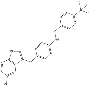
C20H15CLF3N5
|
|---|---|
| 分子量 |
417.81
|
| 精确质量 |
417.096
|
| 元素分析 |
C, 57.49; H, 3.62; Cl, 8.49; F, 13.64; N, 16.76
|
| CAS号 |
1029044-16-3
|
| 相关CAS号 |
Pexidartinib hydrochloride;2040295-03-0
|
| PubChem CID |
25151352
|
| 外观&性状 |
Yellow solid powder
|
| 密度 |
1.5±0.1 g/cm3
|
| 沸点 |
580.0±50.0 °C at 760 mmHg
|
| 闪点 |
304.6±30.1 °C
|
| 蒸汽压 |
0.0±1.6 mmHg at 25°C
|
| 折射率 |
1.662
|
| LogP |
4.77
|
| tPSA |
66.49
|
| 氢键供体(HBD)数目 |
2
|
| 氢键受体(HBA)数目 |
7
|
| 可旋转键数目(RBC) |
5
|
| 重原子数目 |
29
|
| 分子复杂度/Complexity |
537
|
| 定义原子立体中心数目 |
0
|
| SMILES |
ClC1C([H])=NC2=C(C=1[H])C(=C([H])N2[H])C([H])([H])C1=C([H])N=C(C([H])=C1[H])N([H])C([H])([H])C1=C([H])N=C(C(F)(F)F)C([H])=C1[H]
|
| InChi Key |
JGWRKYUXBBNENE-UHFFFAOYSA-N
|
| InChi Code |
InChI=1S/C20H15ClF3N5/c21-15-6-16-14(10-28-19(16)29-11-15)5-12-2-4-18(26-7-12)27-9-13-1-3-17(25-8-13)20(22,23)24/h1-4,6-8,10-11H,5,9H2,(H,26,27)(H,28,29)
|
| 化学名 |
5-[(5-chloro-1H-pyrrolo[2,3-b]pyridin-3-yl)methyl]-N-[[6-(trifluoromethyl)pyridin-3-yl]methyl]pyridin-2-amine
|
| 别名 |
Pexidartinib; CML-261; FP-113; PLX3397; PLX 3397; CML 261; CML261; PLX-3397;FP 113; FP113; Pexidartinib (PLX3397); CML-261; Pexidartinib [INN]; trade name: Turalio
|
| HS Tariff Code |
2934.99.9001
|
| 存储方式 |
Powder -20°C 3 years 4°C 2 years In solvent -80°C 6 months -20°C 1 month |
| 运输条件 |
Room temperature (This product is stable at ambient temperature for a few days during ordinary shipping and time spent in Customs)
|
| 溶解度 (体外实验) |
|
|||
|---|---|---|---|---|
| 溶解度 (体内实验) |
配方 1 中的溶解度: 5 mg/mL (11.97 mM) in 5% DMSO + 40% PEG300 + 5% Tween80 + 50% Saline (这些助溶剂从左到右依次添加,逐一添加), 悬浮液;超声助溶。
*生理盐水的制备:将 0.9 g 氯化钠溶解在 100 mL ddH₂O中,得到澄清溶液。 配方 2 中的溶解度: ≥ 2.08 mg/mL (4.98 mM) (饱和度未知) in 10% DMSO + 40% PEG300 + 5% Tween80 + 45% Saline (这些助溶剂从左到右依次添加,逐一添加), 澄清溶液。 例如,若需制备1 mL的工作液,可将 100 μL 20.8 mg/mL澄清的DMSO储备液加入到400 μL PEG300中,混匀;再向上述溶液中加入50 μL Tween-80,混匀;然后加入450 μL生理盐水定容至1 mL。 *生理盐水的制备:将 0.9 g 氯化钠溶解在 100 mL ddH₂O中,得到澄清溶液。 View More
配方 3 中的溶解度: ≥ 2.08 mg/mL (4.98 mM) (饱和度未知) in 10% DMSO + 90% (20% SBE-β-CD in Saline) (这些助溶剂从左到右依次添加,逐一添加), 澄清溶液。 配方 4 中的溶解度: ≥ 2.08 mg/mL (4.98 mM) (饱和度未知) in 10% DMSO + 90% Corn Oil (这些助溶剂从左到右依次添加,逐一添加), 澄清溶液。 例如,若需制备1 mL的工作液,您可以将 100 μL 20.8 mg/mL 澄清 DMSO 储备液加入到 900 μL 玉米油中并混合均匀。 配方 5 中的溶解度: 10% DMSO+40% PEG 300+ddH2O:15 mg/mL 1、请先配制澄清的储备液(如:用DMSO配置50 或 100 mg/mL母液(储备液)); 2、取适量母液,按从左到右的顺序依次添加助溶剂,澄清后再加入下一助溶剂。以 下列配方为例说明 (注意此配方只用于说明,并不一定代表此产品 的实际溶解配方): 10% DMSO → 40% PEG300 → 5% Tween-80 → 45% ddH2O (或 saline); 假设最终工作液的体积为 1 mL, 浓度为5 mg/mL: 取 100 μL 50 mg/mL 的澄清 DMSO 储备液加到 400 μL PEG300 中,混合均匀/澄清;向上述体系中加入50 μL Tween-80,混合均匀/澄清;然后继续加入450 μL ddH2O (或 saline)定容至 1 mL; 3、溶剂前显示的百分比是指该溶剂在最终溶液/工作液中的体积所占比例; 4、 如产品在配制过程中出现沉淀/析出,可通过加热(≤50℃)或超声的方式助溶; 5、为保证最佳实验结果,工作液请现配现用! 6、如不确定怎么将母液配置成体内动物实验的工作液,请查看说明书或联系我们; 7、 以上所有助溶剂都可在 Invivochem.cn网站购买。 |
| 制备储备液 | 1 mg | 5 mg | 10 mg | |
| 1 mM | 2.3934 mL | 11.9672 mL | 23.9343 mL | |
| 5 mM | 0.4787 mL | 2.3934 mL | 4.7869 mL | |
| 10 mM | 0.2393 mL | 1.1967 mL | 2.3934 mL |
1、根据实验需要选择合适的溶剂配制储备液 (母液):对于大多数产品,InvivoChem推荐用DMSO配置母液 (比如:5、10、20mM或者10、20、50 mg/mL浓度),个别水溶性高的产品可直接溶于水。产品在DMSO 、水或其他溶剂中的具体溶解度详见上”溶解度 (体外)”部分;
2、如果您找不到您想要的溶解度信息,或者很难将产品溶解在溶液中,请联系我们;
3、建议使用下列计算器进行相关计算(摩尔浓度计算器、稀释计算器、分子量计算器、重组计算器等);
4、母液配好之后,将其分装到常规用量,并储存在-20°C或-80°C,尽量减少反复冻融循环。
计算结果:
工作液浓度: mg/mL;
DMSO母液配制方法: mg 药物溶于 μL DMSO溶液(母液浓度 mg/mL)。如该浓度超过该批次药物DMSO溶解度,请首先与我们联系。
体内配方配制方法:取 μL DMSO母液,加入 μL PEG300,混匀澄清后加入μL Tween 80,混匀澄清后加入 μL ddH2O,混匀澄清。
(1) 请确保溶液澄清之后,再加入下一种溶剂 (助溶剂) 。可利用涡旋、超声或水浴加热等方法助溶;
(2) 一定要按顺序加入溶剂 (助溶剂) 。
| NCT Number | Recruitment | interventions | Conditions | Sponsor/Collaborators | Start Date | Phases |
| NCT02975700 | Active Recruiting |
Drug: PLX3397 | Melanoma | Daiichi Sankyo Co., Ltd. | January 2017 | Not Applicable |
| NCT04488822 | Active Recruiting |
Drug: Pexidartinib | Tenosynovial Giant Cell Tumor | Daiichi Sankyo Co., Ltd. | September 25, 2020 | Phase 3 |
| NCT04703322 | Recruiting | Drug: Pexidartinib | Tenosynovial Giant Cell Tumor | Daiichi Sankyo Co., Ltd. | March 15, 2021 | Phase 2 |
| NCT04635111 | Recruiting | Drug: TURALIO™ | Hepatotoxicity Tenosynovial Giant Cell Tumor |
Daiichi Sankyo, Inc. | January 7, 2021 | |
| NCT02390752 | Recruiting | Drug: Turalio | Sarcoma Neurofibroma, Plexiform |
National Cancer Institute (NCI) |
April 29, 2015 | Phase 1 |
Combined PLX3397 and PTX treatment inhibits metastasis in a CD8-dependent manner. Cancer Discov. 2011 Jun 1; 1: 54–67. |
PTX in combination with PLX3397 induces antitumor T-cell response. Cancer Discov. 2011 Jun 1; 1: 54–67. |
CD68/CD4/CD8 immune signature is an independent prognostic indicator of breast cancer survival. Cancer Discov. 2011 Jun;1(1):54-67. |
 NN-2101
 VEGFR2-IN-84
 Protein kinase inhibitor 10
 Opelkibart
InvivoChem的所有产品仅用于作科学研究,不面向患者销售
Copyright 2020 InvivoChem LLC | All Rights Reserved 粤ICP备20063088号-1


 COA
COA
")
")
")
 463611831
