| 规格 | 价格 | 库存 | 数量 |
|---|---|---|---|
| 10 mM * 1 mL in DMSO |
|
||
| 5mg |
|
||
| 10mg |
|
||
| 25mg |
|
||
| 50mg |
|
||
| 100mg |
|
||
| 250mg |
|
||
| 500mg |
|
||
| 1g |
|
||
| Other Sizes |
|

| 靶点 |
MDM-2/p53; HSP70
The primary target of Pifithrin-μ (NSC-303580) is the mitochondrial interaction interface of p53, specifically inhibiting the binding of p53 to anti-apoptotic Bcl-2 family proteins (Bcl-xL, Bcl-2). - Inhibition of p53-Bcl-xL binding: IC50 = 1.2 μM (fluorescence polarization assay, FP) [1] ; - Inhibition of p53-Bcl-2 binding: IC50 = 2.5 μM (same FP assay as p53-Bcl-xL) [1] ; |
|---|---|
| 体外研究 (In Vitro) |
Pifithrin-μ 干扰 p53 与线粒体的结合,并抑制小鼠胸腺细胞原代细胞培养物响应伽玛辐射的快速 p53 依赖性细胞凋亡。 [1]在急性淋巴细胞白血病 (ALL) 和急性髓系白血病 (AML) 细胞系以及原发性 AML 母细胞中,Pifithrin-μ(一种诱导型 HSP70 抑制剂)可显着降低细胞活力。 IC50 值范围为 2.5 至 12.7 μM。 Pifithrin-μ 在原发性 AML 母细胞中以剂量依赖性方式诱导细胞凋亡和细胞周期停滞,中位 IC50 为 8.9 μM(范围 5.7-37.2 μM)。此外,Pifithrin-μ 会降低 NALM-6 细胞中的 AKT 和 ERK1/2,同时增加 caspase-3 的活性形式。 [2] Pifithrin-μ 促进 TRAIL 诱导的细胞凋亡并阻止癌细胞的生长。它还以半胱天冬酶依赖性和非半胱天冬酶依赖性方式增加膜联蛋白 V(+) 细胞的数量。 [3]
抑制p53介导的线粒体凋亡(文献[1]):1)p53野生型HCT116细胞用Pifithrin-μ(1 μM、2 μM)+依托泊苷(10 μM,p53激活剂)处理24小时:线粒体细胞色素c释放较单独依托泊苷组减少42%(2 μM剂量);2)凋亡率(Annexin V-FITC/PI)从依托泊苷组的58%降至2 μM Pifithrin-μ+依托泊苷组的22%;3)不影响p53核靶基因(p21 mRNA水平无变化,RT-PCR),证实选择性抑制p53线粒体功能。[1] - 白血病细胞抗增殖活性(文献[2]):1)p53野生型K562(慢性髓系白血病)细胞:Pifithrin-μ IC50=3.8 μM(MTT法,72小时);2)p53突变型HL-60细胞:IC50 > 20 μM;3)Western blot显示Cleaved Caspase-9(线粒体凋亡标志物)较对照组减少65%(3 μM Pifithrin-μ)。[2] - 与化疗药在结直肠癌细胞中的协同作用(文献[3]):1)HT29(p53野生型)细胞用Pifithrin-μ(1 μM)+5-氟尿嘧啶(5-FU,5 μM)处理:细胞活力降至28%(单独5-FU组为52%,单独Pifithrin-μ组为78%);2)克隆形成率较单独5-FU组(35%)下降72%(联用组);3)免疫荧光显示联用组p53线粒体定位减少(阳性细胞从65%降至18%)。[3] |
| 体内研究 (In Vivo) |
Pifithrin-μ(40 mg/kg,腹腔注射)可保护暴露于 8 或 9 Gy 全身伽玛辐射的 C57B1/6J 小鼠免受 p53 依赖性细胞凋亡。 [1] Pifithrin-μ 显着提高了 TRAIL 在异种移植小鼠模型中抑制 MiaPaca-2 肿瘤生长的能力。 [3]
白血病异种移植模型(文献[2]):携带K562异种移植瘤的NOD/SCID小鼠(6-8周龄,雄性)随机分为3组(每组6只):1)溶媒对照组(5% DMSO + 95%玉米油,灌胃,每日1次);2)Pifithrin-μ 10 mg/kg组(灌胃,每日1次);3)Pifithrin-μ 20 mg/kg组(灌胃,每日1次)。给药21天后:1)肿瘤生长抑制率(TGI)=45%(10 mg/kg)和68%(20 mg/kg);2)外周血白细胞计数较对照组减少38%(20 mg/kg组);3)骨髓免疫组化(IHC)显示Cleaved Caspase-9阳性细胞减少(20 mg/kg组从42%降至15%)。[2] - 结直肠癌异种移植模型(文献[3]):携带HT29异种移植瘤的BALB/c裸鼠(雌性,4-6周龄)随机分为4组(每组5只):1)对照组(溶媒);2)Pifithrin-μ 15 mg/kg组(腹腔注射,每日1次);3)5-FU 20 mg/kg组(腹腔注射,每周1次);4)联用组(Pifithrin-μ+5-FU)。给药28天后:1)TGI=79%(联用组)vs 32%(Pifithrin-μ单药组)和45%(5-FU单药组);2)肿瘤重量:联用组0.28 g vs 对照组0.85 g;3)未观察到小鼠存活时间显著延长,但联用组肿瘤复发延迟10天。[3] |
| 酶活实验 |
p53-Bcl-xL/Bcl-2结合荧光偏振(FP)实验(文献[1]):1)试剂制备:将荧光素标记的p53肽(1-25位氨基酸,含Bcl-xL/Bcl-2结合基序)在实验缓冲液(20 mM Tris-HCl,150 mM NaCl,pH7.5)中稀释至20 nM;2)结合反应:将标记p53肽与重组Bcl-xL(50 nM)或Bcl-2(50 nM)混合,25℃孵育30分钟形成复合物;3)药物处理:将Pifithrin-μ系列稀释(0.1 μM至10 μM)加入复合物,继续孵育60分钟;4)检测:用酶标仪测定FP信号(激发485 nm,发射535 nm);5)数据分析:通过FP信号降低率拟合四参数逻辑模型计算IC50。[1]
|
| 细胞实验 |
通过用 0.5% 亚甲蓝染色并使用 Multiscan Ascent 酶标仪计算光密度,推断贴壁细胞的数量。使用 0.1% 台盼蓝 2 染色或 FACScan 分析膜联蛋白或碘化丙啶 (PI) 阳性细胞,评估原代胸腺细胞短期培养物悬浮液中的细胞活力。
线粒体细胞色素c释放实验(文献[1]):1)将HCT116细胞以5×10⁶个/皿接种于10 cm培养皿,用Pifithrin-μ(1 μM、2 μM)+依托泊苷(10 μM)处理24小时;2)收集细胞,冷PBS洗涤,重悬于线粒体分离缓冲液;3)通过离心(12,000×g,15分钟,4℃)分离线粒体和胞质组分;4)用抗细胞色素c抗体通过Western blot检测胞质组分中的细胞色素c,ImageJ定量灰度值(以β-actin归一化)。[1] - 白血病细胞活力实验(文献[2]):1)将K562/HL-60细胞以4×10³个/孔接种于96孔板,过夜培养;2)加入Pifithrin-μ(0.5 μM至40 μM),细胞在37℃、5% CO₂下培养72小时;3)每孔加入15 μL CCK-8溶液,孵育2小时;4)测定450 nm处吸光度;细胞活力=(处理组/对照组吸光度)×100%;通过GraphPad Prism计算IC50。[2] - 结直肠癌克隆形成实验(文献[3]):1)将HT29细胞以800个/孔接种于6孔板,用Pifithrin-μ(1 μM)+5-FU(5 μM)处理;2)每4天换液1次,培养14天;3)克隆用4%多聚甲醛固定15分钟,0.1%结晶紫染色30分钟;4)计数细胞数>50的克隆;克隆形成率=(处理组克隆数/对照组克隆数)×100%。[3] |
| 动物实验 |
40 mg/kg DMSO
C57B1/6J 小鼠。 K562 白血病异种移植方案(参考文献[2]):1)动物准备:雄性 NOD/SCID 小鼠(6-8 周龄)适应环境 1 周(22±2°C,12 小时光照/黑暗);2)肿瘤接种:将 2×10⁷ 个 K562 细胞悬浮于 200 μL PBS 中,经尾静脉注射至每只小鼠体内;3)分组和给药:当外周血白细胞计数达到 1×10⁹/L 时(接种后第 7 天),将小鼠随机分为 3 组(n=6):对照组(5% DMSO + 95% 玉米油,灌胃,0.2 mL/只,每日一次); Pifithrin-μ 10 mg/kg(溶于对照溶剂,灌胃,每日一次);Pifithrin-μ 20 mg/kg(相同溶剂/给药途径);4)监测:每3天采集外周血进行白细胞计数;21天后,处死小鼠,采集骨髓进行免疫组化染色,并称量脾脏/肝脏重量(以评估肿瘤浸润)。[2] - HT29结肠癌异种移植方案(文献[3]):1)动物准备:雌性BALB/c裸鼠(4-6周龄)适应环境1周;2)肿瘤接种:将5×10⁶个HT29细胞悬浮于100 μL Matrigel:PBS(1:1)混合液中,皮下注射至小鼠右侧腹部; 3) 分组和给药:当肿瘤体积达到 100-150 mm³(接种后第 10 天)时,将小鼠随机分为 4 组(n=5):对照组(5% DMSO + 10% Cremophor EL + 85% 生理盐水,腹腔注射,0.1 mL/只,每日一次);Pifithrin-μ 组(15 mg/kg,溶于对照溶剂,腹腔注射,每日一次);5-FU 组(20 mg/kg,溶于生理盐水,腹腔注射,每周一次);联合用药组(每日注射 Pifithrin-μ + 每周注射 5-FU)。4) 监测:每 3 天测量一次肿瘤体积(长 × 宽² / 2)和体重;28 天后,处死小鼠,取出肿瘤并称重,用 4% 多聚甲醛固定,用于免疫组化染色。[3] |
| 毒性/毒理 (Toxicokinetics/TK) |
在异种移植模型中的体内安全性(文献[2][3]):1)NOD/SCID 小鼠(文献[2]):Pifithrin-μ 20 mg/kg(口服,21 天)导致 6% 的体重减轻(第 18 天恢复),且血清 ALT/AST/BUN/Cr 未见异常(与对照组相比);2)BALB/c 裸鼠(文献[3]):Pifithrin-μ 15 mg/kg(腹腔注射,28 天)+ 5-FU 20 mg/kg(腹腔注射,每周一次)导致 9% 的体重减轻(与单独使用 5-FU 相比为 12%),且肝脏/肾脏/脾脏未见组织病理学损伤。 [2][3]
- 体外对正常细胞的细胞毒性(文献[1]):Pifithrin-μ对正常人结肠上皮细胞(NCM460)的毒性较低:IC50 = 22.5 μM(K562细胞为3.8 μM),表明其对癌细胞具有选择性。[1] |
| 参考文献 | |
| 其他信息 |
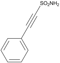
2-苯乙炔磺酰胺属于苯类化合物。
独特的作用机制(文献[1]):与其他p53抑制剂(例如,抑制p53转录活性的Pifithrin-α)不同,Pifithrin-μ通过破坏p53-Bcl-2/Bcl-xL相互作用,选择性地阻断p53的线粒体促凋亡功能。这避免了干扰p53的核肿瘤抑制活性(例如,DNA修复),从而降低了潜在的致癌风险。[1] -治疗潜力(文献[2][3]):Pifithrin-μ有望用于治疗p53野生型血液系统恶性肿瘤(例如,慢性粒细胞白血病)和实体瘤(例如,结肠癌),尤其是在与化疗药物(例如,5-氟尿嘧啶)联合使用时,可减少化疗诱导的正常细胞凋亡。 [2][3] |
| 分子式 |
C8H7NO2S
|
|
|---|---|---|
| 分子量 |
181.21
|
|
| 精确质量 |
181.019
|
|
| 元素分析 |
C, 53.03; H, 3.89; N, 7.73; O, 17.66; S, 17.69
|
|
| CAS号 |
64984-31-2
|
|
| 相关CAS号 |
|
|
| PubChem CID |
327653
|
|
| 外观&性状 |
White to light brown solid powder
|
|
| 密度 |
1.4±0.1 g/cm3
|
|
| 沸点 |
351.7±25.0 °C at 760 mmHg
|
|
| 熔点 |
135.0 to 139.0 °C
|
|
| 闪点 |
166.5±23.2 °C
|
|
| 蒸汽压 |
0.0±0.8 mmHg at 25°C
|
|
| 折射率 |
1.634
|
|
| LogP |
2
|
|
| tPSA |
68.54
|
|
| 氢键供体(HBD)数目 |
1
|
|
| 氢键受体(HBA)数目 |
3
|
|
| 可旋转键数目(RBC) |
1
|
|
| 重原子数目 |
12
|
|
| 分子复杂度/Complexity |
295
|
|
| 定义原子立体中心数目 |
0
|
|
| SMILES |
S(C#CC1C([H])=C([H])C([H])=C([H])C=1[H])(N([H])[H])(=O)=O
|
|
| InChi Key |
ZZUZYEMRHCMVTB-UHFFFAOYSA-N
|
|
| InChi Code |
InChI=1S/C8H7NO2S/c9-12(10,11)7-6-8-4-2-1-3-5-8/h1-5H,(H2,9,10,11)
|
|
| 化学名 |
2-phenylethynesulfonamide
|
|
| 别名 |
|
|
| HS Tariff Code |
2934.99.03.00
|
|
| 存储方式 |
Powder -20°C 3 years 4°C 2 years In solvent -80°C 6 months -20°C 1 month 注意: 请将本产品存放在密封且受保护的环境中(例如氮气保护),避免吸湿/受潮。 |
|
| 运输条件 |
Room temperature (This product is stable at ambient temperature for a few days during ordinary shipping and time spent in Customs)
|
| 溶解度 (体外实验) |
DMSO: ~36 mg/mL (~198.7 mM)
Water: <1 mg/mL (slightly soluble or insoluble) Ethanol: N/A |
|---|---|
| 溶解度 (体内实验) |
配方 1 中的溶解度: ≥ 2.08 mg/mL (11.48 mM) (饱和度未知) in 10% DMSO + 40% PEG300 + 5% Tween80 + 45% Saline (这些助溶剂从左到右依次添加,逐一添加), 澄清溶液。
例如,若需制备1 mL的工作液,可将100 μL 20.8 mg/mL澄清DMSO储备液加入400 μL PEG300中,混匀;然后向上述溶液中加入50 μL Tween-80,混匀;加入450 μL生理盐水定容至1 mL。 *生理盐水的制备:将 0.9 g 氯化钠溶解在 100 mL ddH₂O中,得到澄清溶液。 配方 2 中的溶解度: ≥ 2.08 mg/mL (11.48 mM) (饱和度未知) in 10% DMSO + 90% (20% SBE-β-CD in Saline) (这些助溶剂从左到右依次添加,逐一添加), 澄清溶液。 例如,若需制备1 mL的工作液,可将 100 μL 20.8 mg/mL澄清DMSO储备液加入900 μL 20% SBE-β-CD生理盐水溶液中,混匀。 *20% SBE-β-CD 生理盐水溶液的制备(4°C,1 周):将 2 g SBE-β-CD 溶解于 10 mL 生理盐水中,得到澄清溶液。 View More
配方 3 中的溶解度: ≥ 2.08 mg/mL (11.48 mM) (饱和度未知) in 10% DMSO + 90% Corn Oil (这些助溶剂从左到右依次添加,逐一添加), 澄清溶液。 配方 4 中的溶解度: 1% DMSO +30% polyethylene glycol+1% Tween 80 : 10 mg/mL 1、请先配制澄清的储备液(如:用DMSO配置50 或 100 mg/mL母液(储备液)); 2、取适量母液,按从左到右的顺序依次添加助溶剂,澄清后再加入下一助溶剂。以 下列配方为例说明 (注意此配方只用于说明,并不一定代表此产品 的实际溶解配方): 10% DMSO → 40% PEG300 → 5% Tween-80 → 45% ddH2O (或 saline); 假设最终工作液的体积为 1 mL, 浓度为5 mg/mL: 取 100 μL 50 mg/mL 的澄清 DMSO 储备液加到 400 μL PEG300 中,混合均匀/澄清;向上述体系中加入50 μL Tween-80,混合均匀/澄清;然后继续加入450 μL ddH2O (或 saline)定容至 1 mL; 3、溶剂前显示的百分比是指该溶剂在最终溶液/工作液中的体积所占比例; 4、 如产品在配制过程中出现沉淀/析出,可通过加热(≤50℃)或超声的方式助溶; 5、为保证最佳实验结果,工作液请现配现用! 6、如不确定怎么将母液配置成体内动物实验的工作液,请查看说明书或联系我们; 7、 以上所有助溶剂都可在 Invivochem.cn网站购买。 |
| 制备储备液 | 1 mg | 5 mg | 10 mg | |
| 1 mM | 5.5185 mL | 27.5923 mL | 55.1846 mL | |
| 5 mM | 1.1037 mL | 5.5185 mL | 11.0369 mL | |
| 10 mM | 0.5518 mL | 2.7592 mL | 5.5185 mL |
1、根据实验需要选择合适的溶剂配制储备液 (母液):对于大多数产品,InvivoChem推荐用DMSO配置母液 (比如:5、10、20mM或者10、20、50 mg/mL浓度),个别水溶性高的产品可直接溶于水。产品在DMSO 、水或其他溶剂中的具体溶解度详见上”溶解度 (体外)”部分;
2、如果您找不到您想要的溶解度信息,或者很难将产品溶解在溶液中,请联系我们;
3、建议使用下列计算器进行相关计算(摩尔浓度计算器、稀释计算器、分子量计算器、重组计算器等);
4、母液配好之后,将其分装到常规用量,并储存在-20°C或-80°C,尽量减少反复冻融循环。
计算结果:
工作液浓度: mg/mL;
DMSO母液配制方法: mg 药物溶于 μL DMSO溶液(母液浓度 mg/mL)。如该浓度超过该批次药物DMSO溶解度,请首先与我们联系。
体内配方配制方法:取 μL DMSO母液,加入 μL PEG300,混匀澄清后加入μL Tween 80,混匀澄清后加入 μL ddH2O,混匀澄清。
(1) 请确保溶液澄清之后,再加入下一种溶剂 (助溶剂) 。可利用涡旋、超声或水浴加热等方法助溶;
(2) 一定要按顺序加入溶剂 (助溶剂) 。
 |
|---|
|
InvivoChem的所有产品仅用于作科学研究,不面向患者销售
Copyright 2020 InvivoChem LLC | All Rights Reserved 粤ICP备20063088号-1


 COA
COA

 463611831
