| 规格 | 价格 | 库存 | 数量 |
|---|---|---|---|
| 10mg |
|
||
| 25mg |
|
||
| 50mg |
|
||
| 100mg |
|
||
| 250mg |
|
||
| 500mg |
|
||
| 1g |
|
||
| Other Sizes |
|
| 靶点 |
125 I-CXCL12-CXCR4 ( IC50 = 44 nM ); 125 I-CXCL12-CXCR7; HIV-1 ( EC50 = 1-10 nM ); HIV-2 ( IC50 = 1-10 nM )
CXCR4 receptor (Ki = 4.1 nM, human; IC50 = 7.5 nM for CXCL12 binding inhibition) [1] - CXCR7 receptor (Ki = 35 nM, human; weak agonist activity) [1] - No significant affinity for CXCR1/CXCR2/CXCR3 or CCR5 receptors (Ki > 1000 nM) [1][2] |
|---|---|
| 体外研究 (In Vitro) |
Plerixafor 抑制 CXCL12 介导的趋化作用,其效力略优于其对 CXCR4 的亲和力。 Plerixafor 还拮抗 SDF-1/CXCL12 配体结合,IC50 为 651 nM。 Plerixafor 抑制 SDF-1 介导的 GTP 结合、SDF-1 介导的钙流和 SDF-1 刺激的趋化性,IC50 分别为 27 nM、572 nM 和 51 nM。当用同源配体刺激时,Plerixafor 不会抑制针对表达 CXCR3、CCR1、CCR2b、CCR4、CCR5 或 CCR7 的细胞的钙流,Plerixafor 也不抑制 LTB4 的受体结合。 Plerixafor 本身不会诱导 CCRF-CEM 细胞中的钙流,该细胞表达多种 GPCR,包括 CXCR4、CCR4 和 CCR7。激酶测定:对于针对 CXCR4 的竞争性结合研究,将一定浓度范围的 Plerixafor 在结合缓冲液(含有 5 mM MgCl2、1 mM CaCl2、0.25% BSA,pH 7.4 的 PBS)中与 5 × 10⁵ 一起在 4°C 下孵育 3 小时CCRF-CEM 细胞和 Milipore DuraporeTM 过滤板中的 100 pM 125I-SDF-1α (2200 Ci/mmol)。用冷 50 mM HEPES、0.5 M NaCl pH 7.4 洗涤去除未结合的 125I-SDF-1α。在表达重组 BLT1 的 CHO-S 细胞膜上进行针对 BLT1 的竞争结合测定。通过机械细胞裂解制备膜,然后高速离心,重悬于 50 mm HEPES、5 mM MgCl2 缓冲液中并快速冷冻。将膜制剂与 Plerixafor 在含有 50 mM Tris、pH 7.4、10 mM MgCl2、10 mM CaCl2、4 nM LTB4 与 1 nM 3H-LTB4 (195.0 Ci/mmol) 混合的测定混合物中在室温下孵育 1 小时, 8μg膜。通过在 Millipore GF-C 型过滤板上过滤分离未结合的 3H-LTB4。细胞检测:CXCR4和SDF-1是调节癌细胞侵袭和转移的关键因子,Plerixafor有效阻止SDF-1与CXCR4的结合,抑制癌症转移。
Plerixafor (AMD 3100)(普乐沙福)是选择性小分子CXCR4拮抗剂,对CXCR7有弱结合力,与其他趋化因子受体无交叉反应[1][2] - 在人胶质母细胞瘤(U87)细胞中,Plerixafor(1-100 nM)剂量依赖性阻断CXCL12诱导的跨内皮迁移60-85%,抑制下游ERK1/2磷酸化,且不影响细胞活力[1] - 在人黑色素瘤(A375)细胞中,Plerixafor(0.1-10 μM)通过抑制PI3K/Akt信号通路,减少CXCL12介导的细胞增殖30-50%,下调基质金属蛋白酶-9(MMP-9)表达[2] - 在原代人角质形成细胞中,Plerixafor(1-5 μM)减弱TNF-α诱导的IL-8和CXCL10生成40-60%,抑制趋化因子介导的炎症细胞募集[3] - 在小鼠成骨细胞(MC3T3-E1)中,Plerixafor(0.5-10 μM)促进成骨细胞分化,使碱性磷酸酶(ALP)活性增加2.1-3.3倍,矿化结节形成增加55-70%[4] |
| 体内研究 (In Vivo) |
连续五天给小鼠群组施用 PBS、IGF1、PDGF、SCF 或 VEGF,并在第 5 天施用 Plerixafor。与注射 PDGF、SCF 或 VEGF 加 Plerixafor 治疗的小鼠相比,注射 IGF1 和 Plerixafor 的小鼠集落的数量和大小最高。
在携带U87胶质母细胞瘤异种移植瘤的裸鼠中,腹腔注射Plerixafor(5 mg/kg/天,连续14天)抑制肿瘤血管生成38%,减少肺转移45%[1] - 在接触性超敏反应(CHS)小鼠模型中,Plerixafor(1 mg/kg,腹腔注射,每日一次,连续5天)减少耳肿胀50%,降低表皮厚度,同时减少炎症细胞浸润[3] - 在去卵巢(OVX)骨质疏松小鼠模型中,Plerixafor(2 mg/kg,皮下注射,每周两次,连续8周)使股骨骨密度(BMD)增加18%,骨小梁数量增加25%,改善骨微结构[4] - 在黑色素瘤肺转移小鼠中,Plerixafor(3 mg/kg,静脉注射,每周一次,连续4周)与对照组相比,转移结节数量减少60%[2] |
| 酶活实验 |
对于针对 CXCR4 的竞争性结合研究,将 5 × 10 5 CCRF-CEM 细胞和 100 pM 125I-SDF-1α (2200 Ci/mmol) 在结合缓冲液中于 4 °C 下孵育三小时( Milipore DuraporeTM 过滤板中含有 5 mM MgCl2、1 mM Ca Cl2、0.25% BSA、pH 7.4 的 PBS。用冷 50 mM HEPES 和 0.5 M NaCl pH 7.4 洗涤后,未结合的 125 I-SDF-1α 被消除。在表达重组 BLT1 的 CHO-S 细胞膜上进行竞争结合测定。膜制备涉及的步骤包括机械细胞裂解、高速离心、在含有 5 mM MgCl22 的 50 mm HEPES 缓冲液中重悬以及快速冷冻。检测混合物包含 50 mM Tris,pH 7.4、10 mM MgCl2、10 mM CaCl2、4 nM LTB4 以及 1 nM 3 H-LTB4 (195.0 Ci/mmol) 和 8 μg 膜与 Plerixafor 在室温下孵育一小时。过滤用于在 Millipore GF-C 型滤板上分离未结合的 3 H-LTB4。
CXCR4/CXCR7受体结合实验:制备表达人CXCR4/CXCR7的CHO细胞膜制剂,与[125I]-CXCL12(0.1 nM)及不同浓度的Plerixafor(0.01-1000 nM)在25°C孵育60分钟。在过量未标记CXCL12存在下测定非特异性结合,过滤分离结合态配体,定量放射性强度以计算Ki值[1] - ERK1/2磷酸化实验:U87细胞饥饿12小时后,经Plerixafor(0.1-100 nM)预处理20分钟,再用CXCL12(10 nM)刺激10分钟。Western blot分析细胞裂解物,定量磷酸化ERK1/2与总ERK1/2的比值[1] - PI3K/Akt活性实验:A375细胞经Plerixafor(0.1-10 μM)预处理30分钟后,用CXCL12(10 nM)刺激15分钟。通过免疫沉淀偶联激酶实验,使用特异性底物测定PI3K和Akt的激酶活性[2] |
| 细胞实验 |
将 Peptide R、Plerixafor 或 CXCL12 以 6 ×10 3 细胞密度、200 μL/孔接种到 96 孔板中后,将其应用于 U87MG 细胞。在治疗的最后两个小时内,在 24、48 和 72 小时添加 MTT (5 μg/mL)。除去细胞培养基后,添加 100 μL DMSO,并使用 LT-4000MS 酶标仪测量 595 nm 处的光密度。三个独立实验的测量值一式三份进行。
肿瘤细胞跨内皮迁移实验:人脐静脉内皮细胞(HUVECs)在Transwell小室上培养至融合。经Plerixafor(1-100 nM)预处理30分钟的U87/A375细胞加入上室,下室加入CXCL12(10 nM)。24小时后固定、染色并计数迁移细胞[1][2] - 角质形成细胞炎症实验:原代人角质形成细胞接种于24孔板,经Plerixafor(1-5 μM)预处理1小时后,用TNF-α(10 ng/mL)刺激24小时。ELISA法定量上清液中IL-8和CXCL10水平[3] - 成骨细胞分化实验:MC3T3-E1细胞接种于6孔板,经Plerixafor(0.5-10 μM)处理14-21天。分光光度法测定ALP活性,茜素红S染色并定量矿化结节[4] |
| 动物实验 |
小鼠:实验所用小鼠为6-7周龄、体重20克的雄性C57BL/6小鼠。在22℃、12小时光照/12小时黑暗循环条件下饲养一周后,将小鼠转移至SPF级动物房进行适应性饲养。随后,将小鼠随机分为三组,每组8只:正常组(不进行特殊处理)、UUO+AMD3100组(接受UUO手术并腹腔注射2 mg/kg AMD3100)和UUO+PBS组(接受UUO手术并腹腔注射等量PBS)。每日腹腔注射AMD3100和PBS,直至处死。
大鼠:采用2型糖尿病沙鼠模型,每日腹腔注射溶于水中的CXCR4拮抗剂AMD3100,剂量为6 mg/kg,持续8周。本研究通过补充实验探讨了AMD3100(6 mg/kg/d)CXCR4拮抗作用对调节性T细胞数量的影响。为进行这些研究,AMD3100或载体通过微型泵给药一周。 胶质母细胞瘤异种移植模型:将U87细胞(2×10⁶个细胞/只)皮下接种到雌性裸鼠(18-22 g)体内。当肿瘤体积达到100 mm³时,将溶于生理盐水的普乐沙福以5 mg/kg/天的剂量腹腔注射,持续14天。采用免疫组织化学和组织学方法评估肿瘤血管生成和肺转移[1] - 接触性超敏反应(CHS)模型:雄性BALB/c小鼠(20-25 g)腹部注射2,4-二硝基氟苯(DNFB)致敏,5天后耳部进行激发。激发后,每日腹腔注射溶于生理盐水的普乐沙福(1 mg/kg),连续5天。测量耳肿胀和炎症细胞浸润情况[3] - 骨质疏松(OVX)模型:雌性C57BL/6小鼠(25-30 g)行卵巢切除术。术后两周,每周两次皮下注射溶于生理盐水的普乐沙福(2 mg/kg),连续8周。采用微型CT分析股骨骨密度和骨微结构[4] - 黑色素瘤转移模型:将A375黑色素瘤细胞(1×10⁶个细胞/只小鼠)静脉注射到C57BL/6小鼠(20-25 g)体内。将溶于生理盐水的普乐沙福(3 mg/kg)每周静脉注射一次,持续4周。处死小鼠后计数肺转移结节[2] |
| 药代性质 (ADME/PK) |
吸收、分布和排泄
普乐沙福的药代动力学特征符合双室模型,吸收为一级动力学,在0.04 mg/kg至0.24 mg/kg剂量范围内呈线性动力学。健康受试者的普乐沙福药代动力学特征与接受普乐沙福联合粒细胞集落刺激因子(G-CSF)治疗的非霍奇金淋巴瘤(NHL)和多发性骨髓瘤(MM)患者的药代动力学特征相似。此外,普乐沙福的清除率与肌酐清除率(CLCR)显著相关。群体药代动力学分析表明,随着体重的增加,以mg/kg为单位的剂量会导致普乐沙福暴露量(AUC0-24h)增加。然而,体重<70 kg的非霍奇金淋巴瘤(NHL)患者接受20 mg固定剂量普乐沙福治疗后,其AUC0-10h值比接受0.24 mg/kg普乐沙福治疗的患者高1.43倍。因此,选择83 kg作为合适的体重临界值,以便将患者从固定剂量给药方案过渡到按体重给药方案。皮下注射后,药物浓度峰值(tmax)大约在30-60分钟内达到。在接受4天粒细胞集落刺激因子(G-CSF)预处理后,皮下注射0.24 mg/kg普乐沙福的患者,其Cmax和AUC0-24分别为887 ng/ml和4337 ng·hr/ml。普乐沙福主要经尿液排泄。在肾功能正常的健康志愿者中,给予0.24 mg/kg的普乐沙福后,约70%的原药在24小时内经尿液排出。一项使用MDCKII和MDCKII-MDR1细胞模型进行的体外研究发现,普乐沙福并非P-糖蛋白的底物或抑制剂。普乐沙福的表观分布容积为0.3 L/kg。普乐沙福的总血浆清除率为4.38 L/h,肾清除率为3.15 L/h。普乐沙福不经肝脏代谢,也不是主要细胞色素P450酶(包括1A2、2C9、2C19、2D6和3A4)的代谢依赖性抑制剂。此外,它不诱导细胞色素P450 1A2、2B6或3A4酶。普乐沙福代谢稳定,大鼠和犬的体内研究表明,血浆和尿液中非母体放射性标记成分是普乐沙福的Cu2+络合物。这与普乐沙福中存在两个环胺环相符,这两个环胺环可能作为潜在的螯合位点。 生物半衰期 在肾功能正常的患者中,普乐沙福的分布半衰期为0.3小时,终末群体半衰期为5.3小时。在健康受试者和患者的研究中,血浆中的终末半衰期在3至5小时之间。在非霍奇金淋巴瘤患者中,普乐沙福的终末半衰期为 4.4 小时;在多发性骨髓瘤患者中,终末半衰期为 5.6 小时。 口服生物利用度:在人和啮齿动物中 <5%(由于口服吸收差,需静脉或皮下注射给药)[2] - 血浆蛋白结合率:在人血浆中为 20-25%(浓度范围:0.1-10 μg/mL)[2] - 消除半衰期:在人中为 3-5 小时;在小鼠中为 2-3 小时[2] - 分布:在人体内分布容积 (Vd) = 0.2-0.3 L/kg,主要蓄积于骨髓、淋巴组织和肿瘤基质[2] - 排泄:70-80% 的剂量以原形经尿液排出; <10% 在肝脏中通过极少量的氧化代谢[2] |
| 毒性/毒理 (Toxicokinetics/TK) |
肝毒性
普乐沙福尚未被发现与治疗期间血清酶显著升高或临床上明显的肝损伤病例相关。在多项大型上市前和上市后对照试验中,ALT升高或急性肝损伤均未被提及为不良事件或导致患者退出、提前终止治疗或剂量调整的原因。目前尚无已发表的普乐沙福引起肝损伤的报告,并且普乐沙福已被用作急性肝衰竭动物模型中的潜在治疗手段。因此,普乐沙福引起的临床上明显的肝损伤即使存在,也必定十分罕见。 可能性评分:E(不太可能是临床上明显的肝损伤的原因)。 蛋白结合 普乐沙福的人血浆蛋白结合率高达58%。 急性毒性:小鼠静脉注射LD50 = 200 mg/kg;大鼠剂量为 150 mg/kg [2] - 亚慢性毒性(小鼠皮下注射 28 天):剂量高达 10 mg/kg/天时未见明显的肝毒性或肾毒性;20 mg/kg/天时出现轻度短暂性中性粒细胞减少症(≤10% 减少)[2][4] - 慢性毒性(卵巢切除小鼠皮下注射 8 周):每周两次 2 mg/kg 给药时,血清肌酐、BUN、ALT/AST 或电解质水平未见显著变化 [4] - 血浆蛋白结合率:20-25%(未观察到浓度依赖性结合)[2] - 临床前研究中,未发现与化疗药物或抗炎药存在显著的药物相互作用 [2][3] |
| 参考文献 | |
| 其他信息 |
药效学
普乐沙福是一种双环胺衍生物,它通过与配体结合口袋中的三个酸性残基(Asp171、Asp262 和 Glu288)结合,拮抗 CXC 趋化因子受体 4 (CXCR4)。在健康受试者中,给予 0.24 mg/kg 普乐沙福后,血液中 CD34+ 细胞水平在 6 至 9 小时达到峰值。与粒细胞集落刺激因子 (G-CSF) 联合用药时,外周血中循环 CD34+ 细胞水平在 10 至 14 小时达到峰值。单次剂量高达 0.40 mg/kg 的普乐沙福不会引起 QT/QTc 间期延长。接受普乐沙福治疗的患者曾出现严重的超敏反应,例如过敏性休克。使用普乐沙福还可能导致白血病患者肿瘤细胞动员、脾脏肿大和破裂、胚胎-胎儿毒性以及血液学效应,如白细胞增多症和血小板减少症。当与粒细胞集落刺激因子 (G-CSF) 联合用于造血干细胞动员时,普乐沙福可能导致肿瘤细胞从骨髓中释放,并随后被收集到白细胞分离术产品中。 普乐沙福 (AMD 3100) 是一种选择性 CXCR4 拮抗剂,最初开发用于抗肿瘤和抗炎应用,后来获准用于造血干细胞 (HSC) 动员 [1][2][4] - 其核心机制是阻断 CXCR4-CXCL12 (SDF-1α) 轴,抑制趋化因子介导的细胞迁移、增殖和炎症反应 [1][3] - 研究应用包括抑制肿瘤转移(胶质母细胞瘤、黑色素瘤)、减轻炎症性皮肤病(接触性超敏反应)和调节骨代谢(骨质疏松症)[1][3][4] - 它能增强卵巢切除术后的成骨细胞分化和骨形成。小鼠实验表明其具有治疗绝经后骨质疏松症的潜力[4] - 其对CXCR7的弱激动活性并不对其治疗效果有贡献,其治疗效果主要由CXCR4拮抗作用介导[1] - 已获临床批准,用于将造血干细胞从骨髓动员至外周血,以进行淋巴瘤或骨髓瘤患者的自体移植[2] |
| 分子式 |
C28H54N8
|
|
|---|---|---|
| 分子量 |
502.78
|
|
| 精确质量 |
502.447
|
|
| 元素分析 |
C, 66.89; H, 10.83; N, 22.29
|
|
| CAS号 |
110078-46-1
|
|
| 相关CAS号 |
Plerixafor octahydrochloride; 155148-31-5; Plerixafor-d4; 1246819-87-3
|
|
| PubChem CID |
65015
|
|
| 外观&性状 |
White to off-white solid powder
|
|
| 密度 |
1.0±0.1 g/cm3
|
|
| 沸点 |
657.5±55.0 °C at 760 mmHg
|
|
| 熔点 |
122-125°C
|
|
| 闪点 |
361.8±26.2 °C
|
|
| 蒸汽压 |
0.0±2.0 mmHg at 25°C
|
|
| 折射率 |
1.492
|
|
| LogP |
0.2
|
|
| tPSA |
78.66
|
|
| 氢键供体(HBD)数目 |
6
|
|
| 氢键受体(HBA)数目 |
8
|
|
| 可旋转键数目(RBC) |
4
|
|
| 重原子数目 |
36
|
|
| 分子复杂度/Complexity |
456
|
|
| 定义原子立体中心数目 |
0
|
|
| SMILES |
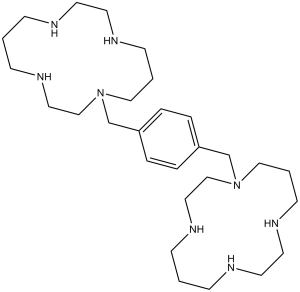
C1(CN2CCCNCCNCCCNCC2)=CC=C(C=C1)CN3CCNCCCNCCNCCC3
|
|
| InChi Key |
YIQPUIGJQJDJOS-UHFFFAOYSA-N
|
|
| InChi Code |
InChI=1S/C28H54N8/c1-9-29-15-17-31-13-3-21-35(23-19-33-11-1)25-27-5-7-28(8-6-27)26-36-22-4-14-32-18-16-30-10-2-12-34-20-24-36/h5-8,29-34H,1-4,9-26H2
|
|
| 化学名 |
1-[[4-(1,4,8,11-tetrazacyclotetradec-1-ylmethyl)phenyl]methyl]-1,4,8,11-tetrazacyclotetradecane
|
|
| 别名 |
|
|
| HS Tariff Code |
2934.99.9001
|
|
| 存储方式 |
Powder -20°C 3 years 4°C 2 years In solvent -80°C 6 months -20°C 1 month |
|
| 运输条件 |
Room temperature (This product is stable at ambient temperature for a few days during ordinary shipping and time spent in Customs)
|
| 溶解度 (体外实验) |
|
|||
|---|---|---|---|---|
| 溶解度 (体内实验) |
配方 1 中的溶解度: ≥ 3 mg/mL (5.97 mM) (饱和度未知) in 10% EtOH + 40% PEG300 + 5% Tween80 + 45% Saline (这些助溶剂从左到右依次添加,逐一添加), 澄清溶液。
例如,若需制备1 mL的工作液,将 100 μL 30.0 mg/mL 澄清乙醇储备液加入到 400 μL PEG300 中,混匀;然后向上述溶液中加入50 μL Tween-80,混匀;加入450 μL生理盐水定容至1 mL。 *生理盐水的制备:将 0.9 g 氯化钠溶解在 100 mL ddH₂O中,得到澄清溶液。 配方 2 中的溶解度: ≥ 3 mg/mL (5.97 mM) (饱和度未知) in 10% EtOH + 90% (20% SBE-β-CD in Saline) (这些助溶剂从左到右依次添加,逐一添加), 澄清溶液。 例如,若需制备1 mL的工作液,可将 100 μL 30.0mg/mL澄清EtOH储备液加入到900μL 20%SBE-β-CD生理盐水中,混匀。 *20% SBE-β-CD 生理盐水溶液的制备(4°C,1 周):将 2 g SBE-β-CD 溶解于 10 mL 生理盐水中,得到澄清溶液。 View More
配方 3 中的溶解度: ≥ 3 mg/mL (5.97 mM) (饱和度未知) in 10% EtOH + 90% Corn Oil (这些助溶剂从左到右依次添加,逐一添加), 澄清溶液。 配方 4 中的溶解度: 30% Propylene glycol , 5% Tween 80 , 65% D5W: 30 mg/mL 1、请先配制澄清的储备液(如:用DMSO配置50 或 100 mg/mL母液(储备液)); 2、取适量母液,按从左到右的顺序依次添加助溶剂,澄清后再加入下一助溶剂。以 下列配方为例说明 (注意此配方只用于说明,并不一定代表此产品 的实际溶解配方): 10% DMSO → 40% PEG300 → 5% Tween-80 → 45% ddH2O (或 saline); 假设最终工作液的体积为 1 mL, 浓度为5 mg/mL: 取 100 μL 50 mg/mL 的澄清 DMSO 储备液加到 400 μL PEG300 中,混合均匀/澄清;向上述体系中加入50 μL Tween-80,混合均匀/澄清;然后继续加入450 μL ddH2O (或 saline)定容至 1 mL; 3、溶剂前显示的百分比是指该溶剂在最终溶液/工作液中的体积所占比例; 4、 如产品在配制过程中出现沉淀/析出,可通过加热(≤50℃)或超声的方式助溶; 5、为保证最佳实验结果,工作液请现配现用! 6、如不确定怎么将母液配置成体内动物实验的工作液,请查看说明书或联系我们; 7、 以上所有助溶剂都可在 Invivochem.cn网站购买。 |
| 制备储备液 | 1 mg | 5 mg | 10 mg | |
| 1 mM | 1.9889 mL | 9.9447 mL | 19.8894 mL | |
| 5 mM | 0.3978 mL | 1.9889 mL | 3.9779 mL | |
| 10 mM | 0.1989 mL | 0.9945 mL | 1.9889 mL |
1、根据实验需要选择合适的溶剂配制储备液 (母液):对于大多数产品,InvivoChem推荐用DMSO配置母液 (比如:5、10、20mM或者10、20、50 mg/mL浓度),个别水溶性高的产品可直接溶于水。产品在DMSO 、水或其他溶剂中的具体溶解度详见上”溶解度 (体外)”部分;
2、如果您找不到您想要的溶解度信息,或者很难将产品溶解在溶液中,请联系我们;
3、建议使用下列计算器进行相关计算(摩尔浓度计算器、稀释计算器、分子量计算器、重组计算器等);
4、母液配好之后,将其分装到常规用量,并储存在-20°C或-80°C,尽量减少反复冻融循环。
计算结果:
工作液浓度: mg/mL;
DMSO母液配制方法: mg 药物溶于 μL DMSO溶液(母液浓度 mg/mL)。如该浓度超过该批次药物DMSO溶解度,请首先与我们联系。
体内配方配制方法:取 μL DMSO母液,加入 μL PEG300,混匀澄清后加入μL Tween 80,混匀澄清后加入 μL ddH2O,混匀澄清。
(1) 请确保溶液澄清之后,再加入下一种溶剂 (助溶剂) 。可利用涡旋、超声或水浴加热等方法助溶;
(2) 一定要按顺序加入溶剂 (助溶剂) 。
Gene Editing For Sickle Cell Disease
CTID: NCT06506461
Phase: Phase 1 Status: Not yet recruiting
Date: 2024-11-08
|
|---|
|
|
 ALX-0651
 LY-2624587
 SDNUM04
 DV1 TFA
InvivoChem的所有产品仅用于作科学研究,不面向患者销售
Copyright 2020 InvivoChem LLC | All Rights Reserved 粤ICP备20063088号-1


 COA
COA
")
")
")
 463611831
