| 规格 | 价格 | 库存 | 数量 |
|---|---|---|---|
| 10 mM * 1 mL in DMSO |
|
||
| 2mg |
|
||
| 5mg |
|
||
| 10mg |
|
||
| 25mg |
|
||
| 50mg |
|
||
| 100mg |
|
||
| 250mg |
|
||
| 500mg |
|
||
| Other Sizes |
|

| 靶点 |
MDM2 (Kd = 11 nM)
The target of RG-7112 (RO-5045337) is MDM2 (murine double minute 2). - In the HTRF (Homogeneous Time-Resolved Fluorescence) MDM2-p53 binding assay, the IC₅₀ value of RG-7112 (RO-5045337) for MDM2 was 11 nM [1] - In the fluorescence polarization (FP) assay measuring MDM2-p53 interaction, the Ki value of RG-7112 (RO-5045337) for MDM2 was 6 nM; no significant binding to MDMX (a homologous protein of MDM2) was observed, with IC₅₀ > 10 μM [1] - In cell-based assays, RG-7112 (RO-5045337) activated p53 signaling, and the EC₅₀ values for inducing p21 (a p53 downstream target) expression were 160 nM in HCT116 (colon cancer cells) and 30 nM in SJSA-1 (osteosarcoma cells) [2] - In MDM2-amplified and TP53 wild-type glioblastoma cell lines (e.g., U87MG, U251MG, LN229), the IC₅₀ values of RG-7112 (RO-5045337) for inhibiting cell growth ranged from 0.1 to 1.0 μM [3] |
|---|---|
| 体外研究 (In Vitro) |
RG7112 是 MDM2 拮抗剂 nutlin 家族的有效且选择性成员,目前正处于 I 期临床研究中。在体外,MDM2 与 p53 的相互作用被 RG7112 与 MDM2 的高度特异性结合(KD 为 10.7 nM)所阻断。 RG7112-MDM2 复合物已结晶,表明该小分子通过与 MDM2 的 p53 口袋结合来模拟关键 p53 氨基酸残基的相互作用。通过激活 p53 通路,RG7112 导致表达野生型 p53 的癌细胞发生细胞周期停滞和凋亡。一组实体瘤细胞系对 RG7112 的抗肿瘤作用敏感。然而,这种药物的凋亡活性差异很大,具有MDM2基因扩增的骨肉瘤细胞表现出最好的反应。 [1]
RG7112是p53-MDM2结合的强效抑制剂[2]。 RG7112在癌症细胞中稳定野生型p53并诱导p53信号传导[2]。 RG7112有效激活癌症细胞中的p53功能[2]。 半胱天冬酶抑制不影响RG7112诱导的细胞死亡的开始[2]。 1. 对癌细胞的抗增殖活性:RG-7112 (RO-5045337) 对TP53野生型癌细胞具有强效抗增殖作用,对TP53突变型癌细胞活性极低。TP53野生型细胞的GI₅₀值:HCT116结肠癌细胞(120 nM)、SJSA-1骨肉瘤细胞(25 nM)、A549肺癌细胞(280 nM)、MCF-7乳腺癌细胞(320 nM);TP53突变型细胞的GI₅₀值:SW480结肠癌细胞(>10 μM)、SK-OV-3卵巢癌细胞(>10 μM) [1] 2. p53信号通路的激活:用RG-7112 (RO-5045337)(100–1000 nM)处理HCT116细胞24小时,western blot检测显示p53蛋白水平呈剂量依赖性升高;同时,p53下游靶蛋白(p21、MDM2)的蛋白水平(western blot)和mRNA水平(RT-PCR)也均呈剂量依赖性上调 [2] 3. 诱导SJSA-1细胞凋亡:RG-7112 (RO-5045337)(50–200 nM)处理SJSA-1细胞48小时,可剂量依赖性诱导细胞凋亡。流式细胞术(Annexin V-FITC/PI染色)显示,凋亡细胞比例从对照组的5%升至200 nM处理组的35%;western blot检测到凋亡标志物caspase-3和PARP的切割片段 [2] 4. 抑制胶质母细胞瘤细胞生长:在MDM2扩增且TP53野生型的胶质母细胞瘤细胞(U87MG、U251MG、LN229)中,RG-7112 (RO-5045337)(0.1–5 μM)可剂量依赖性抑制细胞增殖。1 μM浓度下,细胞增殖较对照组降低40–60%;western blot显示p53、p21、MDM2蛋白水平升高,且出现cleaved caspase-3 [3] 5. 抑制HCT116细胞克隆形成:RG-7112 (RO-5045337)(10–100 nM)可显著减少HCT116细胞的克隆形成数量。100 nM浓度下,克隆形成率较对照组降低80% [2] |
| 体内研究 (In Vivo) |
在体内,RG7112 导致肿瘤细胞凋亡并激活 p53 通路。在无毒剂量下,对携带人类异种移植物的小鼠口服 RG7112 会导致增殖/凋亡生物标志物以及肿瘤抑制和消退发生剂量依赖性变化。值得注意的是,雄激素剥夺和 RG7112 在 LNCaP 异种移植肿瘤中具有强大的协同作用。 [1]
以无毒浓度对携带人类异种移植物的小鼠口服RG7112,会引起增殖/凋亡生物标志物以及肿瘤抑制和消退的剂量依赖性变化。值得注意的是,RG7112与LNCaP异种移植物肿瘤中的雄激素剥夺具有高度协同作用。我们的研究结果提供了一个临床前的概念证明,即RG7112在治疗表达野生型p53的实体瘤方面是有效的。[2] RG7112处理的PDCL颅内异种移植物的PK分析表明,该化合物显著穿过血脑和血肿瘤屏障。最重要的是,MDM2扩增/TP53野生型PDCL衍生模型(皮下和原位)的治疗减少了肿瘤生长,具有细胞毒性,并显著提高了生存率。 结论:这些数据有力地支持了MDM2抑制剂的开发,用于MDM2扩增的GBM患者的临床试验。此外,在非MDM2扩增模型的一个子集中具有显著疗效,这表明必须确定对MDM2抑制剂反应的其他标志物。[3] 1. SJSA-1异种移植瘤的消退:对携带SJSA-1骨肉瘤异种移植瘤的裸鼠,口服给予RG-7112 (RO-5045337)(100/200/300 mg/kg,每日1次,连续21天)。300 mg/kg组所有小鼠均实现肿瘤完全消退(体积减少>95%);200 mg/kg组肿瘤生长抑制率(TGI)为80%。在TP53突变型SW480异种移植模型中,即使300 mg/kg剂量也无肿瘤生长抑制作用 [2] 2. 对U87MG胶质母细胞瘤异种移植瘤的疗效:对携带U87MG(MDM2扩增、TP53野生型)胶质母细胞瘤异种移植瘤的裸鼠,口服给予RG-7112 (RO-5045337)(150 mg/kg,每日2次,连续28天)。TGI为70%,治疗组肿瘤平均重量显著低于对照组(p < 0.01);肿瘤组织免疫组化(IHC)显示,p53和p21的染色强度较对照组升高 [3] 3. SJSA-1异种移植瘤中的药效动力学效应:对携带SJSA-1异种移植瘤的小鼠单次口服RG-7112 (RO-5045337)(200 mg/kg),在给药后2/6/12/24小时收集肿瘤组织。western blot显示,p53和p21蛋白水平在2小时开始升高,6小时达峰值,24小时恢复至基线 [2] |
| 酶活实验 |
均匀时间分辨荧光(HTRF)测定测量两种成分在接近时产生的信号。p53-MDM2结合测定使用来源于p53的MDM2结合结构域的生物素化肽和含有p53结合结构域重组人GST标记的MDM2蛋白的截短N端部分。使用编码lacIq阻遏物和稀有tRNAArg[AGA/AGG]的辅助质粒pUBS 520在大肠杆菌BL21菌株中表达用于晶体结构研究的蛋白质。为了结晶,将冷冻的蛋白质解冻,并使用Centricon浓缩器(3000 MW截止)浓缩至9.8mg/mL。然后通过将蛋白质与稍摩尔过量的抑制剂(DMSO中的储备溶液为100 mmol/L)结合形成复合物,并将该溶液在4°C下静置4小时。在布鲁克海文国家实验室的国家同步辐射光源处,使用低温保存的晶体收集光束线X8C的衍射数据[2]。
1. HTRF MDM2-p53结合实验:在384孔板中进行,使用实验缓冲液(50 mM Tris-HCl pH 7.5、150 mM NaCl、0.01% Tween-20、1 mM DTT)配制含MDM2蛋白(20 nM)、生物素化p53肽(5 nM)和系列浓度RG-7112 (RO-5045337)(0.1–1000 nM)的混合液。室温孵育1小时后,加入链霉亲和素偶联的Eu³⁺穴状化合物和抗小鼠IgG-XL665,继续孵育1小时。用酶标仪检测620 nm和665 nm处的FRET信号,根据665 nm/620 nm信号比计算IC₅₀ [1] 2. MDM2荧光偏振(FP)实验:使用实验缓冲液(20 mM Tris-HCl pH 7.4、150 mM NaCl、0.1% BSA、1 mM DTT),在384孔板中混合荧光标记p53肽(5 nM)、MDM2蛋白(10 nM)和RG-7112 (RO-5045337)(0.1–100 nM)。25°C孵育30分钟后,检测FP信号(激发波长485 nm,发射波长535 nm),采用竞争性结合模型计算Ki [1] 3. MDMX-p53结合实验(选择性检测):实验流程与HTRF MDM2-p53实验一致,仅用MDMX蛋白(20 nM)替代MDM2。RG-7112 (RO-5045337)浓度范围为0.1–10000 nM,通过测定对MDMX的IC₅₀评估化合物的选择性 [1] |
| 细胞实验 |
通过四唑蓝(MTT)法评估细胞增殖/存活率。使用IncuCyte活细胞成像系统测量细胞生长动力学。对于细胞周期分析,将细胞在T75烧瓶中用适当的生长培养基(10 mL中106个细胞/条件)培养,并在37°C下孵育过夜。它们与测试化合物一起孵育,并如前所述进行处理。使用GuavaNexin凋亡检测试剂盒通过Annexin V测定法测定细胞凋亡,并按照制造商的方案使用Guava个人细胞分析仪测定细胞凋亡百分比[2]。
抗增殖试验[3] 对于队列#1细胞系的药物敏感性测定,在37°C下用10μg/mL层粘连蛋白涂覆96孔板1小时。然后将三千个细胞/孔进行电镀RG7112作为10mM DMSO储备溶液重新悬浮,并在镀覆后24小时加入。添加药物72小时后,根据制造商的说明添加WST-1试剂。WST-1盐在活细胞中通过NAD(P)H依赖性反应裂解为可溶性甲氮染料。将板温育3小时,并在450nm波长下通过分光光度法读取。对于队列#2细胞系,细胞以384孔格式铺板,并使用针转移机器人将化合物溶液转移到每个孔中,每种条件有3个重复。通过CellTiter-Glo发光测定法在连续药物暴露72小时后测量细胞存活率。使用GraphPad®Prism 6通过最小二乘曲线拟合确定IC75、IC99和IC100(分别导致细胞存活率降低75%、99%和100%的浓度)。[3] 1. 抗增殖实验(GI₅₀测定):将癌细胞接种于96孔板(1000–3000个细胞/孔),过夜孵育后加入系列浓度的RG-7112 (RO-5045337)(0.01–100 μM),继续孵育72小时。使用MTT或CellTiter-Glo试剂检测细胞活力,通过酶标仪读取吸光度或发光值,GI₅₀定义为抑制细胞生长50%的药物浓度 [1, 2] 2. p53及下游靶蛋白western blot实验:将细胞接种于6孔板,培养至70–80%汇合度,加入RG-7112 (RO-5045337)(0.1–5 μM)孵育24–48小时。用含蛋白酶抑制剂的RIPA缓冲液裂解细胞,裂解液经SDS-PAGE分离后转移至PVDF膜。膜用5%脱脂牛奶封闭,4°C下与一抗(p53、p21、MDM2、caspase-3、PARP、β-actin)孵育过夜,再与HRP偶联的二抗孵育,最后通过ECL发光法显示蛋白条带 [2, 3] 3. p53靶基因mRNA RT-PCR实验:用RG-7112 (RO-5045337)(0.5–2 μM)处理细胞12–24小时,提取总RNA,通过逆转录酶和随机引物合成cDNA。使用p21、MDM2和内参基因GAPDH的特异性引物进行PCR扩增,扩增产物经琼脂糖凝胶电泳分离后,定量条带强度以计算相对mRNA水平 [2] 4. 流式细胞术凋亡检测:用RG-7112 (RO-5045337)(50–200 nM)处理SJSA-1细胞48小时,收集细胞并用PBS洗涤,加入Annexin V-FITC和PI染色。通过流式细胞术分析染色细胞,定量凋亡细胞(Annexin V阳性/PI阴性或阳性)比例 [2] 5. 克隆形成实验:将HCT116细胞接种于6孔板(100–500个细胞/孔),过夜贴壁后加入RG-7112 (RO-5045337)(10–100 nM),孵育14天(每3–4天换液一次)。用甲醇固定克隆,结晶紫染色后,计数含>50个细胞的克隆,计算克隆形成率(相对于对照组) [2] 6. 胶质母细胞瘤细胞增殖实验:将MDM2扩增且TP53野生型的胶质母细胞瘤细胞接种于96孔板(2000个细胞/孔),过夜孵育后加入RG-7112 (RO-5045337)(0.01–10 μM),孵育72小时。通过WST-1实验检测细胞活力,计算IC₅₀ [3] |
| 动物实验 |
1% Klucel LF/0.1% Tween 80;200 mg/kg;口服 SJSA-1、SJSA-1luc2 和 MHM 异种移植 Balb/c 裸鼠 对于 SJSA-1、SJSA-1luc2 和 MHM 异种移植研究,将 5 × 10⁶ 个细胞悬浮于 0.2 mL 的 Matrigel:PBS 1:1 混合物中,皮下植入雌性 Balb/c 裸鼠右侧腹部。对于激素依赖性 LNCaP 异种移植研究,在皮下接种 1 × 10⁷ 个细胞(悬浮于 0.2 mL Matrigel:PBS 中)前 5 天,将 12.5 mg 缓释睾酮颗粒植入去势雄性 Balb/c 裸鼠体内。当平均肿瘤体积达到约 150 至 400 mm³ 时,将小鼠随机分为各治疗组(每组 n = 10)。在所有研究中,小鼠分别接受赋形剂(1% Klucel LF/0.1% Tween 80)或 RG7112,RG7112 以口服混悬液的形式给药,剂量为所示剂量(25–200 mg/kg)。为了评估雄激素剥夺治疗联合 RG7112 对 LNCaP 异种移植瘤小鼠的疗效,在氯胺酮/赛拉嗪麻醉下取出睾酮植入物。使用游标卡尺测量肿瘤体积,并每周记录 2 至 3 次体重。肿瘤体积(mm³)的计算方法如前所述[2]。
对于蛋白质印迹分析,将已建立SJSA-1皮下异种移植瘤的小鼠单次口服载体或50、100或200 mg/kg的RG7112,并在给药后4小时和8小时收集肿瘤组织。使用含有蛋白酶抑制剂的1×放射免疫沉淀分析缓冲液匀浆提取肿瘤组织中的蛋白质。将等量的总蛋白在4%至12%的NuPAGE梯度凝胶上进行电泳分离,并按照制造商推荐的抗体稀释度进行印迹。使用增强型化学发光Plus试剂盒生成化学发光信号,并使用Fujifilm LAS-3000成像仪进行检测。使用Multi Gauge软件对特定条带进行密度定量分析。完整的实验方法见在线补充信息。[2] 对于异位(皮下)模型,将2×10⁶个细胞重悬于Hank's缓冲盐溶液中,与等体积的Matrigel混合,注射到8周龄NU/NU小鼠的两侧腹部。当肿瘤体积达到200 mm³时,将动物随机分配至治疗组或载体组。对于原位和异位模型,均采用灌胃法,每天一次,每周5天,持续3周,给予动物100 mg/kg的RG7112制剂(100 mg/mL RG7112、2%羟丙基纤维素、0.1% Tween 80、0.09%对羟基苯甲酸甲酯和0.01%对羟基苯甲酸丙酯溶于水)或载体。仅为评估胶质母细胞瘤(GBM)血脑屏障(BBB)完整性,在处死小鼠前,静脉注射(iv)1.2 mg溶于PBS的Hoechst 33342。当小鼠出现肿瘤相关疾病症状或皮下肿瘤负荷达到最大值之前,通过窒息处死小鼠。[3] 药代动力学研究[3] 如下所述,将GBM细胞接种到无胸腺裸鼠脑内,当生物发光信号达到1.108光子/秒时,将动物分配到不同的药代动力学时间点。选择此阈值是为了确保肿瘤体积尽可能大,同时又不引起疼痛或疾病症状。将RG7112(100 mg/mL RG7112)配制成溶剂,溶剂由2%羟丙基纤维素、0.1%吐温80、0.09%对羟基苯甲酸甲酯和0.01%对羟基苯甲酸丙酯的水溶液组成。分别于灌胃后0、1小时、2小时、4小时、8小时、24小时和48小时处死小鼠(每个时间点3只小鼠)。使用肝素化注射器通过活体心脏穿刺采集血液至聚乙烯管中。立即将样本以5000 rpm离心15分钟,分离血浆并储存于-80°C直至分析。收集全脑,用0.9%氯化钠溶液冲洗。分别取出左右脑半球,并分别标记为肿瘤半球和对照半球,冷冻于-80°C。采用经验证的液相色谱-串联质谱法测定小鼠血浆和脑组织中的RG7112水平。[3] 1. SJSA-1骨肉瘤异种移植模型:将5×10⁶个SJSA-1细胞(0.2 mL PBS/Matrigel,1:1)皮下注射到6-8周龄雌性无胸腺裸鼠右侧腹部。当肿瘤体积达到100-150 mm³时,将小鼠随机分为4组(每组n=6):对照组(载体)、100/200/300 mg/kg RG-7112(RO-5045337)组。药物溶于0.5%甲基纤维素+0.2% Tween-80水溶液中,每日口服一次,连续21天。每周两次测量肿瘤体积(V = 长×宽²/2)和体重[2] 2. SW480结肠癌异种移植模型(TP53突变型):该方案与SJSA-1模型类似。将5×10⁶个SW480细胞注射到小鼠体内,当肿瘤体积达到100–150 mm³时,用RG-7112(RO-5045337)治疗小鼠(300 mg/kg,口服,每日一次,持续21天)。监测肿瘤体积和体重[2] 3. U87MG胶质母细胞瘤异种移植模型:将1×10⁷个U87MG细胞(0.2 mL PBS/基质胶,1:1)皮下注射到6-8周龄雄性无胸腺裸鼠的右侧腹部。当肿瘤体积达到 150–200 mm³ 时,将小鼠随机分为两组(每组 n=8):对照组(载体)和 150 mg/kg RG-7112 (RO-5045337) 组(制剂如上所述,口服,每日两次,持续 28 天)。每周测量两次肿瘤体积和体重。研究结束时,处死小鼠,并收集肿瘤组织进行免疫组化 (IHC) 分析 [3]。 4. SJSA-1 异种移植瘤的药效学研究:荷 SJSA-1 异种移植瘤的小鼠(肿瘤体积 200–250 mm³)单次口服 RG-7112 (RO-5045337) 组(200 mg/kg,制剂如上所述)。小鼠分别于 2/6/12/24 小时处死(每个时间点 n=3),肿瘤组织置于液氮中冷冻。对肿瘤裂解液进行蛋白质印迹分析,以检测 p53 和 p21 [2] |
| 药代性质 (ADME/PK) |
1. 体外肝微粒体代谢:将RG-7112 (RO-5045337)与人肝微粒体 (HLM) 或小鼠肝微粒体 (MLM) 在 NADPH 存在下孵育。在 HLM 中:t₁/₂ = 45 分钟,固有清除率 (CLint) = 35 μL/min/mg 蛋白;在 MLM 中:t₁/₂ = 60 分钟,CLint = 28 μL/min/mg 蛋白。LC-MS/MS 鉴定出单羟基化衍生物为主要代谢产物 [1]
2. 小鼠口服生物利用度:小鼠分别经灌胃(100 mg/kg)或静脉注射(10 mg/kg)给予RG-7112 (RO-5045337)。采用 LC-MS/MS 测定血浆浓度。口服生物利用度 (F) = 35%;口服 Cmax = 2.8 μM,Tmax = 1 小时,末端半衰期 (t₁/₂) = 3.5 小时 [1] 3. 血浆蛋白结合率:采用平衡透析法测定蛋白结合率。RG-7112 (RO-5045337) 与人血浆蛋白 (>95%) 和小鼠血浆蛋白 (>90%) 均显示出较高的结合率 [1] |
| 毒性/毒理 (Toxicokinetics/TK) |
1. 异种移植模型体内毒性:在SJSA-1和U87MG异种移植研究中,RG-7112 (RO-5045337)(口服剂量高达300 mg/kg)未引起显著的体重下降(300 mg/kg SJSA-1组最大体重下降5%,3天内恢复)。血清ALT、AST(肝功能指标)和BUN(肾功能指标)水平与对照组无显著差异[2, 3]
2. CYP酶抑制:体外测试了RG-7112 (RO-5045337)对人CYP酶(1A2、2C9、2C19、2D6、3A4)的抑制作用。所有酶的IC₅₀ > 10 μM,表明通过CYP抑制不存在显著的药物相互作用风险[1] |
| 参考文献 |
|
| 其他信息 |
RO-5045337 正在进行临床试验 NCT01164033(RO5045337 在实体瘤患者中的研究)。
MDM2 拮抗剂 RO5045337 是一种 MDM2(双微体-2 的人类同源物;HDM2)拮抗剂,具有潜在的抗肿瘤活性。RO5045337 与 MDM2 结合,从而阻止 MDM2 蛋白与肿瘤抑制蛋白 p53 的转录激活结构域结合。通过阻止 MDM2-p53 的相互作用,蛋白酶体介导的 p53 酶促降解受到抑制,p53 的转录活性得以恢复,这可能导致 p53 信号传导的恢复,从而恢复 p53 介导的肿瘤细胞凋亡。MDM2 是一种锌指蛋白,是 p53 通路的负调控因子; MDM2 常在癌细胞中过度表达,与癌细胞增殖和存活密切相关。 1. 背景:RG-7112 (RO-5045337) 是一种处于临床开发阶段的小分子 MDM2 抑制剂。其设计目的是靶向 MDM2-p53 相互作用,因为 MDM2 过度表达(常见于癌症)会抑制 p53 肿瘤抑制活性 [1] 2. 作用机制:RG-7112 (RO-5045337) 与 MDM2 高亲和力结合,阻断 MDM2 介导的 p53 泛素化和降解。这可以稳定p53,激活下游靶基因(例如p21、Bax),并诱导TP53野生型癌细胞发生细胞周期阻滞和凋亡[2] 3.适应症潜力:临床前数据支持RG-7112 (RO-5045337)作为治疗MDM2扩增和TP53野生型癌症(包括骨肉瘤和胶质母细胞瘤)的候选药物[2, 3] |
| 分子式 |
C38H48CL2N4O4S
|
|
|---|---|---|
| 分子量 |
727.78
|
|
| 精确质量 |
726.277
|
|
| 元素分析 |
C, 62.71; H, 6.65; Cl, 9.74; N, 7.70; O, 8.79; S, 4.41
|
|
| CAS号 |
939981-39-2
|
|
| 相关CAS号 |
|
|
| PubChem CID |
57406853
|
|
| 外观&性状 |
White to off-white solid powder
|
|
| 密度 |
1.2±0.1 g/cm3
|
|
| 沸点 |
790.4±70.0 °C at 760 mmHg
|
|
| 闪点 |
431.8±35.7 °C
|
|
| 蒸汽压 |
0.0±2.8 mmHg at 25°C
|
|
| 折射率 |
1.598
|
|
| LogP |
6.67
|
|
| tPSA |
90.9
|
|
| 氢键供体(HBD)数目 |
0
|
|
| 氢键受体(HBA)数目 |
6
|
|
| 可旋转键数目(RBC) |
10
|
|
| 重原子数目 |
49
|
|
| 分子复杂度/Complexity |
1260
|
|
| 定义原子立体中心数目 |
2
|
|
| SMILES |
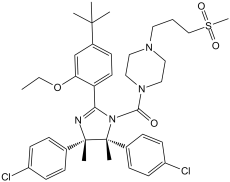
C[C@]1([C@@](C2C=CC(Cl)=CC=2)(C)N=C(C2C=CC(C(C)(C)C)=CC=2OCC)N1C(N1CCN(CCCS(=O)(=O)C)CC1)=O)C1C=CC(Cl)=CC=1
|
|
| InChi Key |
QBGKPEROWUKSBK-QPPIDDCLSA-N
|
|
| InChi Code |
InChI=1S/C38H48Cl2N4O4S/c1-8-48-33-26-29(36(2,3)4)14-19-32(33)34-41-37(5,27-10-15-30(39)16-11-27)38(6,28-12-17-31(40)18-13-28)44(34)35(45)43-23-21-42(22-24-43)20-9-25-49(7,46)47/h10-19,26H,8-9,20-25H2,1-7H3/t37-,38+/m0/s1
|
|
| 化学名 |
[(4S,5R)-2-(4-tert-butyl-2-ethoxyphenyl)-4,5-bis(4-chlorophenyl)-4,5-dimethylimidazol-1-yl]-[4-(3-methylsulfonylpropyl)piperazin-1-yl]methanone
|
|
| 别名 |
|
|
| HS Tariff Code |
2934.99.9001
|
|
| 存储方式 |
Powder -20°C 3 years 4°C 2 years In solvent -80°C 6 months -20°C 1 month |
|
| 运输条件 |
Room temperature (This product is stable at ambient temperature for a few days during ordinary shipping and time spent in Customs)
|
| 溶解度 (体外实验) |
|
|||
|---|---|---|---|---|
| 溶解度 (体内实验) |
配方 1 中的溶解度: ≥ 10 mg/mL (13.74 mM) (饱和度未知) in 10% DMSO + 40% PEG300 + 5% Tween80 + 45% Saline (这些助溶剂从左到右依次添加,逐一添加), 澄清溶液。
例如,若需制备1 mL的工作液,可将100 μL 100.0 mg/mL澄清DMSO储备液加入400 μL PEG300中,混匀;然后向上述溶液中加入50 μL Tween-80,混匀;加入450 μL生理盐水定容至1 mL。 *生理盐水的制备:将 0.9 g 氯化钠溶解在 100 mL ddH₂O中,得到澄清溶液。 配方 2 中的溶解度: ≥ 10 mg/mL (13.74 mM) (饱和度未知) in 10% DMSO + 90% Corn Oil (这些助溶剂从左到右依次添加,逐一添加), 澄清溶液。 例如,若需制备1 mL的工作液,可将 100 μL 100.0 mg/mL 澄清 DMSO 储备液加入到 900 μL 玉米油中并混合均匀。 View More
配方 3 中的溶解度: ≥ 5 mg/mL (6.87 mM) (饱和度未知) in 5% DMSO + 40% PEG300 + 5% Tween80 + 50% Saline (这些助溶剂从左到右依次添加,逐一添加), 澄清溶液。 配方 4 中的溶解度: ≥ 2.5 mg/mL (3.44 mM) in 5% DMSO + 95% (20% SBE-β-CD in Saline) (这些助溶剂从左到右依次添加,逐一添加), 澄清溶液。 *20% SBE-β-CD 生理盐水溶液的制备(4°C,1 周):将 2 g SBE-β-CD 溶解于 10 mL 生理盐水中,得到澄清溶液。 配方 5 中的溶解度: 1% CMC Na : 14mg/mL 1、请先配制澄清的储备液(如:用DMSO配置50 或 100 mg/mL母液(储备液)); 2、取适量母液,按从左到右的顺序依次添加助溶剂,澄清后再加入下一助溶剂。以 下列配方为例说明 (注意此配方只用于说明,并不一定代表此产品 的实际溶解配方): 10% DMSO → 40% PEG300 → 5% Tween-80 → 45% ddH2O (或 saline); 假设最终工作液的体积为 1 mL, 浓度为5 mg/mL: 取 100 μL 50 mg/mL 的澄清 DMSO 储备液加到 400 μL PEG300 中,混合均匀/澄清;向上述体系中加入50 μL Tween-80,混合均匀/澄清;然后继续加入450 μL ddH2O (或 saline)定容至 1 mL; 3、溶剂前显示的百分比是指该溶剂在最终溶液/工作液中的体积所占比例; 4、 如产品在配制过程中出现沉淀/析出,可通过加热(≤50℃)或超声的方式助溶; 5、为保证最佳实验结果,工作液请现配现用! 6、如不确定怎么将母液配置成体内动物实验的工作液,请查看说明书或联系我们; 7、 以上所有助溶剂都可在 Invivochem.cn网站购买。 |
| 制备储备液 | 1 mg | 5 mg | 10 mg | |
| 1 mM | 1.3740 mL | 6.8702 mL | 13.7404 mL | |
| 5 mM | 0.2748 mL | 1.3740 mL | 2.7481 mL | |
| 10 mM | 0.1374 mL | 0.6870 mL | 1.3740 mL |
1、根据实验需要选择合适的溶剂配制储备液 (母液):对于大多数产品,InvivoChem推荐用DMSO配置母液 (比如:5、10、20mM或者10、20、50 mg/mL浓度),个别水溶性高的产品可直接溶于水。产品在DMSO 、水或其他溶剂中的具体溶解度详见上”溶解度 (体外)”部分;
2、如果您找不到您想要的溶解度信息,或者很难将产品溶解在溶液中,请联系我们;
3、建议使用下列计算器进行相关计算(摩尔浓度计算器、稀释计算器、分子量计算器、重组计算器等);
4、母液配好之后,将其分装到常规用量,并储存在-20°C或-80°C,尽量减少反复冻融循环。
计算结果:
工作液浓度: mg/mL;
DMSO母液配制方法: mg 药物溶于 μL DMSO溶液(母液浓度 mg/mL)。如该浓度超过该批次药物DMSO溶解度,请首先与我们联系。
体内配方配制方法:取 μL DMSO母液,加入 μL PEG300,混匀澄清后加入μL Tween 80,混匀澄清后加入 μL ddH2O,混匀澄清。
(1) 请确保溶液澄清之后,再加入下一种溶剂 (助溶剂) 。可利用涡旋、超声或水浴加热等方法助溶;
(2) 一定要按顺序加入溶剂 (助溶剂) 。
| NCT Number | Recruitment | interventions | Conditions | Sponsor/Collaborators | Start Date | Phases |
| NCT00623870 | Completed | Drug: RO5045337 | Hematologic Neoplasms | Hoffmann-La Roche | May 2008 | Phase 1 |
| NCT00559533 | Completed | Drug: RO5045337 | Neoplasms | Hoffmann-La Roche | December 2007 | Phase 1 |
 ReACp53 scrambled peptide TFA
 p53-hDM2 cyclic peptide inhibitor 16e
 MMRi6
MMRi6
 dsP53-285 saRNA
InvivoChem的所有产品仅用于作科学研究,不面向患者销售
Copyright 2020 InvivoChem LLC | All Rights Reserved 粤ICP备20063088号-1


 COA
COA


 463611831
