| 规格 | 价格 | 库存 | 数量 |
|---|---|---|---|
| 5mg |
|
||
| 10mg |
|
||
| 50mg |
|
||
| 100mg |
|
||
| 250mg |
|
||
| Other Sizes |
|
| 靶点 |
GAT-1 (IC50 = 0.28 μM); GAT-2 (IC50 = 137.34 μM); GAT-3 (IC50 = 202.8 μM)
|
|---|---|
| 体外研究 (In Vitro) |
SKF89976A 显示出较弱的抗异常疼痛效果。 SKF89976A 轻度抑制中国仓鼠卵巢 (CHO) 细胞中的去甲肾上腺素转运蛋白 (NET)、多巴胺转运蛋白 (DAT) 和血清素转运蛋白 (SERT),根据底物摄取实验确定,这些细胞是每种转运蛋白的稳定转导物。 3514、202.13 和 728.8 分别是 IC50 值 [1]。 GABA 传输阻断剂是 SKF89976-A。 GABA (1 mM) 产生的内向电流不受 100 μM 印防己毒素的影响,但被 GABA 转运抑制剂噻加宾 (10 μM) 和 SKF89976A (100 μM) 完全抑制。众所周知,100 μM SKF 89976-A 通过阻断 GABA 转运到细胞中来完全、可逆地阻断 GABA 诱导的电流 [2]。 GAT-1 被 SKF89976A 不可转移地阻断。此外,SKF89976-A 还可抑制强直性 GAT 的背景 GABA 激活可能导致的基线内向电流。在所研究的每个细胞中,SKF89976A (100 μM) 可逆地将 GAT 电流降低 67.9±4.4% (n= 19)。 GABA 诱导的 GAT 电流逐渐降低并被细胞内输注 20 μM SKF89976-A 所抑制,而 GABAAR 介导的电流 (n=4) 并未被阻止 [3]。
GAT抑制剂抑制效力的比较为了分析GAT抑制剂对[3H]GABA摄取的影响,我们之前使用CHO细胞建立了稳定表达GAT亚型的细胞系。使用这些细胞系和稳定表达大鼠单胺转运蛋白SERT、NET和DAT的CHO细胞来比较抑制GAT亚型的效力,我们分析了GAT抑制剂对GABA和单胺类摄取的影响。抑制剂SKF89976A和(S)-SNAP5114分别对GAT-1(IC50:0.28 mM)和GAT-3(IC50:5.31 mM)表现出亚型选择性(表1)。然而,NNC05-2090能有效抑制GAT-1(IC50:29.62 mM),BGT-1(10.60 mM)的IC50值低2.5倍以上。NNC05-2090对GAT-2(45.29 mM)和GAT-3(22.51 mM)的IC50值也明显较高。这些结果表明,在所有检查的GAT抑制剂中,NNC05-2090在GAT抑制剂之间抑制BGT-1的效力最高。NNC05-2090还抑制SERT、NET和DAT(IC50:5.29 mM、7.91 mM和4.08 mM),这些IC50值与BGT-1的IC50值相似[1]。 尽管已经在体外研究了胶质细胞GABA的摄取和释放,但切片中胶质细胞中的GABA转运蛋白(GATs)尚未得到表征。从大鼠小脑切片中的Bergmann胶质细胞获得全细胞膜片钳记录,以表征载体介导的GABA流入和流出。GABA在-70 mV下诱导内向电流,在药理学上可分为GABA(A)受体和GAT电流。在GABA(A/B/C)受体阻断剂存在的情况下,-70 mV下测得的平均GABA诱导电流为-48 pA,在-70和+50 mV之间向内整流,受到外部Na(+)去除的抑制,并因外部Cl(-)的减少而减少。GAT-1的不可转运阻断剂(SKF89976A和NNC-711)和所有GAT亚型的可运输阻断剂(尼泊苷酸)分别可逆地将GABA诱导的转运电流降低了68%和100%。BGT-1阻断剂(甜菜碱)没有效果。SKF89976-A和NNC-711也抑制了基线内向电流,这可能是由背景GABA激活的紧张性GAT引起的。底物激动剂,尼泊苷酸和β-丙氨酸,而不是甜菜碱,诱导了电压和钠(+)依赖性电流。在记录过程中,当Na(+)和GABA进入贴片移液管或细胞内GABA灌注时,SKF89976A阻断了在-60 mV时激活的基线外向电流,并且随着去极化电位的增加而增加。这种载体介导的GABA外排诱导了记录细胞上GABA(a)受体激活检测到的细胞外GABA的局部积累。总体而言,这些结果表明Bergmann胶质细胞表达GAT-1,其被环境GABA激活。此外,胶质细胞中的GAT-1可以反向工作,释放足够的GABA来激活附近的GABA受体[3]。 |
| 体内研究 (In Vivo) |
静脉注射 SKF89976A (0.3 mg/kg) 可引起轻微的镇痛作用。在PSL模型小鼠中,注射SKF89976A可以以剂量依赖的方式改善戒断阈值的降低[1]。
GAT抑制剂对PSL小鼠模型中机械性异常性疼痛的影响部分坐骨神经结扎(PSL)模型小鼠同侧的机械性戒断阈值明显低于假手术动物(图1)。对侧的机械性戒断阈值没有降低(图2B、3B、4和5)。PSL小鼠模型中的机械性异常性疼痛不受盐水治疗的影响(图1B)。腹腔注射0.1mg/kg NNC05-2090 3小时后,PSL模型小鼠的机械戒断阈值有所增加(图1B)。另一方面,0.3 mg/kg的SKF89976A腹腔注射对PSL模型小鼠的戒断阈值没有产生任何显著影响(图1B)。静脉注射BGT-1抑制剂NNC05-2090显著逆转了PSL模型小鼠的机械性异常性疼痛(图2)。静脉注射NNC05-2090在0.01-0.1 mg/kg范围内显示出剂量依赖性反应。i.t.注射BGT-1抑制剂NNC05-2090也显著逆转了PSL模型小鼠的机械性异常性疼痛,并且通过i.t.向PSL模型鼠注射NNC05-2009,在15-150pmole范围内也显示出剂量依赖性反应(图3)。除静脉注射0.01mg/kg外,NNC05-2090的抗痛觉过敏作用在注射后1小时内达到峰值(图2和图3)。NNC05-2090给药后,对侧的戒断阈值没有显著差异。静脉注射(0.3 mg/kg)时,SKF89976A产生微弱的抗痛觉过敏反应(图4A)。如图5A所示,腹腔注射SKF89976A剂量依赖性地改善了PSL模型小鼠戒断阈值的降低。我们还注射了GAT-3抑制剂(S)-SNAP5114,以检测其对PSL小鼠机械性异常性疼痛的抗异常性疼痛作用。静脉注射和腹腔注射(S)-SNAP5114均未对PSL模型小鼠戒断阈值的降低产生显著影响(图4B和5B)[1]。 |
| 细胞实验 |
记录过程中GABA转运蛋白阻断剂的细胞内灌注。[3]
GABA转运蛋白阻断剂的细胞内灌注如其他人之前报道的那样,用于单药或多药应用(Tang等人,1990)。我们使用了带有灌注口的直移液管支架(EH-U2,E.W.Wright)。通过灌注口,将聚乙烯管(内径0.86mm,外径1.27mm)引入足够远的地方,以很好地到达贴片移液管溶液中。在记录过程中,将装有LY填充的细胞内溶液的1ml注射器通过细长的塑料移液管尖端连接到聚乙烯管上。在添加贴片移液管之前,手动施加正压,将试管充满到底,去除气泡,并观察溶液的流出情况。然后,在施加负压以防止任何溶液泄漏但不在管端添加气泡后,将贴片移液管插入支架中。为了灌注含有GABA或SKF89976A的LY填充溶液,手动施加正压以添加足够的溶液,使贴片移液管中的体积加倍(约20μl)。GABA和SKF89976A的浓度加倍,以获得细胞中预期的最终浓度。 |
| 动物实验 |
药物制备和给药[1]
神经结扎后约2周(11-16天)进行实验。动物分别接受NNC05-2090、SKF89976A、(S)-SNAP5114或阿米替林给药。NNC05-2090、SKF89976A和(S)-SNAP5114溶于二甲基亚砜(DMSO),并用人工脑脊液(ACSF)或生理盐水适当稀释(DMSO的最终浓度低于0.5%)。人工脑脊液(ACSF)的成分(单位:mM)为:142 mM NaCl、5 mM KCl、2 mM CaCl₂、2 mM MgCl₂、1.25 mM NaH₂PO₄、10 mM D-葡萄糖、10 mM HEPES 和 0.05% 无脂肪酸牛血清白蛋白(pH 7.4)。腹腔注射(ip)给药的剂量为 0.1 ml/10 g 体重。静脉注射(iv)时,将溶液经尾静脉注射,剂量为 0.1 ml/10 g 体重。鞘内注射(it)时,将小鼠头部置于塑料帽内,用一只手固定小鼠身体。将连接 Hamilton 微量注射器的 27 号针头插入清醒小鼠 L5 和 L6 椎骨之间的蛛网膜下腔,并缓慢注射 5 ml 药物溶液,如 Hylden 和 Wilcox (31) 所述。通过快速“甩动”小鼠尾巴来确认针头的准确位置,如 Honda 等人所述。 |
| 参考文献 |
|
| 其他信息 |
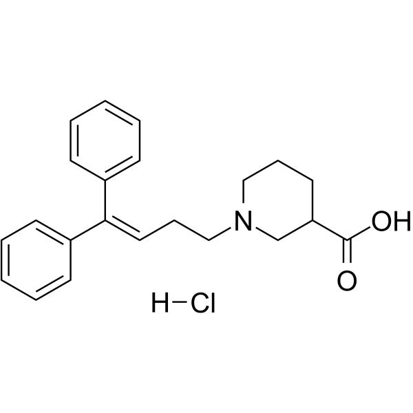
1-(4,4-二苯基丁-3-烯基)-3-哌啶羧酸是一种二芳基甲烷。
脊髓中的GABA能系统已被证实参与多种动物模型中的神经性疼痛。GABA转运蛋白(GATs)在控制GABA的突触清除中发挥作用;然而,它们在神经性疼痛中的作用尚不明确。在本研究中,我们利用坐骨神经结扎小鼠模型,比较了甜菜碱/GABA转运蛋白(BGT-1)与其他GAT亚型,以确定其在神经性疼痛中的作用。1-(3-(9H-咔唑-9-基)-1-丙基)-4-(2-甲氧基苯基)-4-哌啶醇(NNC05-2090)是一种对BGT-1具有中等选择性的抑制剂,通过鞘内和静脉给药途径均对模型小鼠表现出抗痛觉过敏作用。另一方面,选择性GAT-1抑制剂SKF89976A的抗痛觉过敏作用较弱,而选择性GAT-3抑制剂(S)-SNAP5114则无抗痛觉过敏作用。对这些化合物在稳定表达BGT-1的CHO细胞中GABA摄取情况进行系统分析发现,NNC05-2090不仅抑制BGT-1,还抑制5-羟色胺、去甲肾上腺素和多巴胺转运蛋白。在稳定表达各转运蛋白的CHO细胞中进行的底物摄取实验表明,NNC05-2090的IC50值分别为5.29、7.91和4.08 μM。这些值与BGT-1的IC50值(10.6 μM)相近。这些结果表明,NNC05-2090的抗痛觉过敏作用是由于其对BGT-1和单胺转运蛋白的抑制作用所致。[1] 氨基酸GABA向神经元和胶质细胞的转运在调节GABA对脊椎动物视网膜的作用中起着关键作用。我们研究了谷氨酸(脊椎动物感光细胞的可能神经递质)对视网膜水平细胞GABA诱导的转运电流的调节作用。我们采用全细胞电压钳技术检测了经酶解分离的鳐鱼外侧水平细胞。GABA(1 mM)诱导的内向电流可被GABA转运抑制剂噻加宾(10 μM)和SKF89976A(100 μM)完全抑制,但不受100 μM苦味素的影响。预先施加100 μM谷氨酸可显著降低GABA诱导的电流。谷氨酸抑制了GABA的剂量反应曲线,但并未使曲线发生横向移动或改变电流的电压依赖性。离子型谷氨酸受体激动剂红藻氨酸和AMPA也降低了GABA诱发的电流,而谷氨酸和红藻氨酸的作用可被离子型谷氨酸受体拮抗剂6-氰基-7-硝基喹喔啉阻断。NMDA既不诱发电流,也不改变GABA诱发的电流,代谢型谷氨酸类似物也无作用。当去除细胞外钙离子或在记录电极中加入高浓度钙螯合剂BAPTA时,谷氨酸和红藻氨酸对GABA诱发电流的抑制作用减弱。已知能改变细胞内钙离子水平的咖啡因(5 mM)和毒胡萝卜素(2 nM)也降低了GABA诱发的电流,但单纯去极化引起的钙离子浓度升高则没有这种作用。我们的数据表明,谷氨酸通过钙依赖性过程调节视网膜水平细胞中的GABA转运,并暗示这些细胞中钙通透性谷氨酸受体与GABA转运蛋白之间存在密切的物理联系。[2] 尽管神经胶质细胞对GABA的摄取和释放已在体外进行了研究,但尚未在脑片中的神经胶质细胞中对GABA转运蛋白(GATs)进行表征。我们利用全细胞膜片钳技术记录了大鼠小脑脑片中伯格曼胶质细胞的电生理活动,以表征载体介导的GABA内流和外流。GABA在-70 mV时诱导内向电流,该电流可通过药理学方法分离为GABA(A)受体电流和GAT电流。在 GABA(A/B/C) 受体阻滞剂存在的情况下,在 -70 mV 时测得的平均 GABA 诱导电流为 -48 pA,在 -70 至 +50 mV 之间呈内向整流特性,可被胞外 Na⁺ 去除抑制,并可被胞外 Cl⁻ 降低所减弱。GAT-1 的非转运性阻滞剂(SKF89976A 和 NNC-711)以及所有 GAT 亚型的可转运性阻滞剂(尼泊酸)分别可逆地将 GABA 诱导的转运电流降低了 68% 和 100%。BGT-1 阻滞剂(甜菜碱)无此作用。SKF89976-A 和 NNC-711 还抑制了可能由背景 GABA 持续激活 GAT 引起的基线内向电流。底物激动剂尼泊酸和β-丙氨酸(而非甜菜碱)可诱导电压依赖性和钠离子依赖性电流。在膜片钳电极内加入钠离子和GABA,或在记录过程中进行细胞内GABA灌注时,SKF89976A可阻断在-60 mV激活并随去极化电位增加而增强的基线外向电流。这种载体介导的GABA外流导致细胞外GABA在局部积累,可通过记录细胞上的GABAA受体激活来检测。总的来说,这些结果表明伯格曼胶质细胞表达GAT-1,且GAT-1可被周围GABA激活。此外,胶质细胞中的GAT-1还可以反向发挥作用,释放足够的GABA来激活附近的GABA受体。[3] |
| 分子式 |
C22H25NO2.HCL
|
|---|---|
| 分子量 |
371.90034
|
| 精确质量 |
371.165
|
| 元素分析 |
C, 71.05; H, 7.05; Cl, 9.53; N, 3.77; O, 8.60
|
| CAS号 |
85375-15-1
|
| 相关CAS号 |
85375-15-1 ( free base);85375-85-5 (HCl);
|
| PubChem CID |
92409
|
| 外观&性状 |
Light yellow to khaki solid powder
|
| 沸点 |
531.4ºC at 760 mmHg
|
| 闪点 |
275.2ºC
|
| LogP |
5.044
|
| tPSA |
40.54
|
| 氢键供体(HBD)数目 |
1
|
| 氢键受体(HBA)数目 |
3
|
| 可旋转键数目(RBC) |
6
|
| 重原子数目 |
25
|
| 分子复杂度/Complexity |
427
|
| 定义原子立体中心数目 |
0
|
| SMILES |
Cl.O=C(C1CCCN(CC/C=C(\C2C=CC=CC=2)/C2C=CC=CC=2)C1)O
|
| InChi Key |
TXQKSMSLZVKQBI-UHFFFAOYSA-N
|
| InChi Code |
InChI=1S/C22H25NO2/c24-22(25)20-13-7-15-23(17-20)16-8-14-21(18-9-3-1-4-10-18)19-11-5-2-6-12-19/h1-6,9-12,14,20H,7-8,13,15-17H2,(H,24,25)
|
| 化学名 |
1-(4,4-diphenylbut-3-enyl)piperidine-3-carboxylic acid
|
| 别名 |
SKF89976A HCl; 85375-85-5; Skf 89,976A; SKF89,976A; 1-(4,4-diphenylbut-3-en-1-yl)piperidine-3-carboxylic acid; N-(4,4-Diphenyl-3-butenyl)nipecotic acid; N-(4,4-Diphenyl-3-butenyl)homo-beta-proline; SKF-89976; CHEMBL38686; SKF 89976-A; SKF-89976-A; SKF89976A hydrochloride
|
| HS Tariff Code |
2934.99.9001
|
| 存储方式 |
Powder -20°C 3 years 4°C 2 years In solvent -80°C 6 months -20°C 1 month 注意: 请将本产品存放在密封且受保护的环境中,避免吸湿/受潮。 |
| 运输条件 |
Room temperature (This product is stable at ambient temperature for a few days during ordinary shipping and time spent in Customs)
|
| 溶解度 (体外实验) |
DMSO : ~100 mg/mL (~268.89 mM)
H2O : ~20 mg/mL (~53.78 mM) |
|---|---|
| 溶解度 (体内实验) |
配方 1 中的溶解度: ≥ 2.5 mg/mL (6.72 mM) (饱和度未知) in 10% DMSO + 40% PEG300 + 5% Tween80 + 45% Saline (这些助溶剂从左到右依次添加,逐一添加), 澄清溶液。
例如,若需制备1 mL的工作液,可将100 μL 25.0 mg/mL澄清DMSO储备液加入到400 μL PEG300中,混匀;然后向上述溶液中加入50 μL Tween-80,混匀;加入450 μL生理盐水定容至1 mL。 *生理盐水的制备:将 0.9 g 氯化钠溶解在 100 mL ddH₂O中,得到澄清溶液。 配方 2 中的溶解度: ≥ 2.5 mg/mL (6.72 mM) (饱和度未知) in 10% DMSO + 90% (20% SBE-β-CD in Saline) (这些助溶剂从左到右依次添加,逐一添加), 澄清溶液。 例如,若需制备1 mL的工作液,可将 100 μL 25.0 mg/mL澄清DMSO储备液加入900 μL 20% SBE-β-CD生理盐水溶液中,混匀。 *20% SBE-β-CD 生理盐水溶液的制备(4°C,1 周):将 2 g SBE-β-CD 溶解于 10 mL 生理盐水中,得到澄清溶液。 View More
配方 3 中的溶解度: ≥ 2.5 mg/mL (6.72 mM) (饱和度未知) in 10% DMSO + 90% Corn Oil (这些助溶剂从左到右依次添加,逐一添加), 澄清溶液。 配方 4 中的溶解度: 25 mg/mL (67.22 mM) in PBS (这些助溶剂从左到右依次添加,逐一添加), 澄清溶液; 超声助溶. 1、请先配制澄清的储备液(如:用DMSO配置50 或 100 mg/mL母液(储备液)); 2、取适量母液,按从左到右的顺序依次添加助溶剂,澄清后再加入下一助溶剂。以 下列配方为例说明 (注意此配方只用于说明,并不一定代表此产品 的实际溶解配方): 10% DMSO → 40% PEG300 → 5% Tween-80 → 45% ddH2O (或 saline); 假设最终工作液的体积为 1 mL, 浓度为5 mg/mL: 取 100 μL 50 mg/mL 的澄清 DMSO 储备液加到 400 μL PEG300 中,混合均匀/澄清;向上述体系中加入50 μL Tween-80,混合均匀/澄清;然后继续加入450 μL ddH2O (或 saline)定容至 1 mL; 3、溶剂前显示的百分比是指该溶剂在最终溶液/工作液中的体积所占比例; 4、 如产品在配制过程中出现沉淀/析出,可通过加热(≤50℃)或超声的方式助溶; 5、为保证最佳实验结果,工作液请现配现用! 6、如不确定怎么将母液配置成体内动物实验的工作液,请查看说明书或联系我们; 7、 以上所有助溶剂都可在 Invivochem.cn网站购买。 |
| 制备储备液 | 1 mg | 5 mg | 10 mg | |
| 1 mM | 2.6889 mL | 13.4445 mL | 26.8889 mL | |
| 5 mM | 0.5378 mL | 2.6889 mL | 5.3778 mL | |
| 10 mM | 0.2689 mL | 1.3444 mL | 2.6889 mL |
1、根据实验需要选择合适的溶剂配制储备液 (母液):对于大多数产品,InvivoChem推荐用DMSO配置母液 (比如:5、10、20mM或者10、20、50 mg/mL浓度),个别水溶性高的产品可直接溶于水。产品在DMSO 、水或其他溶剂中的具体溶解度详见上”溶解度 (体外)”部分;
2、如果您找不到您想要的溶解度信息,或者很难将产品溶解在溶液中,请联系我们;
3、建议使用下列计算器进行相关计算(摩尔浓度计算器、稀释计算器、分子量计算器、重组计算器等);
4、母液配好之后,将其分装到常规用量,并储存在-20°C或-80°C,尽量减少反复冻融循环。
计算结果:
工作液浓度: mg/mL;
DMSO母液配制方法: mg 药物溶于 μL DMSO溶液(母液浓度 mg/mL)。如该浓度超过该批次药物DMSO溶解度,请首先与我们联系。
体内配方配制方法:取 μL DMSO母液,加入 μL PEG300,混匀澄清后加入μL Tween 80,混匀澄清后加入 μL ddH2O,混匀澄清。
(1) 请确保溶液澄清之后,再加入下一种溶剂 (助溶剂) 。可利用涡旋、超声或水浴加热等方法助溶;
(2) 一定要按顺序加入溶剂 (助溶剂) 。
InvivoChem的所有产品仅用于作科学研究,不面向患者销售
Copyright 2020 InvivoChem LLC | All Rights Reserved 粤ICP备20063088号-1


 463611831
