| 规格 | 价格 | 库存 | 数量 |
|---|---|---|---|
| 100mg |
|
||
| 250mg |
|
||
| 500mg |
|
||
| 1g |
|
||
| Other Sizes |
|
| 靶点 |
DNA Alkylator
|
|---|---|
| 体外研究 (In Vitro) |
孵育后,大鼠肝脏切片显示出硫代 TEPA 的烷基化活性。在 2、5 和 10 mM 的所有剂量下,thio-TEPA 不会在大鼠肝脏切片中积聚,并且对其活力没有影响[1]。
在精密切割的大鼠肝薄片孵育实验中,Thiotepa被代谢为其氧代类似物TEPA (N,N',N''-三乙烯磷酰胺)。该转化遵循一级动力学。 [1] 用Thiotepa(浓度范围5.2至104 µM)孵育肝薄片导致缓冲液中4-(硝基苄基)-吡啶 (NBP) 烷基化活性增加30%–50%,且与药物浓度无关。这种增加与Thiotepa的孵育时间呈显著相关,但与TEPA无关,提示可能形成了除TEPA之外的未知活性代谢物。 [1] |
| 体内研究 (In Vivo) |
在前10周内,硫代TEPA(20 mg/kg,腹膜内)联合全身照射(TBI)增强了供体型血液嵌合性,但并不显着高于单独TBI组。在小鼠中,单独的 thio-TEPA 可以增强短期和长期的植入[2]。
|
| 酶活实验 |
使用精确切割的大鼠肝脏切片研究烷化剂N,N',N'-三乙烯硫代磷酰胺(thio-TEPA)的代谢。暴露于高浓度(1-10mM)硫代TEPA 6小时未证明对肝切片有毒性,这表明细胞中钾的泄漏微不足道。在孵育过程中,硫代TEPA(初始浓度,5.2微M)从缓冲液中消失的时间过程遵循一级动力学。N,N’N’-三乙烯基磷酰胺(TEPA)的形成显然是硫代TEPA消除的原因。苯巴比妥预处理大鼠可显著提高反应速率。相反,用细胞色素P-450抑制剂烯丙基异丙酰胺预处理显著降低了代谢率。硫代TEPA的消除和TEPA的形成与硫代TEPA浓度无关,其范围为5.2至104微M。硫TEPA的氧代类似物TEPA是唯一鉴定的代谢产物,没有进一步代谢。然而,在用硫代TEPA孵育肝切片后,观察到4-(硝基苄基)-吡啶(NBP)烷基化活性随时间显著增加,但在用TEPA孵育后没有。这可能表明存在未知的活性代谢物[1]。
|
| 细胞实验 |
肝薄片活力实验(钾泄漏测定): 用浓度为1、2、5和10 mM的Thiotepa孵育精密切割的大鼠肝薄片,时间长达24小时。通过火焰光度法测定细胞内钾 (K⁺) 含量,并相对于DNA含量进行标准化。通过细胞K⁺泄漏程度评估活力。 [1]
肝薄片药物代谢实验: 在Krebs-Henseleit缓冲液中用Thiotepa孵育肝薄片。使用气相色谱法在不同时间点测定孵育介质中Thiotepa及其代谢物TEPA的浓度。采用比色法测定介质中的NBP烷基化活性。 [1] |
| 动物实验 |
噻替哌 (TT) 长期以来一直被考虑纳入临床骨髓移植 (BMT) 预处理方案,以期预防同种异体移植排斥反应和白血病复发。早期的鼠类实验表明,在全身照射 (TBI) 的基础上添加 TT 可显著提高同种异体骨髓移植的成功率,这为相关研究提供了支持。研究者认为,TT 可通过清除受体体内竞争性干细胞来增强供体型嵌合体。本研究旨在重新评估 TT 对不同干细胞亚群(包括具有长期重建能力的原始细胞)的血液学毒性,并评估 TT 与 TBI 联合应用如何影响同基因 (B6-Gpi-1a → B6-Gpi-1b) 和 H-2 相容的同种异体 (BALB.B10 → B6) BMT 模型中供体型移植的进展。在给予 TT(20 mg/kg)24 小时后,通过测定瞬时重建细胞和更原始的铺路石样区域形成细胞(CAFC)在基质支持的培养物中的生长频率,确定股骨中不同干细胞亚群的含量。该检测显示,TT 诱导了培养第 7 天早期克隆的大量耗竭(存活率 2%),但后期出现的、由更原始的 CAFC 亚群发育而来的克隆的存活率逐渐恢复正常。这些原始干细胞的保留体现在接受 TT 治疗后进行同基因骨髓移植的受体中,供体骨髓重建水平无法检测。在全身照射(TBI)中添加 TT 并未显著改善同基因骨髓的长期植入,但这种联合疗法在异基因骨髓移植中具有显著效果,可预防同种异体移植排斥反应。在这方面,TT 与环磷酰胺具有相似的特性,表明同种异体干细胞移植的显著改善归因于 TT 的免疫抑制特性,而非其对宿主原始干细胞的毒性[2]。
大鼠肝片研究的预处理:使用雄性 Sprague-Dawley 大鼠(250-350 g)。为诱导细胞色素 P-450 活性,大鼠在处死前 3 天每日腹腔注射苯巴比妥(80 mg/kg)。为抑制细胞色素 P-450 活性,大鼠在处死前 1 小时腹腔注射烯丙基异丙基乙酰胺(AIA,100 mg/kg)。大鼠断头处死,立即取出肝脏进行切片制备。[1] 肝片制备和孵育:将取出的肝脏置于冰冷的通气缓冲液中。使用机械切片机制备精确切割的肝片(干重 7.5–10 mg)。将两片肝片置于装有 1.7 ml 孵育培养基(Krebs-Henseleit 缓冲液或 Waymouth's MB 752/1 培养基)的 20 ml 闪烁瓶内的滚轮筛网上,培养基中添加了噻替哌。闪烁瓶用 95% O₂/5% CO₂ 混合气体充入气体,密封后,在 37°C 下以 4 rpm 的转速水平旋转。在将样品转移至含药培养基进行实验孵育之前,先在不含药物的培养基中预孵育 30 分钟。[1] |
| 药代性质 (ADME/PK) |
吸收、分布和排泄
一名34岁患有转移性盲肠癌的患者,静脉注射0.3 mg/kg剂量的14C标记的噻替哌及其代谢物后,尿排泄率为63%。 446 +/- 63 mL/min [45至84岁患有晚期卵巢癌的女性患者,在后续疗程中,每隔4周接受60 mg和80 mg噻替哌静脉输注] 在人体中,静脉注射或局部注射到肿瘤中的14C标记的噻替哌,50%在最初6小时内排出体外,48小时后仅残留低水平。口服 (14)C-噻替哌的吸收情况不一:尿液中回收的注射剂量中,未代谢药物的比例不足 1%。 ……在 Sprague-Dawley 大鼠静脉或动脉注射标记的噻替哌 5 分钟后,与其它器官相比,血浆、心脏、肾脏和肺中的放射性水平略高;静脉注射的放射性物质有 94-98% 在 8.5 小时内经尿液排出。尿液中大部分放射性物质为未代谢的噻替哌。三(1-氮丙啶基)氧化膦 (tepa) 约占放射性的 30%。 在 Sprague-Dawley 大鼠腹腔注射 9.3 mg/kg 体重的噻替哌后 1 小时,在血浆 (5.4%)、腹腔液 (26%)、尿液 (1.9%)、肾脏 (0.7%)、肝脏 (3.8%)、肺 (0.6%) 和肌肉 (25.9%) 中均检测到放射性。 在肾功能正常的患者中,噻替哌、三乙烯磷酰胺 (TEPA) 和具有烷化活性的未鉴定代谢物均经尿液排泄。在给药后的 24-48 小时内,噻替哌、TEPA 和具有烷化活性的未鉴定代谢物的尿排泄量分别约为给药剂量的 0.1-1.5%、4% 和 13-24%。原药在给药后 6-8 小时内即可完全经尿液排泄。尚未研究该药物及其代谢物的粪便排泄情况。静脉注射大剂量噻替哌后,该药物似乎会大量经汗液排泄。 有关噻替哌(共 14 项)的更多吸收、分布和排泄(完整)数据,请访问 HSDB 记录页面。 代谢/代谢物 大鼠、兔和犬单次静脉注射 (32)P-噻替哌后,尿液中的主要代谢物是替哌,替哌也是一种烷化剂。然而,小鼠尿液中的大部分放射性物质以无机磷酸盐的形式回收。 ……在Sprague-Dawley大鼠静脉或动脉注射标记的噻替哌5分钟后,与其它器官相比,血浆、心脏、肾脏和肺脏中的放射性水平略高;静脉注射的放射性物质有94-98%在8.5小时内随尿液排出。尿液中的大部分放射性物质与未代谢的噻替哌相关;三(1-氮丙啶基)氧化膦(替哌)约占放射性物质的30%。 在小鼠中,噻替哌迅速代谢为三(1-氮丙啶基)氧化膦(替哌):30分钟后,尿液和血浆中仅检测到替哌和无机磷酸盐。小鼠具有将药物完全降解为无机磷酸盐的特殊能力。 噻替哌似乎主要通过细胞色素P-450微粒体酶系统在肝脏中广泛代谢,主要途径是氧化脱硫生成三乙烯磷酰胺(TEPA)。尽管TEPA是血浆中唯一检测和鉴定的代谢物,但有证据表明还生成了其他未鉴定的代谢物。在肾功能和肝功能正常的成年人中,快速静脉注射噻替哌后,血浆药物浓度呈双相下降,初始阶段的半衰期约为6-12分钟,终末阶段的半衰期为1.2-2.9小时。 TEPA的血浆消除半衰期约为10-21小时。 本研究测定了接受硫代TEPA联合环磷酰胺和卡铂高剂量化疗方案的患者尿液中N,N',N"-三乙烯硫代磷酰胺(硫代TEPA)及其代谢物N,N',N"-三乙烯磷酰胺(TEPA)、N,N'-二乙烯,N"-2-氯乙基磷酰胺(单氯TEPA)和硫代TEPA-巯基尿酸盐的排泄量。硫代TEPA的剂量为40或60 mg/m²,短时静脉输注,每日两次,持续4天。在给药期间,每天每次排尿后收集尿液样本,直至最后一次硫代TEPA输注后24-48小时。采用气相色谱法测定硫代TEPA、TEPA和单氯TEPA的浓度,采用液相色谱-质谱法测定硫代TEPA-巯基尿酸盐的浓度。采用直接进样法。输注后30分钟,尿液中即可检测到硫替哌(ThioTEPA),且在末次输注后18小时仍可排出。所有代谢物均在输注后1小时的尿液中被检测到。肌酐清除率高于140 mL/min的患者,其替哌(TEPA)的排泄量高于肌酐清除率低于140 mL/min的患者(12.8% vs. 4.9%,p=0.01)。尿液pH值越低,单氯替哌(monochloroTEPA)的排泄量占替哌(TEPA)排泄量的比例越高。接受60 mg/m²剂量治疗的患者,其硫替哌-巯基尿酸盐(thioTEPA-mercapturate)的排泄量占剂量的比例更高。硫替哌(thioTEPA)和单氯替哌(monochloroTEPA)的排泄量均仅占剂量的0.5%,而替哌(TEPA)和硫替哌-巯基尿酸盐(thioTEPA-mercapturate)的排泄量均占剂量的11.1%。 排泄途径:尿液排泄一名34岁患有转移性盲肠癌的患者接受了0.3 mg/kg静脉注射,其体内14C标记的噻替哌及其代谢物的检出率为63%。 半衰期:1.5至4.1小时 生物半衰期 1.5至4.1小时 在一项I期研究中,共有15例一线全身化疗后残留卵巢癌且局限于腹膜腔的患者接受了噻替哌治疗。共进行了50个疗程的噻替哌腹腔注射,剂量范围为30至80 mg/m²。……腹腔液浓度呈一级动力学迅速下降,半衰期为0.96±0.1小时。…… 静脉注射噻替哌后最初几小时内,血浆中噻替哌浓度呈双指数下降。在Swiss-Webster小鼠中,以5 mg/kg体重注射噻替哌后,第一相半衰期为0.21分钟,第二相半衰期为9.62分钟。 在肾功能和肝功能正常的成年人中,快速静脉注射噻替哌后,药物的血浆浓度呈双相下降,初始相半衰期约为6-12分钟,终末相半衰期为1.2-2.9小时。三乙烯磷酰胺(TEPA)的血浆消除半衰期约为10-21小时。 在大鼠肝片孵育实验中,使用未经处理的大鼠肝片,噻替哌从缓冲液中的消除半衰期(t₁/₂)为2.23 ± 0.15小时。苯巴比妥预处理显著缩短了半衰期至0.91 ± 0.05小时,而预先使用烯丙基异丙基乙酰胺 (AIA) 显著延长了其作用时间至 3.23 ± 0.27 小时。[1] 在所测试的浓度范围 (5.2 至 104 µM) 内,噻替哌 向 TEPA 的转化符合一级动力学。[1] 代谢物 TEPA 在肝切片系统中未进一步代谢。[1] 该研究未提供 噻替哌 的全身药代动力学参数(吸收、分布、排泄、体内半衰期、生物利用度)。[1] |
| 毒性/毒理 (Toxicokinetics/TK) |
毒性概述
识别和用途:噻替哌形成晶体或细小的白色结晶片。噻替哌广泛用于大剂量化疗。人体暴露和毒性:基于充分的人体研究证据,噻替哌已被证实为人类致癌物。噻替哌的主要不良反应是血液毒性,通常与剂量相关且具有累积性。不良的造血作用包括白细胞减少症、贫血、血小板减少症和全血细胞减少症,后者有时可致命。接受噻替哌治疗的患者必须密切监测血液系统状况。虽然每周一次静脉注射噻替哌后,白细胞最低值可能在10-14天出现,但对骨髓的初始影响可能在长达30天后才会显现。由于噻替哌可通过浆膜吸收,腔内和膀胱内灌注噻替哌可产生不同程度的全身性不良反应;膀胱内灌注噻替哌后,曾有因药物全身吸收导致骨髓抑制而死亡的病例报道。在高剂量噻替哌联合自体骨髓移植治疗中,可能出现严重的中枢神经系统紊乱、肝损伤、感染、恶心、呕吐、腹泻、黏膜炎、皮疹、出血性膀胱炎和心肌病。过量用药后可出现造血系统毒性,表现为白细胞计数和/或血小板计数下降。红细胞计数并非噻替哌毒性的准确指标。患者可能出现出血症状,更容易感染,且抵抗感染的能力下降。即使在推荐治疗剂量范围内或略高于推荐治疗剂量,噻替哌也可能导致危及生命的造血系统毒性。噻替哌对造血系统具有剂量依赖性的毒性作用。用噻替哌处理培养的人类淋巴细胞可显著增加染色体畸变的频率。噻替哌用于孕妇时可能对胎儿造成损害。动物研究:据报道,在兔眼玻璃体内注射浓度为 8 mg/mL 的噻替哌,未观察到过度炎症或视网膜电图改变,且耐受性良好。断奶大鼠连续六周使用 1:445 或 1:1000 的噻替哌滴眼液治疗后,出现角膜血管化和白内障。每周一次,以 1 mg/kg 体重的剂量给大鼠注射噻替哌,持续 52 周。30% 的治疗组动物出现恶性肿瘤;对照组中,6% 的动物出现恶性肿瘤。在妊娠小鼠中,腹腔注射单剂量0.5-30 mg/kg体重的噻替哌可导致畸形。最小致畸剂量为1 mg/kg体重;注射10 mg/kg体重后,所有胎儿均出现畸形。大鼠注射5 mg/kg体重的噻替哌后,胎儿出现明显的发育异常和骨骼缺陷。噻替哌可干扰仓鼠和小鼠的精子发生。在体内,噻替哌可诱导啮齿动物出现显性致死突变、染色体畸变、微核和姐妹染色单体交换。在体外,噻替哌可诱导啮齿动物细胞出现姐妹染色单体交换和染色体畸变。在体外,噻替哌对中国仓鼠细胞和宿主介导的小鼠淋巴瘤细胞均具有致突变性。噻替哌可诱导果蝇发生性连锁隐性致死突变,引起植物细胞的姐妹染色单体交换和染色体畸变,并在体外和宿主介导的试验中对真菌和细菌具有致突变性。 烷基连接在DNA的鸟嘌呤碱基上,位于咪唑环的7号氮原子处。它们通过交联DNA双螺旋链中的鸟嘌呤核碱基来抑制肿瘤生长,直接攻击DNA。这使得DNA链无法解旋和分离。由于DNA复制需要解旋和分离,因此细胞无法分裂。这些药物的作用是非特异性的。 肝毒性 噻替哌治疗期间血清酶升高发生率较高,但通常程度较轻且可自行消退,无需调整剂量。有报道称,噻替哌可引起罕见的临床表现明显的急性肝损伤,尤其是在高剂量使用时。在大多数情况下,噻替哌与其他已知可引起肝损伤的药物联合使用,噻替哌的具体作用尚不明确。噻替哌常与其他烷化剂联合用于骨髓清除的预处理方案中,以准备造血干细胞移植,因此与肝窦阻塞综合征的发生有关。肝窦阻塞综合征通常在清髓性或大剂量治疗后1至3周内发作,其特征是突然出现腹痛、肝肿大、体重增加和腹水,随后出现黄疸。血清酶升高通常表现为肝细胞性,血清转氨酶和乳酸脱氢酶水平显著升高,而碱性磷酸酶升高幅度较小。在严重病例中,凝血酶原时间延长,并出现进行性肝功能衰竭。免疫过敏和自身免疫特征不常见。死亡率高。肝活检显示小叶中心坏死和充血,小静脉阻塞,肝窦内可见红细胞。 可能性评分:D(可能是临床上明显的肝损伤的罕见原因)。 妊娠和哺乳期影响 ◉ 哺乳期用药概述 大多数资料认为,母亲在接受抗肿瘤药物治疗期间,尤其是使用噻替哌等烷化剂时,应避免哺乳。药品说明书建议,母亲在治疗期间以及最后一次服用噻替哌后1周内不应哺乳。化疗可能会对母乳的正常微生物群和化学成分产生不利影响。妊娠期接受化疗的女性更容易出现哺乳困难。 ◉ 对母乳喂养婴儿的影响 截至修订日期,未找到相关已发表的信息。 ◉ 对泌乳和母乳的影响 截至修订日期,未找到相关已发表的信息。 相互作用 甲基黄嘌呤类药物可增强烷化剂对人类癌细胞的杀伤作用,这种现象归因于其抑制了DNA修复。己酮可可碱是一种无毒的甲基黄嘌呤类药物,临床上用于治疗间歇性跛行。本研究利用体外培养的人类癌细胞或小鼠异种移植模型,考察了烷化剂与己酮可可碱或其他甲基黄嘌呤类药物联合治疗的效果。在体外培养的人类膀胱癌细胞中,经己酮可可碱、己酮可可碱的主要临床代谢产物或咖啡因后处理,噻替哌的细胞毒性可提高至10倍(p<0.01);所需的己酮可可碱浓度(0.4-1.0 mM)在无毒口服剂量后,膀胱内可达到临床可达到的浓度。在改良的肾下包膜下移植瘤模型中,使用人膀胱癌或乳腺癌异种移植瘤,也观察到体内经己酮可可碱后处理可增强噻替哌的疗效。相反,这些联合用药并未增加动物正常组织的毒性,无论是通过体重、死亡率还是正常膀胱尿路上皮的组织学变化来评估。 ... 在培养的人类淋巴细胞中,2 mM 维生素 C 可增强噻替哌或 L-乙硫氨酸诱导的姐妹染色单体交换频率。然而,当维生素 C 浓度为 0.02 mM 和 0.2 mM 时,则观察到其对噻替哌或 L-乙硫氨酸诱导的姐妹染色单体交换率具有一定的保护作用。2 mM 维生素 C 可导致噻替哌或 L-乙硫氨酸处理的细胞培养物中细胞分裂延迟。在 0.02 mM 或 0.2 mM 维生素 C 存在下,噻替哌或 L-乙硫氨酸引起的细胞分裂延迟被逆转。在 2 mM 维生素 C 存在下,噻替哌或 L-乙硫氨酸处理的细胞培养物中的有丝分裂指数持续受到抑制。然而,0.02 mM 维生素 C 可逆转 L-乙硫氨酸或噻替哌引起的有丝分裂指数抑制。这些发现揭示了维生素C在生物系统中相互作用的复杂性,并表明不同浓度的维生素C既可以引起遗传毒性,也可以抑制遗传毒性。 研究人员在中国仓鼠中测试了食物成分叶绿素、β-胡萝卜素和α-亚麻酸(以其甲酯形式存在)对强效诱变剂硫替哌的抗诱变活性。每种天然保护性化合物均能抑制70-85%的诱变剂诱导的染色体断裂效应。当同时使用2种或3种抗诱变剂时,未观察到叠加效应。在实验条件下,α-亚麻酸甲酯是最有效的抗诱变剂。 五名患有转移性乳腺腺癌的女性在接受N,N',N"-三乙烯硫代磷酰胺(噻替哌)和环磷酰胺静脉注射后,出现了局限于被含粘合剂材料覆盖的皮肤的色素沉着过度。研究人员测定了被覆盖和未被覆盖的皮肤、血浆、含粘合剂的绷带以及含有汗液的纱布中的噻替哌浓度。结果表明,这种烷化剂通过汗液分泌到皮肤表面,积聚在含粘合剂的绷带和心电图电极片下方,并产生局部毒性作用,导致色素沉着过度。 非人类毒性值 小鼠静脉注射LD50:14,500 μg/kg 小鼠皮下注射LD50:19,500 μg/kg ug/kg 小鼠口服LD50 38 mg/kg 大鼠静脉注射LD50 9400 ug/kg 大鼠腹腔注射LD50 8 mg/kg 噻替哌浓度高达10 mM时,在6小时孵育期间未引起大鼠肝切片中明显的钾离子(K⁺)泄漏,表明在这些条件下无急性细胞毒性。[1] 孵育12小时后,在最高浓度(10 mM)下出现明显的K⁺泄漏。24小时后,观察到浓度依赖性的K⁺泄漏。然而,在1 mM浓度下(远高于代谢研究中使用的浓度),在整个孵育期间,K⁺泄漏与对照组无显著差异。[1] 该研究未报告噻替哌的经典体内毒性参数(LD50、器官毒性、蛋白结合、药物相互作用)。 [1] |
| 参考文献 | |
| 其他信息 |
治疗用途
抗肿瘤药物,烷化剂;骨髓清除激动剂 /临床试验/ ClinicalTrials.gov 是一个注册库和结果数据库,收录了全球范围内由公共和私人机构资助的人体临床研究。该网站由美国国家医学图书馆 (NLM) 和美国国立卫生研究院 (NIH) 维护。ClinicalTrials.gov 上的每条记录都包含研究方案的摘要信息,包括:疾病或病症;干预措施(例如,正在研究的医疗产品、行为或程序);研究的标题、描述和设计;参与要求(资格标准);研究开展地点;研究地点的联系方式;以及其他健康网站相关信息的链接,例如 NLM 的 MedlinePlus(用于患者健康信息)和 PubMed(用于医学领域学术文章的引文和摘要)。噻替哌已收录于数据库中。 注射用噻替哌(USP)已在多种肿瘤性疾病的姑息治疗中进行了尝试,但疗效不一。然而,在以下肿瘤中观察到了最稳定的疗效:1. 乳腺腺癌;2. 卵巢腺癌;3. 用于控制由各种浆膜腔弥漫性或局限性肿瘤性疾病引起的腔内积液;4. 用于治疗膀胱浅表性乳头状癌。尽管噻替哌目前已被其他疗法广泛取代,但它对其他淋巴瘤(如淋巴肉瘤和霍奇金淋巴瘤)也有效。/美国产品标签中包含/ 噻替哌曾被用作眼用滴剂,以预防翼状胬肉手术切除后的复发;然而,术后β射线照射通常被优先选择作为预防性治疗,因为它复发率低且相对容易操作。许多临床医生建议将噻替哌的使用限制在术后β射线照射后复发的翼状胬肉的治疗中。/未包含在美国产品标签中/ 有关噻替哌(共6种)的更多治疗用途(完整)数据,请访问HSDB记录页面。 药物警告 噻替哌眼内滴注偶尔可能引起刺激或眼周皮肤色素脱失;色素脱失通常在停药6个月或更长时间后出现。噻替哌鞘内注射与部分患者出现下肢无力、疼痛以及脊髓脱髓鞘有关;鞘内注射该药物高渗溶液后,也曾出现短暂性下肢感觉异常。 其他已报道的噻替哌不良反应包括注射部位疼痛、头痛、头晕、视力模糊、结膜炎、排尿困难、尿潴留、闭经和咽喉紧缩感。一些症状,例如高尿酸血症或发热反应以及皮下病变渗出,可能是由于肿瘤组织分解所致。据报道,部分患者膀胱内注射噻替哌后会出现下腹痛、膀胱刺激、血尿,罕见情况下还会出现出血性化学性膀胱炎。 接受噻替哌治疗的患者曾出现超敏反应,包括过敏反应、皮疹、荨麻疹、喉头水肿、哮喘、过敏性休克和喘息。此外,也有接触性皮炎和脱发的报道。局部使用该药物后曾有皮肤色素脱失的报道。 噻替哌给药后偶有恶心、呕吐、腹痛和厌食的发生。也有口腔炎和肠黏膜溃疡的报道。 有关噻替哌(共15条)的更多药物警告(完整)数据,请访问HSDB记录页面。 药效学 不稳定的氮碳基团与DNA烷基化,造成不可修复的DNA损伤。它们通过交联DNA双螺旋链中的鸟嘌呤核碱基来阻止肿瘤生长,直接攻击DNA。这使得DNA链无法解旋和分离。由于DNA复制需要解旋和分离,细胞将无法分裂。这些药物的作用是非特异性的。 噻替哌是一种烷化剂。 [1] 其主要代谢产物是TEPA,由肝细胞色素P-450酶(特别是苯巴比妥诱导型酶)催化的氧化脱硫反应生成。[1] 肝切片与噻替哌孵育后观察到的NBP烷基化活性增加,超出噻替哌和TEPA所能解释的范围,提示可能生成了其他未鉴定的活性代谢产物。[1] 肝切片模型维持了组织完整性,在实验条件下可用于研究噻替哌的代谢,且未观察到明显的药物毒性。[1] |
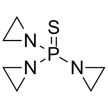
| 分子式 |
C6H12N3PS
|
|---|---|
| 分子量 |
189.2183
|
| 精确质量 |
189.048
|
| 元素分析 |
C, 38.09; H, 6.39; N, 22.21; P, 16.37; S, 16.95
|
| CAS号 |
52-24-4
|
| 相关CAS号 |
55-98-1 (Busulfan); 299-75-2 (Treosulfan)
|
| PubChem CID |
5453
|
| 外观&性状 |
White to off-white solid powder
|
| 密度 |
1.5±0.1 g/cm3
|
| 沸点 |
270.2±23.0 °C at 760 mmHg
|
| 熔点 |
54-57 °C
|
| 闪点 |
117.2±22.6 °C
|
| 蒸汽压 |
0.0±0.6 mmHg at 25°C
|
| 折射率 |
1.709
|
| LogP |
0.52
|
| tPSA |
50.93
|
| 氢键供体(HBD)数目 |
0
|
| 氢键受体(HBA)数目 |
4
|
| 可旋转键数目(RBC) |
3
|
| 重原子数目 |
11
|
| 分子复杂度/Complexity |
194
|
| 定义原子立体中心数目 |
0
|
| SMILES |
S=P(N1C([H])([H])C1([H])[H])(N1C([H])([H])C1([H])[H])N1C([H])([H])C1([H])[H]
|
| InChi Key |
FOCVUCIESVLUNU-UHFFFAOYSA-N
|
| InChi Code |
InChI=1S/C6H12N3PS/c11-10(7-1-2-7,8-3-4-8)9-5-6-9/h1-6H2
|
| 化学名 |
tri(aziridin-1-yl)phosphine sulfide
|
| 别名 |
NSC-6396; AI3 24916; WR45312; NSC 6396; AI324916; WR 45312; NSC6396; AI3-24916; Girostan; thiophosphoramide; thiophosphamide; THIO-TEPA; Triethylenethiophosphoramide; Thiophosphamide; Thiofozil; Tiofosfamid; triethylene thiophosphoramide. trade names: Girostan; STEPA; TESPA; Thiofozil; Thioplex; Tifosyl. Foreign brand names: Ledertepa; Oncotiotepa; Onco Tiotepa; Tespamin; Tespamine; Thiotef; TioTEF; TSPA; WR45312.
|
| HS Tariff Code |
2934.99.9001
|
| 存储方式 |
Powder -20°C 3 years 4°C 2 years In solvent -80°C 6 months -20°C 1 month 注意: 本产品在运输和储存过程中需避光。 |
| 运输条件 |
Room temperature (This product is stable at ambient temperature for a few days during ordinary shipping and time spent in Customs)
|
| 溶解度 (体外实验) |
DMSO : 50~100 mg/mL ( 264.24 ~528.48 mM )
Water : ~100 mg/mL Ethanol : ~100 mg/mL |
|---|---|
| 溶解度 (体内实验) |
配方 1 中的溶解度: 2.5 mg/mL (13.21 mM) in 10% DMSO + 40% PEG300 + 5% Tween80 + 45% Saline (这些助溶剂从左到右依次添加,逐一添加), 悬浮液;超声助溶。
例如,若需制备1 mL的工作液,可将100 μL 25.0 mg/mL澄清DMSO储备液加入到400 μL PEG300中,混匀;然后向上述溶液中加入50 μL Tween-80,混匀;加入450 μL生理盐水定容至1 mL。 *生理盐水的制备:将 0.9 g 氯化钠溶解在 100 mL ddH₂O中,得到澄清溶液。 配方 2 中的溶解度: 2.5 mg/mL (13.21 mM) in 10% DMSO + 90% (20% SBE-β-CD in Saline) (这些助溶剂从左到右依次添加,逐一添加), 悬浊液; 超声助溶。 例如,若需制备1 mL的工作液,可将 100 μL 25.0 mg/mL澄清DMSO储备液加入900 μL 20% SBE-β-CD生理盐水溶液中,混匀。 *20% SBE-β-CD 生理盐水溶液的制备(4°C,1 周):将 2 g SBE-β-CD 溶解于 10 mL 生理盐水中,得到澄清溶液。 View More
配方 3 中的溶解度: ≥ 2.5 mg/mL (13.21 mM) (饱和度未知) in 10% DMSO + 90% Corn Oil (这些助溶剂从左到右依次添加,逐一添加), 澄清溶液。 1、请先配制澄清的储备液(如:用DMSO配置50 或 100 mg/mL母液(储备液)); 2、取适量母液,按从左到右的顺序依次添加助溶剂,澄清后再加入下一助溶剂。以 下列配方为例说明 (注意此配方只用于说明,并不一定代表此产品 的实际溶解配方): 10% DMSO → 40% PEG300 → 5% Tween-80 → 45% ddH2O (或 saline); 假设最终工作液的体积为 1 mL, 浓度为5 mg/mL: 取 100 μL 50 mg/mL 的澄清 DMSO 储备液加到 400 μL PEG300 中,混合均匀/澄清;向上述体系中加入50 μL Tween-80,混合均匀/澄清;然后继续加入450 μL ddH2O (或 saline)定容至 1 mL; 3、溶剂前显示的百分比是指该溶剂在最终溶液/工作液中的体积所占比例; 4、 如产品在配制过程中出现沉淀/析出,可通过加热(≤50℃)或超声的方式助溶; 5、为保证最佳实验结果,工作液请现配现用! 6、如不确定怎么将母液配置成体内动物实验的工作液,请查看说明书或联系我们; 7、 以上所有助溶剂都可在 Invivochem.cn网站购买。 |
| 制备储备液 | 1 mg | 5 mg | 10 mg | |
| 1 mM | 5.2849 mL | 26.4243 mL | 52.8485 mL | |
| 5 mM | 1.0570 mL | 5.2849 mL | 10.5697 mL | |
| 10 mM | 0.5285 mL | 2.6424 mL | 5.2849 mL |
1、根据实验需要选择合适的溶剂配制储备液 (母液):对于大多数产品,InvivoChem推荐用DMSO配置母液 (比如:5、10、20mM或者10、20、50 mg/mL浓度),个别水溶性高的产品可直接溶于水。产品在DMSO 、水或其他溶剂中的具体溶解度详见上”溶解度 (体外)”部分;
2、如果您找不到您想要的溶解度信息,或者很难将产品溶解在溶液中,请联系我们;
3、建议使用下列计算器进行相关计算(摩尔浓度计算器、稀释计算器、分子量计算器、重组计算器等);
4、母液配好之后,将其分装到常规用量,并储存在-20°C或-80°C,尽量减少反复冻融循环。
计算结果:
工作液浓度: mg/mL;
DMSO母液配制方法: mg 药物溶于 μL DMSO溶液(母液浓度 mg/mL)。如该浓度超过该批次药物DMSO溶解度,请首先与我们联系。
体内配方配制方法:取 μL DMSO母液,加入 μL PEG300,混匀澄清后加入μL Tween 80,混匀澄清后加入 μL ddH2O,混匀澄清。
(1) 请确保溶液澄清之后,再加入下一种溶剂 (助溶剂) 。可利用涡旋、超声或水浴加热等方法助溶;
(2) 一定要按顺序加入溶剂 (助溶剂) 。
 Antibacterial agent 166
 LasB-IN-1
 Ticarcillin monosodium
 G0775
G0775
InvivoChem的所有产品仅用于作科学研究,不面向患者销售
Copyright 2020 InvivoChem LLC | All Rights Reserved 粤ICP备20063088号-1


 COA
COA
 463611831
