| 规格 | 价格 | 库存 | 数量 |
|---|---|---|---|
| 10 mM * 1 mL in DMSO |
|
||
| 1mg |
|
||
| 5mg |
|
||
| 10mg |
|
||
| 25mg |
|
||
| 50mg |
|
||
| 100mg |
|
||
| 250mg |
|
||
| 500mg |
|
||
| 1g |
|
||
| Other Sizes |
|
| 靶点 |
MEK1 (IC50 = 0.92 nM); MEK2 (IC50 = 1.8 nM)
|
|---|---|
| 体外研究 (In Vitro) |
GSK1120212 的 IC50 范围为 0.92 nM 至 3.4 nM,无论 Raf 和 MEK 的同种型如何,都会抑制 MBP 的磷酸化。 c-Raf、B-Raf、ERK1 和 ERK2 不受 GSK1120212 激酶活性的抑制。此外,GSK1120212 并未显着抑制其他 98 种激酶。 GSK1120212 能有效抑制人结直肠癌细胞系。对 GSK1120212 敏感性最高的细胞的 IC50 值分别为 0.48 nM 和 0.52 nM,并且已知在 HT-29 和 COLO205 中具有组成型活性 B-Raf 突变体。具有 K-Ras 突变的细胞系的 IC50 范围为 2.2–174 nM,对 GSK1120212 表现出广泛的敏感性。 B-Raf 和 K-Ras 中的野生型基因均存在于 COLO320 DM 细胞中,即使在 10 μM 浓度下,该细胞也能抵抗 GSK1120212。所有敏感细胞系在用 GSK1120212 处理 24 小时后都会经历细胞周期停滞在 G1 期。在大多数结直肠癌细胞系中,GSK1120212 治疗后 p15INK4b 和/或 p27KIP1 持续上调。 GSK1120212 的 ERK 磷酸化在所有易感细胞系中均受到抑制。 HT-29 和 COLO205 细胞均经历 GSK1120212 诱导细胞凋亡;然而,COLO205 细胞比 HT-29 细胞更容易受到这种诱导。 [1] 外周血单核细胞 (PBMC) 不能产生肿瘤坏死因子或白细胞介素 6,因为 GSK1120212 抑制此过程。 [2]
曲美替尼/JTP-74057可抑制LPS诱导的ERK1/2磷酸化和促炎细胞因子的产生[2] JTP-74057是一种强效的MEK1/2抑制剂,特异性抑制MEK1/2,IC50值约为2nM。众所周知,LPS通过COT/Tpl2-MEK1/2途径诱导单核细胞中ERK1/2磷酸化,因此我们检测了JTP-74057对LPS刺激的人、小鼠或大鼠PBMC中ERK1/2磷酸化的抑制活性。ERK1/2磷酸化在LPS刺激后30分钟内迅速磷酸化,在所有物种中,10 nM的JTP-74057完全抑制了ERK1/2的磷酸化,表明该化合物的抑制活性没有物种差异(图1)。使用来自不同供体的人PBMCs以及在小鼠和大鼠PBMCs的重复实验中获得了相同的结果(数据未显示)。由于在RA患者的滑膜组织中已经报道了MEK-ERK通路的激活,并且这种激活导致TNF-α和IL-6等促炎细胞因子的产生,我们接下来研究了MEK1/2抑制剂对LPS刺激的hPBMCs产生细胞因子的影响。如图2所示,与MEK1/2的抑制活性一致,10 nM的JTP-74057抑制TNF-α的产生约为对照的10%。IL-6的产生也受到抑制;然而,即使在100nM的化合物下,最大抑制作用也约为对照的50%,这意味着除了MEK-ERK途径外,可能还有其他途径可以激活IL-6的产生。 曲美替尼/JTP-74057和来氟米特对抗CII抗体产生和CII反应性T细胞再激活的差异作用[2] 为了研究MEK1/2抑制是否影响自身抗体的产生,在第35天通过ELISA测定血清中的抗CII IgG。来氟米特以剂量依赖的方式抑制抗CII IgG的升高。另一方面,即使在最高剂量下,JTP-74057也不影响抗CII IgG的产生(图6a),这表明MEK1/2在自身抗体的产生中不起作用。接下来,我们研究了MEK1/2抑制剂对CIA小鼠抗原特异性记忆T细胞再激活的影响。在第二次CII免疫接种后5天,从用CIA进行非药物治疗的小鼠中收集淋巴结细胞,然后在有或没有试验药物的情况下,用热降解的II型胶原在体外重新刺激。两天后,通过[3H]胸苷掺入评估LN细胞的增殖。JTP-74057在CII刺激下抑制了LN细胞的增殖(图6b),这意味着MEK1/2抑制剂对CIA发育的抑制作用至少部分是由于阻断了抗原特异性记忆T细胞的再激活。来氟米特的活性代谢产物A77 1726对LN细胞的增殖影响很小(图6b)。这些结果清楚地表明,MEK抑制剂与来氟米特具有不同的疾病改善活性。 |
| 体内研究 (In Vivo) |
当每天口服一次 0.3 mg/kg 或 1 mg/kg 剂量的 GSK1120212,连续 14 天时,可以有效阻止 HT-29 异种移植物的生长。剂量为 1 mg/kg 时,肿瘤生长几乎完全停止。单次口服剂量1 mg/kg GSK1120212完全抑制已形成肿瘤组织中ERK1/2的磷酸化,治疗14天后,蛋白p15INK4b和p27KIP1的水平均升高。即使剂量为 0.3 mg/kg,COLO205 异种移植模型中也可以看到肿瘤消退。接受 1 mg/kg 剂量的六只小鼠中,有四只经历了完全消退,其中肿瘤已消退到不再可检测到其体积的程度。 [1] Lewis 大鼠或 DBA1/J 小鼠的佐剂诱导性关节炎 (AIA) 和 II 型胶原诱导性关节炎 (CIA) 在给予 0.1 mg/kg 的 GSK1120212 后几乎完全被抑制。 [2]
JTP-74057/曲美替尼对大鼠佐剂性关节炎模型的影响[2] 为了证实MEK1/2抑制剂对炎性关节炎发展的药理作用,我们首先采用了大鼠佐剂诱导性关节炎(AIA)模型,该模型被广泛用作RA的模型。在第0天,雄性Lewis大鼠在尾部底部皮内注射含有佐剂的结核分枝杆菌,然后监测后爪的体积。在第21天,对后爪的关节破坏进行了放射学评估。从第0天开始每天口服一次JTP-74057。来氟米特被用作参考药物。如图3所示,曲美替尼/JTP-74057以剂量依赖的方式显著阻断了后爪肿胀,0.1 mg/kg的JTP-74057显示出与10 mg/kg的来氟米特相当的疗效。AIA大鼠在关节炎发展过程中体重减轻,而JTP-74057和来氟米特都抑制了这种体重减轻(数据未显示)。在肉眼观察中,0.1 mg/kg的JTP-74057或10 mg/kg的来氟米特均未发现不良事件的迹象;特别是用0.1mg/kg JTP-74057治疗的大鼠的肝损伤标志物(AST、ALT)或肾损伤标志物的肌酐没有显著变化(数据未显示)。大鼠JTP-74057的最大耐受剂量(MTD)被确认为0.3mg/kg(数据未显示)。在后爪的放射学评估中,在第21天,在受AIA影响的大鼠中检测到骨侵蚀和破坏,尤其是跗骨和踝骨(图4b)。JTP-74057防止了后爪的骨侵蚀和破坏(图4c),表明MEK抑制剂对AIA大鼠既有抗炎作用,也有骨保护作用。 JTP-74057/曲美替尼给药可改善小鼠胶原诱导性关节炎模型中的足肿胀[2] 为了进一步比较曲美替尼/JTP-74057与来氟米特的药理作用,我们在另一种广泛使用的RA模型——小鼠胶原诱导的关节炎模型中测试了这些化合物。在第0天和第21天,将用弗氏完全佐剂乳化的CII皮内注射到DBA1/J小鼠的尾基。在第二次免疫接种后,定期对爪子肿胀进行评分。从第21天至第35天,每天口服一次JTP-74057或来氟米特。如图5a所示,JTP-74057以剂量依赖的方式抑制关节炎的发展,0.3 mg/kg的JTP-74057完全抑制了临床评分的恶化。来氟米特也抑制了关节炎的发展,但即使在10mg/kg的剂量下也没有完全抑制它(图5b)。据报道,JTP-74057在小鼠体内的MTD为3mg/kg。与此一致,在接受0.3mg/kg JTP-74057治疗的组中,没有出现AST升高或体重减轻等不良反应的迹象(数据未显示)。 |
| 酶活实验 |
B-Raf/c-Raf、非磷酸化 MEK1/MEK2 和 EERRK2 以及非磷酸化髓磷脂碱性蛋白 (MBP) 的活性形式在存在以下物质的情况下与含有 12.5 mM MgCl2 和 10 μM ATP 的 MOPS 缓冲液混合不同浓度的 GSK1120212。抗磷酸化MBP抗体可以识别已磷酸化的MBP。
|
| 细胞实验 |
在 96 孔组织培养板中,将指数生长的细胞预培养 24 小时,然后暴露于 GSK1120212。基于磺胺罗丹明 B 的体外毒理学检测试剂盒可测量细胞生长。收集贴壁细胞和漂浮细胞用于细胞凋亡测定并用 70% 乙醇固定。然后用 PBS 洗涤细胞,悬浮在 100 μg/mL RNase 和 25 μg/mL 碘化丙啶 (PI) 中,并在黑暗中加热至 37°C 30 分钟。 Cytomics FC500 或 Guava EasyCyte plus 流式细胞仪用于测量每个细胞的 DNA 含量。
PBMC在添加了10%热灭活胎牛血清(HI-FBS)的RPMI1640中培养,然后在有或没有不同浓度的曲美替尼/JTP-74057的情况下用LPS(人,1μg/ml;小鼠和大鼠,10μg/ml)激活。对于蛋白质印迹分析,在刺激后30分钟裂解细胞,并如前所述通过蛋白质印迹分析ERK1/2的磷酸化。为了分析细胞因子的产生,在刺激过夜后收集细胞上清液,并通过ELISA测定TNF-α和IL-6的浓度。[2] Affymetrix表达分析[3] 在单独用GSK2118436和曲美替尼/GSK1120212复合处理24小时后,选择16R6-4与A375进行比较。数据分析按照补充方法中的描述进行。微阵列数据保存在NCBI的基因表达综合数据库(GEO,http://www.ncbi.nlm.nih.gov/geo/)可通过GEO系列登录号GSE35230访问。 细胞生长试验[4] 将细胞(2.5 E4)铺在96孔板中,并在第二天用浓度递增的药物或等摩尔二甲亚砜(DMSO)处理三次。72小时后,根据制造商的说明,通过荧光光度法,使用Cell Titer Blue Assay测定每种处理相对于单独DMSO处理的氧化还原染料转化率。等摩尔浓度的DMSO载体对所有细胞系的细胞活力没有显著影响。 流式细胞术[4] 对于细胞周期分析,收集培养上清液,并与通过短暂胰蛋白酶处理去除的培养细胞合并。用PBS洗涤细胞两次,并用70%乙醇固定。细胞在-20°C下储存过夜。然后用PBS洗涤细胞两次,在RNA酶A 100ug/mL和20ug/mL碘化丙啶中复溶,并在分析前储存在4°C下。对于凋亡测量,类似地收集细胞,用膜联蛋白V对洗涤后的细胞进行染色,洗涤一次,然后重新悬浮在20 ug/mL碘化丙啶中。在FACs Canto上分析细胞,并使用FlowJo分析数据。细胞指数被确定为S/G2/M期细胞的百分比,与未处理的基线培养物标准化。使用Graphpad Prism将三次重复实验的平均值与重复测量的单因素方差分析和Bonferroni多重比较检验进行比较。显著性表示在95%的置信区间内,p值小于0.05。 |
| 动物实验 |
小鼠:本研究使用BALB/c-nu/nu雌性小鼠。将悬浮于冰冷HBSS(-)中的HT-29细胞或COLO205细胞,于第0天皮下注射至小鼠右侧腹部,注射密度分别为5×10⁶个细胞/100 µL/点或1×10⁶个细胞/100 µL。当平均肿瘤体积达到100 mm³时,将醋酸溶解的曲美替尼(JTP-74057,0.3 mg/kg或1 mg/kg)溶于10% Cremophor EL-10% PEG400溶液中,每日一次口服给药,连续14天。给药两周后,使用微型测量仪测量肿瘤的长度[L(mm)]和宽度[W(mm)],并使用公式肿瘤体积(mm³)=L×W×W/2计算肿瘤体积。
大鼠佐剂诱导关节炎[2] 将0.5 mg结核分枝杆菌溶于100 μl石蜡油中,皮内注射到6周龄雄性Lewis大鼠尾根部(第0天),诱导关节炎。正常未处理的大鼠作为对照组。曲美替尼DMSO溶剂化物和来氟米特研磨后悬浮于0.5%甲基纤维素溶液中,配制成5 ml/kg的体积。第0天,根据体重将大鼠随机分为6组(每组6只)。从第0天到第21天,每天口服一次试验药物。关节炎诱导后,分别于第6、13、16和21天采用排水法,使用大鼠体积测量仪测量后爪体积。于第21天使用X光机拍摄双侧后肢的X光片。 胶原诱导性关节炎[2] 将牛II型胶原蛋白(CII)溶解于0.01 M乙酸中,浓度为2 mg/ml,然后与等体积的弗氏完全佐剂H37Ra乳化。将100 μl CII乳剂经尾根部皮内注射免疫6周龄雄性DBA/1J小鼠。21天后,根据体重将小鼠随机分为7组(每组16只)。所有小鼠均注射相同量的CII乳剂以诱导关节炎。将曲美替尼乙酸溶剂化物溶解于 10% Cremophor EL/10% 聚乙二醇 400 溶液中,配制成 10 ml/kg 的溶液。将来氟米特研磨后悬浮于 0.5% MC 溶液中,配制成 10 ml/kg 的溶液。从第 21 天到第 35 天,每天口服一次试验药物或赋形剂。通过对每条肢体的视觉严重程度进行评分来计算关节炎的临床评分,其中趾和整个爪的肿胀程度按以下方式评分(每条肢体的最高分为 4 分):趾肿胀(0 分,无肿胀;1 分,一个趾肿胀;2 分,两个或更多趾肿胀);整个爪肿胀(0 分,无肿胀;1 分,轻度肿胀;2 分,整个爪严重肿胀)。评分采用盲法进行。每只小鼠的关节炎评分以 4 条肢体的平均评分表示。在第35天,采用夹心ELISA法检测血清中CII特异性抗体。将500 ng CII溶解于100 μl PBS中,加入96孔EIA板,4℃孵育过夜。洗去多余的CII后,用Block Ace封闭1小时。将CIA小鼠血清稀释后加入孔中。室温孵育2小时后,洗孔,然后用辣根过氧化物酶标记的抗小鼠IgG抗体检测CII特异性抗体。 CIA小鼠淋巴结细胞增殖[2] 在第二次免疫后5天,从未经药物治疗的CIA小鼠中收集腹股沟淋巴结(LN)细胞,并以5 × 10⁵个细胞/孔的浓度接种于96孔培养板中,培养基为含青霉素-链霉素、2-巯基乙醇和10% HI-FBS的RPMI1640培养基。在有或无测试化合物Trametinib或A77 1726的情况下,向细胞中加入终浓度为10 μg/ml的CII溶液。在37°C、5% CO₂条件下孵育42小时后,向每个孔中加入0.5 μCi的[³H]胸苷,并继续培养6小时。使用TopCount微孔板闪烁计数器测量放射性掺入量。 |
| 药代性质 (ADME/PK) |
吸收、分布和排泄
口服后,曲美替尼吸收迅速且易于吸收。本研究在实体瘤和BRAF V600突变阳性转移性黑色素瘤患者中考察了曲美替尼的吸收情况。每日服用0.125 mg(相当于成人推荐剂量的0.0625倍)至4 mg(相当于成人推荐剂量的2倍)曲美替尼片剂后,Cmax和AUC均呈剂量比例增加。稳态时AUC和Cmax的个体间变异性分别为22%和28%。每日重复给药可使曲美替尼蓄积,每日一次2 mg剂量时的平均蓄积比为6.0。第15天达到稳态。口服片剂的平均绝对生物利用度为72%,口服溶液的平均绝对生物利用度为81%。Tmax为1.5小时。与空腹状态相比,高脂肪、高热量餐(约 1000 卡路里)使曲美替尼的 AUC 降低 24%,Cmax 降低 70%,Tmax 延迟约 4 小时。 口服 [14C]-曲美替尼后,超过 80% 的放射性物质从粪便中排出,而不到 20% 的放射性物质从尿液中排出,其中母体分子占排泄剂量的比例不到 0.1%。 表观分布容积 (Vc/F) 为 214 L。 表观清除率为 4.9 L/h。 代谢/代谢物 曲美替尼主要通过羧酸酯酶(例如羧酸酯酶 1b/c 和 1b/c)介导的脱乙酰化作用代谢。 2) 以及其他水解酶。脱乙酰代谢物可能进一步发生葡萄糖醛酸化。体外研究表明,脱乙酰化可能伴随单加氧、羟基化和葡萄糖醛酸化。CYP3A4 介导的氧化是次要途径。在晚期癌症患者中已鉴定出四种代谢物(M1/2/3/4)。体外研究表明,M1 和 M3 代谢物的磷酸化 MEK1 抑制活性与母体化合物大致相同或低 10 倍。单次注射 [14C]-曲美替尼后,循环放射性中约 50% 为母体化合物。根据曲美替尼重复给药后代谢物分析的结果,血浆中未代谢的母体药物占药物相关物质的75%或以上。 生物半衰期 估计消除半衰期为3.9至4.8天。 |
| 毒性/毒理 (Toxicokinetics/TK) |
肝毒性
在大型临床试验中,常规肝功能检查异常较为常见,接受曲美替尼治疗的患者中,39%至60%出现血清转氨酶升高,24%至67%出现碱性磷酸酶升高。然而,ALT升高超过正常值上限5倍的情况并不常见,发生率仅为0%至5%,且通常可通过暂时停药或调整剂量迅速恢复正常。在曲美替尼联合或不联合达拉非尼的上市前对照试验中,未报告临床上明显的急性肝损伤或肝功能衰竭病例。目前尚未有已发表的、归因于曲美替尼的临床上明显的肝毒性病例报告。然而,它仅短期使用过。 可能性评分:E(未经证实但怀疑是临床上明显的肝损伤的原因)。 妊娠和哺乳期影响 ◉ 哺乳期用药概述 目前尚无关于曲美替尼在哺乳期临床应用的信息。由于曲美替尼与血浆蛋白的结合率高达97%,因此其在乳汁中的含量可能很低。然而,其半衰期为3.9至4.8天,可能会在婴儿体内蓄积。制造商建议在接受曲美替尼治疗期间以及末次给药后 4 个月内停止母乳喂养。 ◉ 对母乳喂养婴儿的影响 截至修订日期,未找到相关的已发表信息。 ◉ 对泌乳和母乳的影响 截至修订日期,未找到相关的已发表信息。 蛋白结合 曲美替尼与人血浆蛋白的结合率为 97.4%。 |
| 参考文献 | |
| 其他信息 |
药效学
曲美替尼在体外和体内均能抑制多种BRAF V600突变阳性肿瘤细胞的生长。曲美替尼常与BRAF抑制剂达拉非尼联合使用。在BRAF突变型结直肠癌中,EGFR介导的MAPK通路再激活已被确定为BRAF抑制剂的内在耐药机制。 MAPK通路是新型抗癌药物研发中最重要的通路之一。我们对诱导p15INK4b表达的化合物进行了高通量筛选,并鉴定出JTP-74057(GSK1120212),该化合物目前正在进行I期、II期和III期临床试验。我们对其体外和体内抗肿瘤活性进行了表征。JTP-74057能强效抑制MEK1/2激酶活性,但对其他98种激酶活性无抑制作用。 JTP-74057 处理可抑制大多数受试结直肠癌细胞系的生长,并伴有 p15INK4b 和/或 p27KIP1 的上调。每日口服 JTP-74057 14 天可抑制裸鼠体内 HT-29 和 COLO205 异种移植瘤的生长。值得注意的是,仅在 COLO205 异种移植瘤中观察到肿瘤消退,且 COLO205 细胞在体外对 JTP-74057 诱导的细胞凋亡比 HT-29 细胞更为敏感。Akt 抑制剂可增强 JTP-74057 诱导的 HT-29 细胞凋亡。最后,JTP-74057 与标准治疗药物 5-氟尿嘧啶、奥沙利铂或 SN-38 联合用药表现出叠加或协同效应。 JTP-74057 是一种高特异性和强效的 MEK1/2 抑制剂,在体外和体内均表现出良好的抗肿瘤活性。对 JTP-74057 诱导的细胞凋亡的敏感性可能是评估其体内疗效的重要因素,而 Akt 抑制剂可增强这种敏感性。这些结果表明 JTP-74057 在结直肠癌患者的治疗应用中具有潜在价值。[1] 目的和设计:探讨丝裂原活化蛋白激酶/细胞外信号调节激酶激酶 1/2 抑制剂 JTP-74057 对炎症性关节炎发展的影响,并将其抗关节炎作用与来氟米特进行比较。材料:采用人、小鼠和大鼠外周血单核细胞 (PBMC)。本研究采用Lewis大鼠和DBA/1J小鼠作为动物模型。治疗:体外实验中,JTP-74057的浓度范围为0.1-100 nM。体内实验中,分别口服给予JTP-74057(0.01-0.3 mg/kg)和来氟米特(2-10 mg/kg)。方法:用脂多糖刺激外周血单核细胞(PBMC)。分别在Lewis大鼠和DBA1/J小鼠中诱导佐剂诱导性关节炎(AIA)和II型胶原诱导性关节炎(CIA)。结果:JTP-74057抑制PBMC中肿瘤坏死因子-α和白细胞介素-6的产生。0.1 mg/kg的JTP-74057或10 mg/kg的来氟米特几乎完全抑制了AIA和CIA的发生发展。在CIA模型中,JTP-74057(而非来氟米特)可抑制体外胶原反应性T细胞增殖,而来氟米特(而非JTP-74057)可抑制抗胶原抗体的产生。结论:JTP-74057具有强效的抗关节炎作用,其作用机制与来氟米特不同,提示JTP-74057可能作为一种新的治疗药物用于治疗类风湿性关节炎。[2] 近期BRAF抑制剂GSK2118436(达拉非尼)和PLX4032(维莫非尼)的临床试验结果显示出令人鼓舞的缓解率;然而,缓解持续时间有限。为了鉴定获得性耐药 GSK2118436 的决定因素以及克服耐药性的策略,我们从 A375 BRAF(V600E) 和 YUSIT1 BRAF(V600K) 黑色素瘤细胞系中分离出 GSK2118436 耐药克隆。这些克隆对变构丝裂原活化蛋白/细胞外信号调节激酶 (MEK) 抑制剂 GSK1120212(曲美替尼)的敏感性也降低。对这些克隆进行基因表征发现,在 BRAF(V600E) 背景下,存在 MEK1 的框内缺失 (MEK1(K59del)) 或 NRAS 突变 (NRAS(Q61K) 和/或 NRAS(A146T)),伴或不伴 MEK1(P387S);在 BRAF(V600K) 背景下,存在 NRAS(Q61K)。利用短发夹RNA稳定敲低NRAS可部分恢复突变NRAS克隆对GSK2118436的敏感性,而A375亲代细胞中NRAS(Q61K)或NRAS(A146T)的表达则降低了其对GSK2118436的敏感性。类似地,MEK1(K59del)的表达(而非MEK1(P387S)的表达)降低了A375细胞对GSK2118436的敏感性。GSK2118436与GSK1120212的联合应用可有效抑制耐药克隆的细胞生长,降低ERK磷酸化水平,降低细胞周期蛋白D1的表达,并增加p27(kip1)蛋白的表达。此外,GSK2118436 或 GSK1120212 与磷脂酰肌醇 3-激酶/mTOR 抑制剂 GSK2126458 联用可增强细胞生长抑制并降低这些克隆中 S6 核糖体蛋白的磷酸化水平。我们的结果表明,NRAS 和/或 MEK 突变是体外 BRAF 抑制剂耐药的致病因素,而 GSK2118436 与 GSK1120212 联用可克服这种耐药性。此外,这些耐药克隆对 GSK2126458 与 GSK2118436 或 GSK1120212 联用也有反应。目前正在进行或计划开展临床试验以测试这些组合疗法。[3] |
| 分子式 |
C26H23FIN5O4
|
|---|---|
| 分子量 |
615.39
|
| 精确质量 |
615.077
|
| 元素分析 |
C, 50.74; H, 3.77; F, 3.09; I, 20.62; N, 11.38; O, 10.40
|
| CAS号 |
871700-17-3
|
| 相关CAS号 |
Trametinib (DMSO solvate);1187431-43-1;Trametinib-d4;Trametinib-13C6;Trametinib-13C,d3;2712126-59-3
|
| PubChem CID |
11707110
|
| 外观&性状 |
white solid powder
|
| 密度 |
1.7±0.1 g/cm3
|
| 折射率 |
1.734
|
| LogP |
2.68
|
| tPSA |
110.62
|
| 氢键供体(HBD)数目 |
2
|
| 氢键受体(HBA)数目 |
6
|
| 可旋转键数目(RBC) |
5
|
| 重原子数目 |
37
|
| 分子复杂度/Complexity |
1090
|
| 定义原子立体中心数目 |
0
|
| SMILES |
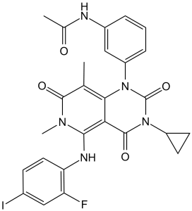
O=C(C)NC1C=C(N2C3C(=C(N(C)C(C=3C)=O)NC3C(F)=CC(I)=CC=3)C(=O)N(C3CC3)C2=O)C=CC=1
|
| InChi Key |
LIRYPHYGHXZJBZ-UHFFFAOYSA-N
|
| InChi Code |
InChI=1S/C26H23FIN5O4/c1-13-22-21(23(31(3)24(13)35)30-20-10-7-15(28)11-19(20)27)25(36)33(17-8-9-17)26(37)32(22)18-6-4-5-16(12-18)29-14(2)34/h4-7,10-12,17,30H,8-9H2,1-3H3,(H,29,34)
|
| 化学名 |
N-[3-[3-cyclopropyl-5-(2-fluoro-4-iodoanilino)-6,8-dimethyl-2,4,7-trioxopyrido[4,3-d]pyrimidin-1-yl]phenyl]acetamide
|
| 别名 |
JTP-74057; GSK 1120212; GSK1120212; GSK-1120212; JTP74057; Trametinib. Trade name: Mekinist
|
| HS Tariff Code |
2934.99.9001
|
| 存储方式 |
Powder -20°C 3 years 4°C 2 years In solvent -80°C 6 months -20°C 1 month |
| 运输条件 |
Room temperature (This product is stable at ambient temperature for a few days during ordinary shipping and time spent in Customs)
|
| 溶解度 (体外实验) |
|
|||
|---|---|---|---|---|
| 溶解度 (体内实验) |
配方 1 中的溶解度: ≥ 2.5 mg/mL (4.06 mM) (饱和度未知) in 10% DMSO + 40% PEG300 + 5% Tween80 + 45% Saline (这些助溶剂从左到右依次添加,逐一添加), 澄清溶液。
例如,若需制备1 mL的工作液,可将100 μL 25.0 mg/mL澄清DMSO储备液加入到400 μL PEG300中,混匀;然后向上述溶液中加入50 μL Tween-80,混匀;加入450 μL生理盐水定容至1 mL。 *生理盐水的制备:将 0.9 g 氯化钠溶解在 100 mL ddH₂O中,得到澄清溶液。 配方 2 中的溶解度: ≥ 2.5 mg/mL (4.06 mM) (饱和度未知) in 10% DMSO + 90% Corn Oil (这些助溶剂从左到右依次添加,逐一添加), 澄清溶液。 例如,若需制备1 mL的工作液,可将 100 μL 25.0 mg/mL 澄清 DMSO 储备液添加到 900 μL 玉米油中并混合均匀。 View More
配方 3 中的溶解度: 4% DMSO+corn oil: 3mg/mL 配方 4 中的溶解度: 6.67 mg/mL (10.84 mM) in 0.5%HPMC 1%Tween80 (这些助溶剂从左到右依次添加,逐一添加), 悬浊液; 超声助溶。 1、请先配制澄清的储备液(如:用DMSO配置50 或 100 mg/mL母液(储备液)); 2、取适量母液,按从左到右的顺序依次添加助溶剂,澄清后再加入下一助溶剂。以 下列配方为例说明 (注意此配方只用于说明,并不一定代表此产品 的实际溶解配方): 10% DMSO → 40% PEG300 → 5% Tween-80 → 45% ddH2O (或 saline); 假设最终工作液的体积为 1 mL, 浓度为5 mg/mL: 取 100 μL 50 mg/mL 的澄清 DMSO 储备液加到 400 μL PEG300 中,混合均匀/澄清;向上述体系中加入50 μL Tween-80,混合均匀/澄清;然后继续加入450 μL ddH2O (或 saline)定容至 1 mL; 3、溶剂前显示的百分比是指该溶剂在最终溶液/工作液中的体积所占比例; 4、 如产品在配制过程中出现沉淀/析出,可通过加热(≤50℃)或超声的方式助溶; 5、为保证最佳实验结果,工作液请现配现用! 6、如不确定怎么将母液配置成体内动物实验的工作液,请查看说明书或联系我们; 7、 以上所有助溶剂都可在 Invivochem.cn网站购买。 |
| 制备储备液 | 1 mg | 5 mg | 10 mg | |
| 1 mM | 1.6250 mL | 8.1249 mL | 16.2499 mL | |
| 5 mM | 0.3250 mL | 1.6250 mL | 3.2500 mL | |
| 10 mM | 0.1625 mL | 0.8125 mL | 1.6250 mL |
1、根据实验需要选择合适的溶剂配制储备液 (母液):对于大多数产品,InvivoChem推荐用DMSO配置母液 (比如:5、10、20mM或者10、20、50 mg/mL浓度),个别水溶性高的产品可直接溶于水。产品在DMSO 、水或其他溶剂中的具体溶解度详见上”溶解度 (体外)”部分;
2、如果您找不到您想要的溶解度信息,或者很难将产品溶解在溶液中,请联系我们;
3、建议使用下列计算器进行相关计算(摩尔浓度计算器、稀释计算器、分子量计算器、重组计算器等);
4、母液配好之后,将其分装到常规用量,并储存在-20°C或-80°C,尽量减少反复冻融循环。
计算结果:
工作液浓度: mg/mL;
DMSO母液配制方法: mg 药物溶于 μL DMSO溶液(母液浓度 mg/mL)。如该浓度超过该批次药物DMSO溶解度,请首先与我们联系。
体内配方配制方法:取 μL DMSO母液,加入 μL PEG300,混匀澄清后加入μL Tween 80,混匀澄清后加入 μL ddH2O,混匀澄清。
(1) 请确保溶液澄清之后,再加入下一种溶剂 (助溶剂) 。可利用涡旋、超声或水浴加热等方法助溶;
(2) 一定要按顺序加入溶剂 (助溶剂) 。
Platform Study of JDQ443 in Combinations in Patients With Advanced Solid Tumors Harboring the KRAS G12C Mutation
CTID: NCT05358249
Phase: Phase 1/Phase 2 Status: Active, not recruiting
Date: 2024-11-15
|
|
|
|
InvivoChem的所有产品仅用于作科学研究,不面向患者销售
Copyright 2020 InvivoChem LLC | All Rights Reserved 粤ICP备20063088号-1


 COA
COA


 463611831
