| 规格 | 价格 | 库存 | 数量 |
|---|---|---|---|
| 25mg |
|
||
| 50mg |
|
||
| 100mg |
|
||
| 250mg |
|
||
| 500mg |
|
||
| 1g |
|
||
| Other Sizes |
|
| 靶点 |
Nucleoside analogue; Influenza virus
|
|---|---|
| 体外研究 (In Vitro) |
Triazavirin 对蜱传脑炎病毒的有效性是在敏感的细胞培养物中测量的。 Triazavirin 在 SKEV 细胞培养物中的浓度为 128 mcg/mL,可有效抑制蜱传脑炎病毒(Sofiin 株)的繁殖[2]。
在第一阶段,我们研究了三氮唑核苷钠水合物(Triazavirin sodium hydrate,Riamilovir)和乙酰水杨酸对体外血小板聚集的影响。ADP诱导的兔全血对照阻抗为8.3 Ω。100 μM浓度的参比药物将血小板聚集幅度降至3.4 Ω,相当于相对于对照显著抑制了59.8%的血小板功能活性(表1)。将乙酰水杨酸浓度降至10和1 μM后,血小板聚集幅度分别降至5.5和6.6 Ω。因此,在上述浓度下,乙酰水杨酸分别抑制了23.7%和11.6%的血小板聚集。参比药物的IC50为57.5 μM。 100 μM浓度的三氮唑核苷钠(Riamilovir)将ADP诱导的血小板聚集幅度降至6.9 Ω,即抑制了17.1%的聚集过程(表1)。在全血与LPS孵育时,血小板聚集幅度从8.3显著增加至11.9 Ω,表明血小板止血系统在巨噬细胞激活后活性增强(表2)。研究药物的抗血小板活性评估基于ADP诱导的完整血小板与LPS处理血小板聚集水平差异范围。 100 μM浓度的乙酰水杨酸将血小板聚集幅度显著降至8.91 Ω,即抑制了83.2%的聚集过程(表2,图1)。10和1 μM浓度下,乙酰水杨酸分别抑制了52.4%和3.2%的活性,血小板聚集幅度分别降至10.02和11.8 Ω。乙酰水杨酸的IC50值为12.4 μM(表2)。因此,在LPS刺激巨噬细胞后,参比药物的活性比在完整血液中提高了4.6倍。 三氮唑核苷钠(Riamilovir)在LPS存在下显示出高抗血小板活性:100 μM浓度下抑制了96.3%的血小板聚集(图1),并将该过程幅度降至8.43 Ω(表2)。当riamilovir浓度降至10和1 μM时,血小板聚集幅度分别降至9.9和10.9 Ω,即分别抑制了56.1%和26.8%的血小板聚集(图1)。riamilovir的IC50为5.2 μM。因此,体外实验表明,在LPS刺激巨噬细胞的条件下,riamilovir的IC50值比乙酰水杨酸高2.4倍。[4] |
| 体内研究 (In Vivo) |
研究了三氮唑核苷治疗白化小鼠实验性森林泉脑炎的有效性。研究结果表明,高剂量(200-400 mg/kg)的三氮杂韦林可以适度保护受感染的动物。测试组的动物寿命显着延长(从 4.1 天延长至 4.8 天),并且靶器官中病毒积累量显着下降[3]。
第二阶段旨在验证三氮唑核苷钠(Riamilovir)在体内实验是否具有相同效应。对照组大鼠ADP诱导的血小板聚集幅度为7.9Ω。20mg/kg剂量的抗病毒药物Riamilovir使血小板聚集幅度降至5.57Ω,血小板功能活性抑制率达29.4%(表3),证实该药物在体内具有抗血小板作用。 静脉注射LPS的大鼠,其ADP诱导的血小板聚集幅度较空白对照组显著升高(达10.9Ω),表明高细胞因子血症可激活血小板。Riamilovir将血小板聚集幅度降至8.98Ω,在细胞因子中毒条件下的抗血小板活性较正常动物提高2.2倍。[4] 两种治疗方案中,三氮唑核苷钠(Riamilovir)均能缩短住院周期。每日高剂量给药组患者住院时间最短。该药物可减轻疾病全身感染症状的持续时间和严重程度,其中每日1250mg、持续5天给药方案的患者发热总时长和呼吸道综合征持续时间最短,且未记录不良反应。该剂量组在治疗第6天实现100%的急性呼吸道病毒感染病原体清除率。 结论:三氮唑核苷钠(Riamilovir)在两种治疗方案中均表现出临床有效性和良好安全性。每日1250mg给药方案能产生更显著的临床效果,研究组在住院第6天即实现病原体完全清除。[5] TZV/三氮唑病毒(利阿昔洛韦)在动物模型中的抗流感活性。[6] 如表3所示,当根据治疗和预防方案给药时(感染前24和1小时以及感染后24、48和72小时),TZV保护小鼠免受A型和B型流感病毒引起的死亡。TZV以1至200mg/kg体重的剂量范围通过i.g.途径给药。确定最佳有效剂量为50至100mg/kg体重。从数据中可以明显看出,TZV和金刚乙胺对感染血清型a流感病毒(a/Aichi/2/68[H3N2])的小鼠提供了相似水平的保护,但感染B型(B/Lee/40)并以大约相同剂量治疗的动物的存活率是金刚乙胺的三到四倍。因此,TZV可以保护65%至75%的感染A或B病毒的小鼠。当应用TZV给药的治疗(+24小时、+48小时和+72小时)或预防(-24小时和-1小时)方案时,TZV也是有效的(数据未显示)。同样值得注意的是,TZV的毒性较低:小鼠腹腔注射TZV后,LD50为1400±120mg/kg体重,肌肉注射TZV的LD50为2200±96mg/kg体重。潜在抗病毒药物的基本特征是它们的稳定性、代谢转化、药代动力学和生物利用度。 |
| 酶活实验 |
本研究依据现行《新药理学物质临床前研究手册》要求进行。三氮唑核苷钠水合物(Triazavirin sodium hydrate,Riamilovir)作为抗病毒药物被选为研究对象。体外实验中以抗血小板药物乙酰水杨酸作为对照药,选择依据是该药物作为循证等级较高的抗血小板剂被广泛应用。
三氮唑核苷钠水合物(Riamilovir)使用生理盐水溶解;对照药先溶于30μl DMSO,再用生理盐水稀释至所需体积。采用Chrono-Log-700双通道发光聚集仪,通过阻抗法检测药物对血小板聚集的影响。
体外实验用兔血经耳缘静脉自由滴落法采集,用3.8%枸橼酸钠(9:1)抗凝。取450μl恒定体积全血用于研究。将100μM浓度的药物直接加入含全血的比色杯,5μM ADP作为血小板聚集诱导剂。
若显示高抗血小板活性,为计算IC50值(抑制50%血小板聚集的浓度),需追加检测10μM和1μM浓度样本。同时在高细胞因子血症条件下分析抗血小板活性:将终浓度20μM的LPS溶液(大肠杆菌O111:B4)与受试药同步加入全血样本比色杯,孵育5分钟后加入血小板聚集诱导剂。[4]
|
| 细胞实验 |
与活性药物利巴韦林相比,在敏感细胞培养物中评估了三唑韦林对蜱传脑炎病毒的疗效。在128 mcg/ml的浓度下,三唑韦灵通过在SKEV细胞培养物内积累而对蜱传乙脑病毒繁殖(Sofin株)具有抑制活性[2]。
CAM模型中抗病毒活性的体外研究[6] 选取11-13日龄鸡胚绒毛尿囊膜(CAM)剪切成约1mm³碎片,悬浮于含青霉素和硫酸链霉素的Hanks盐溶液中。将含病毒的起始尿囊液按10⁻¹至10⁻⁷梯度稀释后加入单层细胞长满的孔板,每孔悬浮液37℃孵育1小时,随后加入不同浓度的三氮唑核苷(Riamilovir)TZV水溶液。参照文献26方法,经36-37℃孵育48小时后,通过血凝素滴度测定和空斑试验评估抗病毒活性。 三氮唑核苷(Riamilovir)在兔肝匀浆中的稳定性[6] 按类似文献方法制备兔肝匀浆,反应体系含25mg/ml蛋白和500μM TZV,37℃孵育。定时取样后立即加入预冷甲醇至66%(v/v)终止反应,10000×g离心4分钟收集沉淀。上清液经SpeedVac冷冻干燥后,残渣水溶解,通过前述HPLC方法分析产物浓度(按峰面积定量)。 TZV/三氮唑核苷(Riamilovir)在细胞培养中的代谢[6] 将TZV水溶液加入含6×10⁶个HEK 293T肾细胞或Huh7肝细胞单层的培养皿,终浓度1mM。细胞与化合物共孵育1.5小时或24小时后,用PBS缓冲液洗涤三次,等体积PBS重悬并通过三次冻融法裂解。加入等体积6%三氟乙酸离心后取上清,用饱和Na₂CO₃调至中性pH,按上述HPLC方法分析代谢产物。 |
| 动物实验 |
对白化小鼠实验性森林-泉脑炎中,三唑韦林与活性药物利巴韦林®的治疗效果比较研究表明,高剂量(200-400 mg/kg)三唑韦林对感染动物具有中等程度的保护作用。试验组动物的寿命显著延长(从4.1天延长至4.8天),且靶器官(脑)中病毒的积累量显著降低(p ≤ 0.05)[3]。
体内实验采用大鼠,分为4组(每组6只):两组未给予LPS(完整大鼠和灌胃给予三唑韦林钠水合物(利阿米洛韦)的大鼠),两组LPS中毒(对照组大鼠静脉注射LPS,实验组大鼠静脉注射LPS并灌胃利阿米洛韦)。在采血前1小时(对应于血液中药物浓度峰值),使用无创胃探针经胃内灌注给予大鼠20 mg/kg剂量的利米洛韦(相当于人类剂量,该剂量使用种间转换因子计算)。大鼠用水合氯醛(400 mg/kg,腹腔注射)麻醉,并从腹主动脉(如上所述,血液稳定剂)获取研究用生物材料。通过尾静脉注射2 mg/kg脂多糖(LPS)建立细胞因子风暴模型。在LPS给药前1小时经口给予利米洛韦,并在LPS给药后4小时采血。对照组经胃内灌注等体积的蒸馏水。使用GraphPad Prism 8.0软件进行统计分析(单因素方差分析,Bonferroni校正,p<0.05)。使用 Microsoft Excel 2020 内置函数计算各组的 IC50、均值和标准差。[4] 目的:评估不同给药方案下抗病毒药物三氮唑韦林钠水合物(利阿米洛韦)治疗非冠状病毒(SARS-CoV-2)病因引起的急性呼吸道病毒感染(ARVI)患者的临床疗效和安全性。 材料与方法:本研究纳入 150 例年龄在 18-27 岁之间的 ARVI 患者(50 例患者接受三氮唑韦林钠水合物(利阿米洛韦)治疗,剂量为 250 mg,每日 3 次,疗程 5 天;50 例患者接受利阿米洛韦治疗,剂量为 250 mg,每日 5 次,疗程 5 天,属于超适应症用药;50 例患者仅接受抗病毒药物治疗)。治疗)。[5] 三唑韦林(Riamilovir)在动物模型中的抗流感活性。[6] 评估了感染A/Aichi/2/68 (H3N2)或B/Lee/40流感病毒的CBA小鼠的抗病毒活性。每组小鼠包含20只。在轻度乙醚麻醉下,通过鼻内途径给予1和10个半数致死剂量(LD50)的病毒。根据以下三种方案之一,通过胃内灌注(ig)途径给予TZV水溶液(0.2 ml):治疗和预防方案(感染前24小时和1小时[-24和-1 h]以及感染后24、48和72小时[+24、+48和+72 h])、预防方案(-24 h和-1 h)或治疗方案(+24 h、+48 h和+72 h)。以金刚烷胺作为对照。观察动物14天,每日记录对照组和实验组的死亡情况。基于这些数据,计算TZV对动物的保护程度,并与金刚烷胺的保护程度进行比较。 兔体内的药代动力学。 [6] 对兔子进行单次三唑韦林(Riamilovir)/TZV灌胃给药时,将动物(n = 4)用10:1的乙醚-氟烷混合物麻醉,并将聚氨酯胃肠管插入15厘米深。三唑韦林(Riamilovir)/TZV以水溶液(12毫升)的形式给药,剂量为105毫克/公斤体重。对4只兔子(n = 4)进行静脉给药时,按照参考文献11的方法,将TZV(4.3 mg/kg体重)溶于生理盐水(1 ml)中,经耳缘静脉注射1分钟。在给药后24小时内的预定时间点,从耳缘静脉自然采集血样(平均1 ml),置于含有5 μl肝素(5,000 U/ml)的微量离心管中。摇匀后,取出0.5 ml血样,加入1 ml甲醇混匀,并于-24℃保存。对照组血样在给药前采集。进行高效液相色谱(HPLC)分析前,将样品以1,500 × g离心10分钟;上清液真空蒸发;残余物溶于100 μl水中。然后,在上述条件下,采用高效液相色谱法(HPLC)分析样品。 药代动力学参数采用 Kinetica 程序计算。采用 Thermo Kinetica 程序的血管外非房室模型研究了肠内给药后的药代动力学。对于静脉给药,则采用非房室静脉输注模型。测定的参数包括:血浆浓度-时间曲线下面积(AUCtot)、表观消除半衰期(T1/2)、血浆中化合物的最大浓度(Cmax)、达峰时间(Tmax)和平均滞留时间(MRT)。化合物的肠内生物利用度(F)计算公式为 (AUCi.g./dosei.g.)/(AUCi.v./dosei.v.),其中 AUCi.g. 为肠内给药后化合物的 AUC,dosei.g. 为肠内给药剂量,AUCi.v. 为静脉给药后的 AUC,dosei.v. 为静脉给药剂量。是静脉注射剂量。血浆总清除率 (CL) 计算公式为剂量/AUC。TZV 稳态分布容积 (Vss) 计算公式为 CL × MRT。 |
| 药代性质 (ADME/PK) |
TZV/三唑韦林(Riamilovir)单次静脉注射或肌注给药后,在兔体内的药代动力学参数。[6]
为了确定TZV的肌注生物利用度,将该化合物通过肌注和静脉注射途径给予兔子(n = 4)。在给药后24小时内的预定时间点采集血样,并用高效液相色谱法(HPLC)进行分析。肌注给药后10分钟内,在兔血中仅观察到TZV峰,保留时间(Tret)为22.5分钟;而2小时后,出现一个新的峰(M1),其保留时间随时间增加,为27.5分钟。图2A显示了给药(105 mg/kg体重)后12小时内兔血中TZV和M1的浓度。 TZV 的血药浓度峰值 (Cmax) 为 1.1 mg/L,达到时间为 0.40 ± 0.16 小时,消除半衰期为 1.1 小时。TZV 的清除率 (CL) 为 37.0 ± 11.2 L/h·kg,稳态分布容积 (Vss) 为 83.5 ± 19.2 L/kg。代谢物 M1 的浓度在 3 小时内升高,随后在接下来的 5 小时内略有下降。给兔子静脉注射 4.3 mg/kg 剂量的 TZV 后,血浆中 TZV 的浓度迅速下降,半衰期 (T1/2) 为 0.9 小时(图 2B)。与灌胃给药的结果相反,未检测到代谢物 M1。在整个实验期间(24 小时),观察到的唯一产物是 TZV。表4总结了TZV及其代谢物的药代动力学参数值。TZV在兔体内的免疫生物利用度(F)计算为12.5%。 TZV/三唑韦林(利阿米洛韦)代谢物(M1)在兔肝匀浆中的生成。[6] TZV代谢物很可能是在肝脏或肾脏中生成的。为了验证这一假设,将TZV与兔肝匀浆孵育。图3显示了肝匀浆与500 μM TZV孵育后的HPLC分析结果。如图所示,孵育10分钟后出现了一个新的峰,且峰强度随时间增加。该峰的保留时间与兔血中经肠内注射TZV后形成的代谢物的保留时间吻合良好。 TZV/三唑韦林(利阿米洛韦)在HEK 293T肾细胞和Huh7肝细胞培养物中的代谢。[6] 将HEK 293T或Huh7细胞培养物与500 μM TZV孵育不同时间。对细胞提取物进行HPLC分析,结果显示存在TZV及其代谢物,该代谢物的保留时间与兔肝匀浆中形成的M1代谢物以及经肠内注射TZV后在兔血中检测到的代谢物的保留时间一致(图4)。 TZV/三唑韦林(利阿米洛韦)代谢物结构。 [6] 通过比较HPLC分析、紫外光谱和质谱数据的保留时间,确认了在细胞培养物和兔血中发现的代谢物(包括经肠内给药和在兔肝匀浆中孵育TZV后)的身份。图5显示了TZV及其与兔肝匀浆孵育后形成的代谢物的紫外光谱图。TZV和M1的光谱图明显不同:TZV有两个λmax,分别位于257 nm和360 nm,而M1的λmax位于249 nm和320 nm。我们推测TZV的硝基可以被还原生成2-甲基硫代-6-氨基-1,2,4-三唑并[5,1-c]-1,2,4-三嗪-7(4H)-酮(AMTZV)。我们合成了AMTZV,并证实其紫外吸收光谱与M1的紫外吸收光谱相同。为了验证肝匀浆中生成的M1代谢物的结构,我们将其质谱图与合成化合物的质谱图进行了比较(图6A和B)。主要离子为[MH]−,分子量为197,与AMTZV的分子量一致。此外,还出现了TZV的[MH]−离子峰227。对于离子197和227,我们观察到了相应的卫星离子,分别为198 [M + 1-H]−和199 [M + 2-H]−,以及228 [M + 1-H]−和229 [M + 1-H]−。峰213对应的离子结构尚不明确。峰 167 可能与代谢物降解产物有关,通过混合 [M1 + AMTZV] 和 [M1 + 15N-AMTZV] 证实了这一点(数据未显示)。 |
| 毒性/毒理 (Toxicokinetics/TK) |
蛋白质结合
关于三唑病毒素的蛋白质结合数据尚不明确。 |
| 参考文献 |
|
| 其他信息 |
本文阐述了一种新型的带正电荷脂质体包覆改性壳聚糖的方法。脂质体通过孔径分别为0.2 μm和0.1 μm的无机膜(Anotop)逐步挤出制备。壳聚糖衍生物通过Ugi多组分反应合成。制备了多组脂质体,并比较了它们的粒径、多分散指数(PDI)、zeta电位和稳定性等性质。研究了各种添加剂的影响,并确定了脂质膜的最佳组成。添加不带电荷的脂肪酸酯可使挤出法制备的脂质体的粒径减小至145-150 nm,PDI为0.13-0.15。将制备的脂质体负载新型抗病毒药物三唑韦林,并用于测定其释放曲线。三唑韦林以盐的形式包覆在脂质体层中,盐由生物相容性胆碱衍生物和限制性脂肪酸组成。采用合适的脂质组成制备了大量改性壳聚糖包覆的脂质体。结果表明,脂质体与多糖层的适当组合可将胶体稳定性延长至3个月,并展现出广泛的表面修饰功能。[1]
在敏感细胞培养物中,评估了Triazavirin对蜱传脑炎病毒的疗效,并与活性药物利巴韦林进行了比较。结果表明,浓度为128 mcg/ml的Triazavirin可通过在SKEV细胞培养物中的积累抑制蜱传脑炎病毒(Sofiin株)的复制。[2] 在敏感细胞培养物中,评估了Triazavirin对蜱传脑炎病毒的疗效,并与活性药物利巴韦林进行了比较。在浓度为 128 mcg/ml 时,三唑韦林可通过在 SKEV 细胞培养物中的积累,有效抑制蜱传脑炎病毒(Sofiin 株)的复制。[3] 我们研究了抗病毒药物利阿米洛韦在有无 LPS 存在下对 ADP 诱导的血小板聚集的影响。与乙酰水杨酸(参考药物)不同,利阿米洛韦在体外未表现出抗血小板作用。然而,它显著抑制了 LPS 处理的血液样本中的血小板反应性,其 IC50 值是乙酰水杨酸的 2.2 倍。在体内实验中,在细胞因子风暴条件下,利阿米洛韦可抑制大鼠 64% 的血小板聚集。[4] 甲型和乙型流感病毒会在人群中引起周期性大流行。目前获准用于对抗流感病毒感染的抗病毒药物种类有限。我们研究了一种新型有效的甲型和乙型流感病毒抑制剂——三唑韦林[2-甲基硫代-6-硝基-1,2,4-三唑并[5,1-c]-1,2,4-三嗪-7(4I)-酮] (TZV)。TZV能够抑制细胞培养和鸡绒毛尿囊膜中流感病毒的复制,并能保护小鼠免于甲型和乙型流感病毒感染导致的死亡。TZV对金刚烷胺耐药的流感病毒株和禽流感A型病毒H5N1株也有效。在给兔子服用TZV后,我们计算了其药代动力学参数和生物利用度。在灌胃给予TZV后,在IAK 293T和Huh7细胞培养物、肝脏匀浆和兔血中发现了TZV代谢物AMTZV [2-甲基硫代-6-氨基-1,2,4-三唑并[5,1-s]-1,2,4-三嗪(e)-7(4I)-酮]。AMTZV无毒,且在细胞培养中不抑制流感病毒。该代谢物很可能是TZV的消除产物。[6] |
| 分子式 |
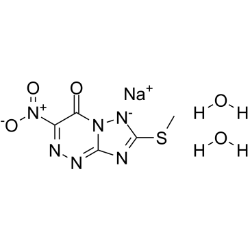
C₅H₇N₆NAO₅S
|
|---|---|
| 分子量 |
286.20
|
| 精确质量 |
286.009
|
| 元素分析 |
C, 20.98; H, 2.47; N, 29.37; Na, 8.03; O, 27.95; S, 11.20
|
| CAS号 |
928659-17-0
|
| 相关CAS号 |
116061-59-7 (sodium);123606-06-4 (free);928659-17-0 (sodium hydrate);
|
| PubChem CID |
52947239
|
| 外观&性状 |
Light yellow to yellow solid powder
|
| LogP |
2.0
|
| tPSA |
152Ų
|
| 氢键供体(HBD)数目 |
2
|
| 氢键受体(HBA)数目 |
10
|
| 可旋转键数目(RBC) |
1
|
| 重原子数目 |
18
|
| 分子复杂度/Complexity |
262
|
| 定义原子立体中心数目 |
0
|
| SMILES |
[Na].O=C1N2C(NC(SC)=N2)=NN=C1[N+](=O)[O-].O
|
| InChi Key |
GDVSBVWTWGUDAW-UHFFFAOYSA-M
|
| InChi Code |
InChI=1S/C5H4N6O3S.Na.2H2O/c1-15-5-6-4-8-7-2(11(13)14)3(12)10(4)9-5;;;/h12H,1H3;;2*1H2/q;+1;;/p-1
|
| 化学名 |
sodium;7-methylsulfanyl-3-nitro-[1,2,4]triazolo[5,1-c][1,2,4]triazin-4-olate;dihydrate
|
| 别名 |
Triazavirin; Riamilovir; TZV; Triazavirin; 928659-17-0; Riamilovir sodium dihydrate; Sodium 7-(methylthio)-3-nitro-4-oxo-4H-[1,2,4]triazolo[5,1-c][1,2,4]triazin-6-ide dihydrate; G1JE34QF2S; sodium;7-methylsulfanyl-3-nitro-[1,2,4]triazolo[5,1-c][1,2,4]triazin-4-olate;dihydrate; [1,2,4]Triazolo[5,1-c][1,2,4]triazin-4(6H)-one, 7-(methylthio)-3-nitro-,sodium salt, hydrate (1:1:2); (1,2,4)Triazolo(5,1-C)(1,2,4)triazin-4(6H)-one, 7-(methylthio)-3-nitro-, sodium salt, hydrate; Riamilovir sodium hydrate
|
| HS Tariff Code |
2934.99.9001
|
| 存储方式 |
Powder -20°C 3 years 4°C 2 years In solvent -80°C 6 months -20°C 1 month 注意: 请将本产品存放在密封且受保护的环境中,避免吸湿/受潮。 |
| 运输条件 |
Room temperature (This product is stable at ambient temperature for a few days during ordinary shipping and time spent in Customs)
|
| 溶解度 (体外实验) |
DMSO: ~125 mg/mL (~436.8 mM)
H2O: ~50 mg/mL (~174.7 mM) |
|---|---|
| 溶解度 (体内实验) |
配方 1 中的溶解度: ≥ 2.08 mg/mL (7.27 mM) (饱和度未知) in 10% DMSO + 40% PEG300 + 5% Tween80 + 45% Saline (这些助溶剂从左到右依次添加,逐一添加), 澄清溶液。
例如,若需制备1 mL的工作液,可将100 μL 20.8 mg/mL澄清DMSO储备液加入400 μL PEG300中,混匀;然后向上述溶液中加入50 μL Tween-80,混匀;加入450 μL生理盐水定容至1 mL。 *生理盐水的制备:将 0.9 g 氯化钠溶解在 100 mL ddH₂O中,得到澄清溶液。 配方 2 中的溶解度: ≥ 2.08 mg/mL (7.27 mM) (饱和度未知) in 10% DMSO + 90% (20% SBE-β-CD in Saline) (这些助溶剂从左到右依次添加,逐一添加), 澄清溶液。 例如,若需制备1 mL的工作液,可将 100 μL 20.8 mg/mL澄清DMSO储备液加入900 μL 20% SBE-β-CD生理盐水溶液中,混匀。 *20% SBE-β-CD 生理盐水溶液的制备(4°C,1 周):将 2 g SBE-β-CD 溶解于 10 mL 生理盐水中,得到澄清溶液。 请根据您的实验动物和给药方式选择适当的溶解配方/方案: 1、请先配制澄清的储备液(如:用DMSO配置50 或 100 mg/mL母液(储备液)); 2、取适量母液,按从左到右的顺序依次添加助溶剂,澄清后再加入下一助溶剂。以 下列配方为例说明 (注意此配方只用于说明,并不一定代表此产品 的实际溶解配方): 10% DMSO → 40% PEG300 → 5% Tween-80 → 45% ddH2O (或 saline); 假设最终工作液的体积为 1 mL, 浓度为5 mg/mL: 取 100 μL 50 mg/mL 的澄清 DMSO 储备液加到 400 μL PEG300 中,混合均匀/澄清;向上述体系中加入50 μL Tween-80,混合均匀/澄清;然后继续加入450 μL ddH2O (或 saline)定容至 1 mL; 3、溶剂前显示的百分比是指该溶剂在最终溶液/工作液中的体积所占比例; 4、 如产品在配制过程中出现沉淀/析出,可通过加热(≤50℃)或超声的方式助溶; 5、为保证最佳实验结果,工作液请现配现用! 6、如不确定怎么将母液配置成体内动物实验的工作液,请查看说明书或联系我们; 7、 以上所有助溶剂都可在 Invivochem.cn网站购买。 |
| 制备储备液 | 1 mg | 5 mg | 10 mg | |
| 1 mM | 3.4941 mL | 17.4703 mL | 34.9406 mL | |
| 5 mM | 0.6988 mL | 3.4941 mL | 6.9881 mL | |
| 10 mM | 0.3494 mL | 1.7470 mL | 3.4941 mL |
1、根据实验需要选择合适的溶剂配制储备液 (母液):对于大多数产品,InvivoChem推荐用DMSO配置母液 (比如:5、10、20mM或者10、20、50 mg/mL浓度),个别水溶性高的产品可直接溶于水。产品在DMSO 、水或其他溶剂中的具体溶解度详见上”溶解度 (体外)”部分;
2、如果您找不到您想要的溶解度信息,或者很难将产品溶解在溶液中,请联系我们;
3、建议使用下列计算器进行相关计算(摩尔浓度计算器、稀释计算器、分子量计算器、重组计算器等);
4、母液配好之后,将其分装到常规用量,并储存在-20°C或-80°C,尽量减少反复冻融循环。
计算结果:
工作液浓度: mg/mL;
DMSO母液配制方法: mg 药物溶于 μL DMSO溶液(母液浓度 mg/mL)。如该浓度超过该批次药物DMSO溶解度,请首先与我们联系。
体内配方配制方法:取 μL DMSO母液,加入 μL PEG300,混匀澄清后加入μL Tween 80,混匀澄清后加入 μL ddH2O,混匀澄清。
(1) 请确保溶液澄清之后,再加入下一种溶剂 (助溶剂) 。可利用涡旋、超声或水浴加热等方法助溶;
(2) 一定要按顺序加入溶剂 (助溶剂) 。
 Deoxyribosyl dihydropyrimido[4,5-c][1,2]oxazin-7-one
 Lobucavir
 CMX-521
 2,6-Bis(4-morpholinyl)-9-bD-ribofuranosyl-9H-purine
InvivoChem的所有产品仅用于作科学研究,不面向患者销售
Copyright 2020 InvivoChem LLC | All Rights Reserved 粤ICP备20063088号-1


 COA
COA
 463611831
