| 规格 | 价格 | 库存 | 数量 |
|---|---|---|---|
| 10 mM * 1 mL in DMSO |
|
||
| 1mg |
|
||
| 5mg |
|
||
| 10mg |
|
||
| 25mg |
|
||
| 50mg |
|
||
| 100mg |
|
||
| 250mg |
|
||
| Other Sizes |
|

| 靶点 |
ERK1/2
|
|---|---|
| 体外研究 (In Vitro) |
Akt 和 ERK1/2 由 Yoda1 以不依赖于 Piezo1 的方式激活(0–6 μM,5 分钟)[2]。 Yoda1(1.5 μM,5 分钟)可抑制 Rac1 激活 [3]。
Piezo1的抑制或激活影响体外ICH模型中HT22细胞的凋亡[1] 根据Wang等人(2022)建立了体外ICH模型。我们首先用不同浓度(0、10、20、40、60、80、100、120或140μM)的氯化血红素溶液处理体外培养的HT22细胞,以找出氯化血红素的IC50。我们发现氯化血红素的IC50为100μM(图6a)。然后,我们使用IC50(100μM)的氯化血红素溶液处理HT22细胞24小时,观察氯化血红素对HT22细胞存活率和HT22细胞中指定基因蛋白质水平的影响。在氯化血红素处理后1小时,表面上就观察到HT22细胞变小变圆的异常形态变化(图6b)。活细胞计数的定量分析显示,氯化血红素处理24小时后,活HT22细胞的数量减少了50%(图6c)Yoda-1(一种Piezo1激活剂)处理进一步减少了用氯化血红素预处理的活HT22细胞的数量。相比之下,GsMTx4(Piezo1阻断剂)治疗显著逆转了氯化血红素预处理后活HT22细胞数量的大幅减少(图6c)。 Yoda-1诱导内皮细胞中Akt和ERK1/2的激活。 钆(Gd3+)可以消除Yoda-1对Akt的激活,但不能激活ERK1/2。 钌红(RR)能有效阻断Yoda1诱导的Akt激活。 GsMTx4是Piezo1的强效阻断剂,不会抑制Yoda1诱导的Akt或ERK1/2磷酸化。[2] Yoda-1对EGF刺激的大颗粒细胞增多的抑制作用依赖于Piezo1。 KCa3.1激活是Yoda-1抑制褶皱形成所必需的。 在这项研究中,我们发现,Yoda-1处理导致Rac1活化受到抑制,从而抑制了外周膜褶皱的形成(图3)。据报道,在癌症细胞中敲低Piezo1导致Rac1激活51。这项先前的研究可能表明Piezo1激活抑制Rac1,尽管其机制尚不清楚。重要的是,我们进一步表明,Yoda1对大白细胞增多的抑制作用取决于通过Piezo1的细胞外Ca2+内流(图4)。即使在Yoda1存在的情况下,对钙激活钾通道KCa3.1的抑制也恢复了EGF刺激的膜褶皱形成(即肌动蛋白重排)(图5A,B)。这表明,在KCa3.1被抑制的条件下,即使在Yoda1存在的情况下,EGF刺激的肌动蛋白重排也可以被诱导。因此,我们提出Piezo1激活后KCa3.1激活可能导致肌动蛋白重排的抑制。之前的一项研究报告称,KCa3.1的激活在膜褶皱闭合中至关重要,这是大细胞吞噬过程的后期21。另一方面,我们发现KCa3.1激活剂和Yoda1也会损害大白细胞增多症(图5C-E)。总之,我们的结果表明,KCa3.1的适当时间激活在大白细胞增多症中很重要,在Yoda1诱导的急性Ca2+内流后,KCa3-1的激活可能会导致肌动蛋白重排的抑制(图5F)[3]。 |
| 体内研究 (In Vivo) |
脑出血后继发性脑损伤是脑出血患者预后不良的主要原因,但其潜在机制尚不清楚。在脑出血小鼠模型中研究了Piezo1在脑出血后脑损伤中的作用。ICH是通过将自体动脉血注入小鼠基底节而建立的。将载体、Piezo1阻断剂、GsMTx4、Piezo-1激活剂、Yoda-1或与甘露醇(尾静脉注射)一起注射到小鼠脑左侧脑室后,通过各种指示方法确定Piezo1水平以及Piezo1在脑出血后神经元损伤、脑水肿和神经功能障碍中的作用。脑出血后24小时,神经元中的Piezo1蛋白水平显著上调(人和小鼠)。作为体外ICH模型,氯化血红素处理后24小时,体外培养的HT22细胞(一种小鼠神经元细胞系)中Piezo1蛋白水平也显著上调。GsMTx4治疗或与甘露醇一起显著下调Piezo1和AQP4水平,显著提高Bcl2水平,维持更多神经元存活,显著恢复脑血流,显著缓解脑水肿,显著降低血清IL-6水平,并几乎完全逆转ICH 24小时组小鼠的神经功能障碍。相比之下,Yoda-1处理达到了相反的效果。综上所述,Piezo1在脑出血后脑损伤的发病机制中起着至关重要的作用,可能成为脑出血临床治疗的靶点。[4]
|
| 酶活实验 |
384-well格式[1]
转染后2天,使用ELx405 CW洗板机用测定缓冲液(1×HBSS,10 mM HEPES,pH7.4)洗涤细胞。将细胞与含有4μM Fluo3和0.04%Pluronic F-127的测定缓冲液一起孵育约60分钟,然后再次用测定缓冲液洗涤。在荧光成像板阅读器(FLIPR)Tetra上监测荧光。为了螯合细胞外钙(1×HBSS含有1.26 mM CaCl2),在2 mM EGTA存在下,在加入指定浓度的Yoda1前1分钟向细胞中加入2 mM乙二醇四乙酸(EGTA)。为了消耗细胞内钙,在加入Yoda1前15分钟加入7.5μM的thapsigargin。使用Yoda1在二甲亚砜(DMSO)中的10mM储备溶液,在测定中产生最多1%的DMSO。使用可变斜率的S形剂量反应拟合浓度反应曲线。[1] 1536-well高通量筛选格式[1] 为了鉴定Piezo1或Piezo2激动剂,我们用等量的mPhoto1和mPhoto2 cDNA共转染细胞。2天后,根据制造商的说明,将转染细胞与Calcium5一起孵育,并在FLIPR Tetra上监测荧光。从LMW诺华筛选库中筛选了约325万种化合物,其中包括公共领域和专有药物样分子,浓度为5μM。在共转染细胞以及单个Piezo1和2转染和对照细胞中,选择约9000次点击(定义为DMSO对照孔上方50%的激活)进行重新测试。由此,Yoda1被确定为潜在的Piezo1激活剂,并被选中进行进一步研究。 |
| 细胞实验 |
蛋白质印迹分析
细胞类型:人冠状动脉内皮细胞 (HCAEC) [2]、A431 细胞 [3] 测试浓度: 0、1.5、 3.0 和 6.0 μM 孵育时间: 5 分钟 实验结果:诱导 Akt 和 ERK1/2 激活,并提高 Akt 和 ERK1/2 的磷酸化水平呈剂量依赖性。抑制 EGF 诱导的 Rac1-GTP 量增加并抑制 Rac1 激活. 体外intracerebral hemorrhage (ICH) 模型[4] 通过氯化血红素处理体外培养的HT22细胞(一种小鼠海马神经元细胞系)来模拟体外ICH模型。根据Wang等人(2022)的研究,HT22细胞与DMEM(10%胎牛血清和1%青霉素-链霉素)预培养12小时。然后,将每种不同浓度(0、10、20、40、60、80、100、120或140μM)的氯化血红素溶液加入细胞培养基中,再培养24小时的HT22细胞。对于GsMTx4或Yoda-1治疗组,在向HT22细胞中添加氯化血红素1小时后添加GsMTx4或Yoda-1。 |
| 动物实验 |
小鼠脑出血(ICH)后Piezo1的抑制或激活[4]
本研究在27只小鼠中探讨了Piezo1阻断剂GsMTx4和Piezo1激活剂Yoda-1对脑出血后脑水肿和神经功能障碍的影响。小鼠被分为五组:(1)对照组(n = 6),(2)脑出血24小时组(n = 6),(3)脑出血72小时组(n = 3),(4)脑出血24小时+GsMTx4(3 μM,n = 6),以及(5)脑出血24小时+Yoda-1(10 μM,n = 6)。详情请参阅补充材料和方法文件(附录A补充数据)。 |
| 参考文献 | |
| 其他信息 |
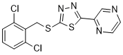
Yoda 1 是一种噻二唑类化合物,其结构为 1,3,4-噻二唑,分别在 2 位和 5 位被吡嗪-2-基和 (2,6-二氯苄基)硫代二基取代。它是机械敏感通道 Piezo1 的选择性激活剂。它既是甘氨酸转运蛋白 2 的抑制剂,又是 Piezo1 的激动剂。Yoda 1 是一种芳香族化合物,属于吡嗪类、噻二唑类、有机硫化物和二氯苯类化合物。
Piezo 离子通道可被多种机械刺激激活,并在脊椎动物和无脊椎动物中发挥生物压力传感器的作用。迄今为止,机械刺激是激活 Piezo 离子通道的唯一途径,是否存在其他激活方式尚不清楚。在本研究中,我们利用基于细胞的荧光检测方法筛选了约325万种化合物,并鉴定出一种名为Yoda1的合成小分子,该分子可作为人源和鼠源Piezo1通道的激动剂。细胞功能研究表明,Yoda1会影响机械诱导反应的敏感性和失活动力学。在人工液滴脂双层中对Yoda1的表征显示,Yoda1可在没有其他细胞成分的情况下激活纯化的Piezo1通道。我们的研究表明,Piezo1通道可被化学方法激活,并提示可能存在内源性Piezo1激动剂。Yoda1将成为研究Piezo1通道调控和功能的重要工具化合物。[1] Piezo1是一种机械敏感性阳离子通道,可在内皮细胞(ECs)中被剪切应力激活。研究表明,Piezo1介导剪切应力诱导的内皮细胞反应,包括钙离子内流增加,以及血管功能,例如血管张力和血压。 Yoda1 是一种选择性 Piezo1 激活剂,已被证实能够模拟内皮细胞 (EC) 中的剪切力诱导反应。由于剪切力诱导的钙离子内流会导致内皮细胞中 Akt 和 ERK1/2 的激活,我们研究了 Yoda1 的作用以及 Piezo1 对其激活的影响。结果表明,Yoda1 能够显著激活内皮细胞中的 Akt 和 ERK1/2。此外,Piezo1 拮抗剂钆和钌红(而非 GsMTx4)能够有效阻断 Yoda1 诱导的 Akt 激活。我们的研究结果表明,Yoda1 诱导的 Akt 和 ERK1/2 激活并不依赖于 Piezo1。[2] 巨胞饮作用是一种伴随肌动蛋白重排驱动的膜变形(例如伪足形成和膜皱褶)的内吞作用,随后形成大型囊泡,即巨胞饮体。 Ras转化的癌细胞通过巨胞饮作用高效获取外源氨基酸以维持生存。因此,抑制巨胞饮作用是一种很有前景的癌症治疗策略。迄今为止,针对巨胞饮作用的特异性抑制剂寥寥无几。本文以机械敏感性离子通道Piezo1为研究对象,发现Piezo1激动剂Yoda1能够有效抑制表皮生长因子(EGF)诱导的巨胞饮作用。Yoda1对细胞膜皱褶形成的抑制作用依赖于Piezo1介导的细胞外Ca2+内流以及钙激活钾通道KCa3.1的激活。这表明Ca2+离子可以调控EGF刺激的巨胞饮作用。我们提出,利用化学方法调控机械敏感性离子通道的活性是抑制巨胞饮作用的潜在途径。[3] |
| 分子式 |
C13H8CL2N4S2
|
|---|---|
| 分子量 |
355.265417098999
|
| 精确质量 |
353.956
|
| 元素分析 |
C, 43.95; H, 2.27; Cl, 19.96; N, 15.77; S, 18.05
|
| CAS号 |
448947-81-7
|
| 相关CAS号 |
448947-81-7;
|
| PubChem CID |
2746822
|
| 外观&性状 |
White to light yellow solid powder
|
| 密度 |
1.6±0.1 g/cm3
|
| 沸点 |
538.4±60.0 °C at 760 mmHg
|
| 闪点 |
279.4±32.9 °C
|
| 蒸汽压 |
0.0±1.4 mmHg at 25°C
|
| 折射率 |
1.714
|
| LogP |
4.89
|
| tPSA |
105
|
| 氢键供体(HBD)数目 |
0
|
| 氢键受体(HBA)数目 |
6
|
| 可旋转键数目(RBC) |
4
|
| 重原子数目 |
21
|
| 分子复杂度/Complexity |
329
|
| 定义原子立体中心数目 |
0
|
| SMILES |
C1=CC(=C(C(=C1)Cl)CSC2=NN=C(S2)C3=NC=CN=C3)Cl
|
| InChi Key |
BQNXBSYSQXSXPT-UHFFFAOYSA-N
|
| InChi Code |
InChI=1S/C13H8Cl2N4S2/c14-9-2-1-3-10(15)8(9)7-20-13-19-18-12(21-13)11-6-16-4-5-17-11/h1-6H,7H2
|
| 化学名 |
2-[5-(2,6-Dichloro-benzylsulfanyl)-[1,3,4]thiadiazol-2-yl]-pyrazine
|
| 别名 |
Yoda1; Yoda-1; GlyT2-IN-1; YODA-1; 2-((2,6-dichlorobenzyl)thio)-5-(pyrazin-2-yl)-1,3,4-thiadiazole; TW6GF9RW6S; 2-[(2,6-dichlorophenyl)methylsulfanyl]-5-pyrazin-2-yl-1,3,4-thiadiazole; Yoda 1
|
| HS Tariff Code |
2934.99.9001
|
| 存储方式 |
Powder -20°C 3 years 4°C 2 years In solvent -80°C 6 months -20°C 1 month |
| 运输条件 |
Room temperature (This product is stable at ambient temperature for a few days during ordinary shipping and time spent in Customs)
|
| 溶解度 (体外实验) |
DMSO : ~15.62 mg/mL (~43.97 mM)
Ethanol : ~5 mg/mL (~14.07 mM) |
|---|---|
| 溶解度 (体内实验) |
配方 1 中的溶解度: ≥ 1.56 mg/mL (4.39 mM) (饱和度未知) in 10% DMSO + 90% (20% SBE-β-CD in Saline) (这些助溶剂从左到右依次添加,逐一添加), 澄清溶液。
例如,若需制备1 mL的工作液,可将100 μL 15.6mg/mL澄清的DMSO储备液加入到900μL 20%SBE-β-CD生理盐水中,混匀。 *20% SBE-β-CD 生理盐水溶液的制备(4°C,1 周):将 2 g SBE-β-CD 溶解于 10 mL 生理盐水中,得到澄清溶液。 配方 2 中的溶解度: ≥ 1.56 mg/mL (4.39 mM) (饱和度未知) in 10% DMSO + 90% Corn Oil (这些助溶剂从左到右依次添加,逐一添加), 澄清溶液。 例如,若需制备1 mL的工作液,可将 100 μL 15.6 mg/mL 澄清 DMSO 储备液添加到 900 μL 玉米油中并混合均匀。 请根据您的实验动物和给药方式选择适当的溶解配方/方案: 1、请先配制澄清的储备液(如:用DMSO配置50 或 100 mg/mL母液(储备液)); 2、取适量母液,按从左到右的顺序依次添加助溶剂,澄清后再加入下一助溶剂。以 下列配方为例说明 (注意此配方只用于说明,并不一定代表此产品 的实际溶解配方): 10% DMSO → 40% PEG300 → 5% Tween-80 → 45% ddH2O (或 saline); 假设最终工作液的体积为 1 mL, 浓度为5 mg/mL: 取 100 μL 50 mg/mL 的澄清 DMSO 储备液加到 400 μL PEG300 中,混合均匀/澄清;向上述体系中加入50 μL Tween-80,混合均匀/澄清;然后继续加入450 μL ddH2O (或 saline)定容至 1 mL; 3、溶剂前显示的百分比是指该溶剂在最终溶液/工作液中的体积所占比例; 4、 如产品在配制过程中出现沉淀/析出,可通过加热(≤50℃)或超声的方式助溶; 5、为保证最佳实验结果,工作液请现配现用! 6、如不确定怎么将母液配置成体内动物实验的工作液,请查看说明书或联系我们; 7、 以上所有助溶剂都可在 Invivochem.cn网站购买。 |
| 制备储备液 | 1 mg | 5 mg | 10 mg | |
| 1 mM | 2.8148 mL | 14.0738 mL | 28.1476 mL | |
| 5 mM | 0.5630 mL | 2.8148 mL | 5.6295 mL | |
| 10 mM | 0.2815 mL | 1.4074 mL | 2.8148 mL |
1、根据实验需要选择合适的溶剂配制储备液 (母液):对于大多数产品,InvivoChem推荐用DMSO配置母液 (比如:5、10、20mM或者10、20、50 mg/mL浓度),个别水溶性高的产品可直接溶于水。产品在DMSO 、水或其他溶剂中的具体溶解度详见上”溶解度 (体外)”部分;
2、如果您找不到您想要的溶解度信息,或者很难将产品溶解在溶液中,请联系我们;
3、建议使用下列计算器进行相关计算(摩尔浓度计算器、稀释计算器、分子量计算器、重组计算器等);
4、母液配好之后,将其分装到常规用量,并储存在-20°C或-80°C,尽量减少反复冻融循环。
计算结果:
工作液浓度: mg/mL;
DMSO母液配制方法: mg 药物溶于 μL DMSO溶液(母液浓度 mg/mL)。如该浓度超过该批次药物DMSO溶解度,请首先与我们联系。
体内配方配制方法:取 μL DMSO母液,加入 μL PEG300,混匀澄清后加入μL Tween 80,混匀澄清后加入 μL ddH2O,混匀澄清。
(1) 请确保溶液澄清之后,再加入下一种溶剂 (助溶剂) 。可利用涡旋、超声或水浴加热等方法助溶;
(2) 一定要按顺序加入溶剂 (助溶剂) 。
|
|
|
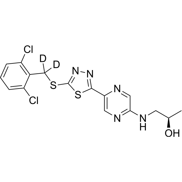 Piezo1 agonist 1-d2
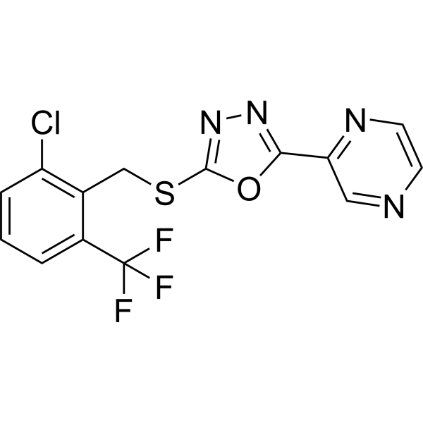 Yaddle1
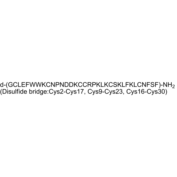 D-GsMTx4
 D-GsMTx4 TFA
InvivoChem的所有产品仅用于作科学研究,不面向患者销售
Copyright 2020 InvivoChem LLC | All Rights Reserved 粤ICP备20063088号-1


 COA
COA


 463611831
