| 规格 | 价格 | 库存 | 数量 |
|---|---|---|---|
| 1mg |
|
||
| 5mg |
|
||
| 10mg |
|
||
| 50mg |
|
||
| Other Sizes |
|

| 靶点 |
FAP (fibroblast activation protein)
|
|---|---|
| 体外研究 (In Vitro) |
FAPI框架的化学修饰导致体外FAP结合增加[2]
为了确定放射性示踪剂的fap结合亲和力(补充表1),使用表达fap的人HT-1080细胞进行了放射性配体结合试验。为了弥补FAP表达率的变化,并允许与导联结构进行直接比较,所有实验都与FAPI-04并行进行。所有化合物都显示出与人FAP的强结合,在孵育1和4小时后的结合值等于或高于FAPI-04(图1)。除FAPI-38外,所有化合物的内化率与FAPI-04相当(24小时后内化率为63.1%;FAPI-04, 97.1%;虽然大多数衍生物在24小时后显示出比FAPI-04更高的结合值,但化合物FAPI-38、-39、-40和-41从表达fap的细胞中被清除的速度要快得多,因此不考虑进行更详细的表征。与FAPI-04类似,所有化合物与结构相关的膜蛋白CD26的结合都可以忽略不计(数据未显示)。 |
| 体内研究 (In Vivo) |
FAPI-4(静脉注射;每只小鼠 30 nmol;一次)在 BALB/c nu/nu 小鼠中表现出优异的肿瘤食物效应 [2]。
成纤维细胞激活蛋白(FAP)在几种肿瘤实体的癌症相关成纤维细胞中过度表达。最近开发的以喹啉为基础的PET示踪剂作为FAP抑制剂(fapi)在临床前和一些临床病例中显示出了令人鼓舞的结果。因此,这些示踪剂目前在我院应用,以修正癌症患者面临标准检查限制的诊断。在这里,我们分析了这类新型PET放射性药物的2个成员的组织生物分布和初步剂量学。方法:采用QDOSE剂量测定软件套装,对2例示踪剂注射后0.2、1和3 h检测的患者进行68Ga-FAPI-2和68Ga-FAPI-4的剂量学初步估计。在注射68Ga-FAPI-2 (n = 25)或68Ga-FAPI-4 (n = 25)后1小时对肿瘤患者进行进一步的PET/CT扫描;6例患者进行了个体内相关的18F-FDG扫描(注射后1小时)。对于16个器官的正常组织,在实质中放置一个2厘米的感兴趣的球体体积;对于肿瘤病变,使用阈值分割感兴趣体积来量化SUVmean和SUVmax。结果:与18F-FDG, 68Ga-DOTATATE和68Ga-PSMA-11的文献值相似,200 MBq的68Ga-FAPI-2或68Ga-FAPI-4的检测相当于大约3-4 mSv的等效剂量。经肾脏快速清除后,正常器官示踪剂摄取较低,在注射后10分钟至3小时之间变化极小。68Ga-FAPI-2注射后1 ~ 3 h的肿瘤摄取减少75%,而68Ga-FAPI-4注射后肿瘤滞留时间延长(25%洗脱)。关于肿瘤与背景比,注射后1小时,两种68Ga-FAPI示踪剂表现相同。与18F-FDG相比,肿瘤摄取几乎相等(平均SUVmax, 18F-FDG为7.41,68Ga-FAPI-2为7.37;无统计学意义);68Ga-FAPI在脑(11.01 vs. 0.32)、肝脏(2.77 vs. 1.69)和口腔/咽粘膜(4.88 vs. 2.57)的背景摄取显著降低。其他器官在18F-FDG和68Ga-FAPI之间没有相关性差异。结论:FAPI PET/CT是一种新的肿瘤诊断方法。与18F-FDG相比,在检查前不需要节食或禁食,在使用示踪剂几分钟后就可以开始图像采集。肿瘤与背景对比度等于甚至优于18F-FDG[1]。 |
| 细胞实验 |
细胞培养[1]
转染人FAP基因的HT-1080细胞,以及小鼠FAP和cd26转染的人胚胎肾细胞,在含有10%胎牛血清的Dulbecco修饰Eagle培养基中,在37°C/5%二氧化碳条件下培养。 对于放射性配体结合研究,将细胞接种于6孔板中,培养48 h,最终汇合率约为80%-90%(每孔120 - 20万个细胞)。用1 mL不含胎牛血清的新鲜培养基代替培养基。放射性标记的化合物添加到细胞培养,培养不同的间隔10分钟至24小时。竞争实验由同时暴露在无标号(10−5到10−10米)和放射性标记的化合物60分钟。细胞流出决心孵化后的细胞示踪60分钟。此后,放射性介质被移除,细胞被洗和孵化与非放射性介质1,2,4,24 h。在所有的实验中,细胞用1ml pH为7.4的磷酸盐缓冲盐水洗涤两次,然后用1.4 mL裂解缓冲液(0.3 m NaOH, 0.2%十二烷基硫酸钠)裂解。在γ-计数器中测定放射性,归一化到100万个细胞,并以施加剂量的百分比计算。每个实验进行3次,每个独立实验进行3次重复。 |
| 动物实验 |
动物/疾病模型: 将 8 周龄 BALB/c nu/nu(裸鼠)接种 HT-1080-FAP 细胞 [2]。
剂量: 每只小鼠 30 nmol。 给药途径: 静脉注射;每只小鼠 30 nmol;单次注射。 实验结果: 显示肿瘤总体摄取量较高(注射后 4 小时为 9.44% ID/g)。 |
| 药代性质 (ADME/PK) |
改善PET的药代动力学和图像对比度[2]
为了评估肿瘤滞留的潜在增加及其药代动力学行为,我们对最有希望的候选药物进行了体内分析。为此,我们对HT-1080-FAP异种移植小鼠进行了小动物PET成像。所有化合物均表现出快速的肿瘤蓄积,总体背景活性较低,且主要经肾脏排泄(补充图4)。FAPI-55的肿瘤摄取最高(60分钟后SUVmax为1.8,120分钟后为1.7),其次是FAPI-36(60分钟后为1.5,120分钟后为1.3)和FAPI-21(60分钟和120分钟后均为1.3)(图2,补充图5)。由于绝对摄取值对放射性示踪剂的比较有限,因此根据时间-活性曲线计算AUC值,代表注射后2小时内累积的放射性。如表1所示,10种化合物中有7种的肿瘤摄取高于FAPI-04,其中FAPI-21、-36、-46和-55的摄取最高。然而,FAPI-36的全身循环时间较长,导致其肿瘤/血液比值不利,且图像对比度低于FAPI-04(补充图4)。尽管FAPI-35的肿瘤/血液比值和肿瘤/肝脏比值与FAPI-04相当,但其肿瘤/肌肉比值略有改善(图3)。FAPI-21和-55在肝脏和肌肉组织中的累积量高于FAPI-04。在所有测试化合物中,FAPI-46 的肿瘤/血液、肿瘤/肌肉和肿瘤/肝脏比值最高。 基于影像学研究的观察结果,我们选择 FAPI-21、-35、-46 和 -55,使用 177Lu 标记的放射性示踪剂进行更详细的生物分布研究。如图 4 所示,所有化合物均表现出显著的肿瘤蓄积,而健康组织的摄取总体较低。由于放射性示踪剂主要经肾脏清除,且活性主要集中在肾盏系统,因此仅在肾脏中检测到中等放射性(注射后 1 小时为 1.8–3.5 %ID/g)。与 FAPI-04 相比,FAPI-21 和 -46 在注射后 1 小时和 4 小时表现出更高的肿瘤蓄积。尽管其他所有化合物在注射后1小时均显示出最高的肿瘤内放射性,但FAPI-21的肿瘤摄取量在注射后1至4小时内持续增加。此外,FAPI-21在注射后24小时的肿瘤滞留率最高(6.03 ± 0.68 %ID/g),其次是FAPI-35(2.47 ± 0.23 %ID/g)和FAPI-46(2.29 ± 0.16 %ID/g),它们的摄取率与FAPI-04(2.86 ± 0.31 %ID/g)相似。相应地,FAPI-21在注射后24小时仍保留了最大肿瘤活性的64%,其次是FAPI-35(37%)、FAPI-46和FAPI-55(均接近20%)。与FAPI-04相比,除FAPI-55外,所有化合物在所有指定时间点的血液放射性水平均相等或略高。FAPI-55在注射后6小时内血液放射性活性最高,但随后逐渐下降,24小时后降至与FAPI-04相似的水平。除FAPI-46外,所有衍生物的肝脏摄取均高于FAPI-04。FAPI-46在注射后6小时内的活性与FAPI-04相当,但在24小时内活性逐渐降低。FAPI-04、-21和-35的肾脏活性相当,而FAPI-46和-55在所有指定时间点的肾脏活性均显著降低。对注射后1至24小时的时间-活性曲线计算得到的AUC进行比较,结果显示FAPI-21的总体肿瘤摄取最高,其次是FAPI-46(表2)。基于总体AUC计算的肿瘤/器官比值表明,FAPI-21和FAPI-46的药代动力学总体上有所改善,除FAPI-35外,其他放射性示踪剂均无显著变化(图5,补充表3)。值得注意的是,FAPI-46的肿瘤/肝脏、肾脏和脑摄取比值显著提高。[2] 生物分布[1] 注射后10分钟至3小时内对2例患者进行检查,结果表明两种FAPI示踪剂均迅速达到稳定的生理生物分布。在正常组织中,注射后10分钟至3小时内的变化极小。使用68Ga-FAPI-2时,肿瘤摄取在注射后1小时至3小时内平均下降了75%;与注射后1至3小时相比,68Ga-FAPI-4的清除率较低,平均仅为25%(即肿瘤滞留时间更长)(图2,底部)。然而,在注射后1小时(该时间点也用于与18F-FDG进行比较),两种68Ga-FAPI示踪剂在肿瘤/背景比值方面表现相当。[1] FAPI PET的定量肿瘤摄取与目前肿瘤PET的参考标准18F-FDG相似(平均SUVmax分别为18F-FDG的7.41和68Ga-FAPI-2的7.37;差异无统计学意义)。在胰腺癌、食管癌、肺癌、头颈癌和结直肠癌中,定量肿瘤摄取不劣于18F-FDG。相反,对18F-FDG摄取呈翻转式变化的去分化甲状腺癌不积聚68Ga-FAPI(图3)。关于背景活性,68Ga-FAPI-2在脑(0.32 vs. 11.01)、肝(1.69 vs. 2.77)和口腔/咽黏膜(2.57 vs. 4.88)中的平均SUVmax显著降低,从而提高了胰腺癌和结直肠癌肝转移的对比度以及食管癌的显像清晰度(图3)。对于所有其他器官,68Ga-FAPI-2与18F-FDG无显著差异(图4A)。 |
| 参考文献 |
|
| 其他信息 |
癌相关成纤维细胞是肿瘤间质的重要组成部分,存在于超过90%的上皮癌中。丝氨酸蛋白酶成纤维细胞活化蛋白(FAP)的过表达使得基于抑制剂的放射性药物(FAPI)能够选择性地靶向多种肿瘤。在这些化合物中,FAPI-04近期被引入作为一种诊疗一体化放射性示踪剂,并已证实其在癌症患者的不同FAP阳性肿瘤中具有较高的摄取率。为了实现更高剂量的递送,从而改善治疗效果,研究人员设计了多种FAPI变体,以进一步提高这些示踪剂在肿瘤中的摄取和滞留。方法:采用有机化学方法合成了新型喹啉类放射性示踪剂,并使用表达FAP的HT-1080细胞进行了放射性配体结合试验。根据其体外性能,对荷HT-1080-FAP肿瘤小鼠进行了小动物PET成像和生物分布研究。最有前景的化合物被用于8名癌症患者的临床PET成像。结果:与FAPI-04相比,15种FAPI衍生物中有11种在体外显示出更高的FAP结合能力。其中,7种化合物在荷瘤小鼠体内表现出更高的肿瘤摄取。此外,大多数化合物的肿瘤/正常器官摄取比值均有所提高,从而获得了对比度更高的图像。值得注意的是,两种放射性示踪剂FAPI-21和FAPI-46的肿瘤/血液、肝脏、肌肉和肠道摄取比值显著提高。在癌症患者中的首次诊断应用显示,两种放射性示踪剂在给药后10分钟内均表现出较高的肿瘤内摄取,但FAPI-21在口腔黏膜、唾液腺和甲状腺中的摄取更高。结论:FAPI骨架的化学修饰增强了大多数衍生物的FAP结合能力和药代动力学性质,从而获得了高对比度的图像。此外,可以在最大限度减少对健康组织损害的情况下输送更高剂量的放射性物质,这可能会改善治疗效果。[2]
|
| 分子式 |
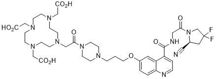
C40H54F2N10O10
|
|---|---|
| 分子量 |
872.93
|
| 精确质量 |
872.4
|
| 元素分析 |
C, 55.04; H, 6.24; F, 4.35; N, 16.05; O, 18.33
|
| CAS号 |
2374782-02-0
|
| PubChem CID |
138454803
|
| 外观&性状 |
White to off-white solid powder
|
| 密度 |
1.46±0.1 g/cm3(Predicted)
|
| 沸点 |
1144.1±65.0 °C(Predicted)
|
| LogP |
-6.8
|
| tPSA |
244
|
| 氢键供体(HBD)数目 |
4
|
| 氢键受体(HBA)数目 |
19
|
| 可旋转键数目(RBC) |
16
|
| 重原子数目 |
62
|
| 分子复杂度/Complexity |
1580
|
| 定义原子立体中心数目 |
1
|
| SMILES |
FC1(C([H])([H])N(C(C([H])([H])N([H])C(C2C([H])=C([H])N=C3C([H])=C([H])C(=C([H])C=23)OC([H])([H])C([H])([H])C([H])([H])N2C([H])([H])C([H])([H])N(C(C([H])([H])N3C([H])([H])C([H])([H])N(C([H])([H])C(=O)O[H])C([H])([H])C([H])([H])N(C([H])([H])C(=O)O[H])C([H])([H])C([H])([H])N(C([H])([H])C(=O)O[H])C([H])([H])C3([H])[H])=O)C([H])([H])C2([H])[H])=O)=O)[C@]([H])(C#N)C1([H])[H])F
|
| InChi Key |
ICWDAESAANBIGG-LJAQVGFWSA-N
|
| InChi Code |
InChI=1S/C40H54F2N10O10/c41-40(42)21-29(22-43)52(28-40)34(53)23-45-39(61)31-4-5-44-33-3-2-30(20-32(31)33)62-19-1-6-46-15-17-51(18-16-46)35(54)24-47-7-9-48(25-36(55)56)11-13-50(27-38(59)60)14-12-49(10-8-47)26-37(57)58/h2-5,20,29H,1,6-19,21,23-28H2,(H,45,61)(H,55,56)(H,57,58)(H,59,60)/t29-/m0/s1
|
| 化学名 |
(S)-2,2',2''-(10-(2-(4-(3-((4-((2-(2-cyano-4,4-difluoropyrrolidin-1-yl)-2-oxoethyl)carbamoyl)quinolin-6-yl)oxy)propyl)piperazin-1-yl)-2-oxoethyl)-1,4,7,10-tetraazacyclododecane-1,4,7-triyl)triacetic acid
|
| 别名 |
FAPI-04; FAPI 04; FAPI04; FAPI-4; FAPI-4; 2374782-02-0; DOTA-fapi-04; (S)-2,2',2''-(10-(2-(4-(3-((4-((2-(2-cyano-4,4-difluoropyrrolidin-1-yl)-2-oxoethyl)carbamoyl)quinolin-6-yl)oxy)propyl)piperazin-1-yl)-2-oxoethyl)-1,4,7,10-tetraazacyclododecane-1,4,7-triyl)triacetic acid; D7X5DKW7N9; FAPI-04; UNII-D7X5DKW7N9; SCHEMBL21257058; FAPI 4; FAPI4;
|
| HS Tariff Code |
2934.99.9001
|
| 存储方式 |
Powder -20°C 3 years 4°C 2 years In solvent -80°C 6 months -20°C 1 month 注意: 请将本产品存放在密封且受保护的环境中(例如氮气保护),避免吸湿/受潮。 |
| 运输条件 |
Room temperature (This product is stable at ambient temperature for a few days during ordinary shipping and time spent in Customs)
|
| 溶解度 (体外实验) |
DMSO : ~100 mg/mL (~114.56 mM)
|
|---|---|
| 溶解度 (体内实验) |
配方 1 中的溶解度: ≥ 2.5 mg/mL (2.86 mM) (饱和度未知) in 10% DMSO + 40% PEG300 + 5% Tween80 + 45% Saline (这些助溶剂从左到右依次添加,逐一添加), 澄清溶液。
例如,若需制备1 mL的工作液,可将100 μL 25.0 mg/mL澄清DMSO储备液加入到400 μL PEG300中,混匀;然后向上述溶液中加入50 μL Tween-80,混匀;加入450 μL生理盐水定容至1 mL。 *生理盐水的制备:将 0.9 g 氯化钠溶解在 100 mL ddH₂O中,得到澄清溶液。 配方 2 中的溶解度: ≥ 2.5 mg/mL (2.86 mM) (饱和度未知) in 10% DMSO + 90% (20% SBE-β-CD in Saline) (这些助溶剂从左到右依次添加,逐一添加), 澄清溶液。 例如,若需制备1 mL的工作液,可将 100 μL 25.0 mg/mL澄清DMSO储备液加入900 μL 20% SBE-β-CD生理盐水溶液中,混匀。 *20% SBE-β-CD 生理盐水溶液的制备(4°C,1 周):将 2 g SBE-β-CD 溶解于 10 mL 生理盐水中,得到澄清溶液。 View More
配方 3 中的溶解度: ≥ 2.5 mg/mL (2.86 mM) (饱和度未知) in 10% DMSO + 90% Corn Oil (这些助溶剂从左到右依次添加,逐一添加), 澄清溶液。 配方 4 中的溶解度: 4 mg/mL (4.58 mM) in Saline (这些助溶剂从左到右依次添加,逐一添加), 澄清溶液; 超声助溶. *生理盐水的制备:将 0.9 g 氯化钠溶解在 100 mL ddH₂O中,得到澄清溶液。 1、请先配制澄清的储备液(如:用DMSO配置50 或 100 mg/mL母液(储备液)); 2、取适量母液,按从左到右的顺序依次添加助溶剂,澄清后再加入下一助溶剂。以 下列配方为例说明 (注意此配方只用于说明,并不一定代表此产品 的实际溶解配方): 10% DMSO → 40% PEG300 → 5% Tween-80 → 45% ddH2O (或 saline); 假设最终工作液的体积为 1 mL, 浓度为5 mg/mL: 取 100 μL 50 mg/mL 的澄清 DMSO 储备液加到 400 μL PEG300 中,混合均匀/澄清;向上述体系中加入50 μL Tween-80,混合均匀/澄清;然后继续加入450 μL ddH2O (或 saline)定容至 1 mL; 3、溶剂前显示的百分比是指该溶剂在最终溶液/工作液中的体积所占比例; 4、 如产品在配制过程中出现沉淀/析出,可通过加热(≤50℃)或超声的方式助溶; 5、为保证最佳实验结果,工作液请现配现用! 6、如不确定怎么将母液配置成体内动物实验的工作液,请查看说明书或联系我们; 7、 以上所有助溶剂都可在 Invivochem.cn网站购买。 |
| 制备储备液 | 1 mg | 5 mg | 10 mg | |
| 1 mM | 1.1456 mL | 5.7278 mL | 11.4557 mL | |
| 5 mM | 0.2291 mL | 1.1456 mL | 2.2911 mL | |
| 10 mM | 0.1146 mL | 0.5728 mL | 1.1456 mL |
1、根据实验需要选择合适的溶剂配制储备液 (母液):对于大多数产品,InvivoChem推荐用DMSO配置母液 (比如:5、10、20mM或者10、20、50 mg/mL浓度),个别水溶性高的产品可直接溶于水。产品在DMSO 、水或其他溶剂中的具体溶解度详见上”溶解度 (体外)”部分;
2、如果您找不到您想要的溶解度信息,或者很难将产品溶解在溶液中,请联系我们;
3、建议使用下列计算器进行相关计算(摩尔浓度计算器、稀释计算器、分子量计算器、重组计算器等);
4、母液配好之后,将其分装到常规用量,并储存在-20°C或-80°C,尽量减少反复冻融循环。
计算结果:
工作液浓度: mg/mL;
DMSO母液配制方法: mg 药物溶于 μL DMSO溶液(母液浓度 mg/mL)。如该浓度超过该批次药物DMSO溶解度,请首先与我们联系。
体内配方配制方法:取 μL DMSO母液,加入 μL PEG300,混匀澄清后加入μL Tween 80,混匀澄清后加入 μL ddH2O,混匀澄清。
(1) 请确保溶液澄清之后,再加入下一种溶剂 (助溶剂) 。可利用涡旋、超声或水浴加热等方法助溶;
(2) 一定要按顺序加入溶剂 (助溶剂) 。
 ikB
ikB
 2-PMDQ
 GW590735
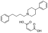 4-PPBP maleate
InvivoChem的所有产品仅用于作科学研究,不面向患者销售
Copyright 2020 InvivoChem LLC | All Rights Reserved 粤ICP备20063088号-1


 463611831
