| 规格 | 价格 | 库存 | 数量 |
|---|---|---|---|
| 100mg |
|
||
| 250mg |
|
||
| 500mg |
|
||
| 1g |
|
||
| Other Sizes |
|

| 靶点 |
- Human dermal fibroblasts: enhances elastic fiber and collagen deposition via sodium-dependent vitamin C transporters (SVCTs), reduction of intracellular ROS, activation of c-Src kinase, and enhancement of IGF-1 receptor phosphorylation. [1]
- Neuronal T-type calcium channels: selectively inhibits Cav3.2 (α1H) subtype via metal-catalyzed oxidation of histidine 191 in domain I, with no effect on Cav3.1 or Cav3.3. [3] Cav3.2 T-type Calcium Channels: Ascorbate selectively inhibits Cav3.2 T-type calcium channels (Cav3.2). (IC50 = 6.5 ± 3.9 μM in rat dorsal root ganglion neurons) [3] |
|---|---|
| 体外研究 (In Vitro) |
抗坏血酸(L-抗坏血酸钠)(50–200 μM) 显著刺激正常人真皮成纤维细胞和脂肪来源成纤维细胞培养72小时后免疫可检测的弹性纤维和胶原纤维的沉积。更高浓度 (400 μM) 并未进一步刺激,而 800 μM 则抑制弹性蛋白生成。氯化钠或氯化钠与抗坏血酸 (AA) 的混合物均无此作用。[1] - 100 μM SA 与脯氨酰羟化酶抑制剂 DMOG 联用可抑制胶原蛋白沉积,但不会减弱增强的弹性蛋白生成;每日添加 SA 培养7天后,弹性蛋白含量增加,SA+DMOG 进一步增加了弹性纤维和不溶性弹性蛋白的含量。 [1]
- SA (100 μM) 上调原弹性蛋白 mRNA (18 h)、细胞内原弹性蛋白 (24 h) 和不溶性弹性蛋白 (72 h)。SVCT 抑制剂丙磺舒 (400 μM) 消除了这些弹性蛋白生成作用。[1] - SA (100 μM,2 h) 显著降低了成纤维细胞内活性氧 (ROS) 水平,通过 CM-H₂DCFDA 荧光探针和流式细胞术检测。丙磺舒阻断了这种作用。[1] - SA 仅在含 5% FBS(含 IGF-1)的培养基中增强弹性蛋白生成。在无血清培养基中,SA 单独作用不诱导弹性蛋白生成,但 SA 增强了 IGF-1 诱导的原弹性蛋白合成。SA 增强了 IGF-1 受体磷酸化; c-Src抑制剂PP2或IGF-1R激酶抑制剂PPP可阻断这一过程。SA不增强胰岛素受体磷酸化。[1] - 在真皮妊娠纹成纤维细胞中,SA(200 μM)上调胶原蛋白和弹性纤维沉积,而AA选择性抑制弹性蛋白生成。[1] - 抗坏血酸(抗坏血酸盐)抑制急性分离的大鼠背根神经节(DRG)神经元中天然T型Ca²⁺电流,IC₅₀为6.5 ± 3.9 μM,最大抑制率为70.2 ± 2.1%(Hill系数0.56 ± 0.12)。抗坏血酸使激活电压依赖性向更去极化的电位移动(V₅₀ 从 -49.0 mV 移至 -44.1 mV),使稳态失活电压依赖性向更超极化的电位移动(V₅₀ 从 -75.0 mV 移至 -80.4 mV),并减缓了激活和失活动力学。[3] - 抗坏血酸(100–300 μM)可逆地抑制在 HEK293 细胞中表达的重组人 Cav3.2 T 型通道,但对 Cav3.1 或 Cav3.3 通道无影响。这种抑制作用呈浓度依赖性。[3] - 定点诱变实验发现,I 结构域 S3 和 S4 之间的胞外环中的组氨酸 191 (H191) 对抗坏血酸敏感性至关重要。 H191Q 或 H191C 突变消除了抗坏血酸的抑制作用。H191Q 突变还降低了 Cu²⁺ 的敏感性(>40 倍)。[3] - 金属螯合剂 DTPA、H₂O₂ 分解酶过氧化氢酶和 ROS 清除剂 c-PTIO 可阻止抗坏血酸的抑制作用。添加 300 nM Cu²⁺ 会增强抗坏血酸的抑制作用。 [3] B16F10 细胞条件培养基中活性成分的相对分子质量小于 5,000,并且能显著降低 L-抗坏血酸钠 (10 mM) 诱导的细胞凋亡 [4]。 选择性抑制 Cav3.2 通道:抗坏血酸能快速且可逆地抑制急性分离的大鼠背根神经节 (DRG) 神经元中的 T 型钙电流,IC50 为 6.5 ± 3.9 μM,最大抑制率为 70.2 ± 2.1%。该抑制作用具有电压依赖性,使激活的电压依赖性向更去极化的电位移动(V50 从 -49.0 ± 0.3 mV 移至 -44.1 ± 0.9 mV),使稳态失活的电压依赖性向更超极化的电位移动(V50 从 -75.0 ± 0.3 mV 移至 -80.4 ± 0.4 mV)。[3] - 亚型特异性:抗坏血酸 (1 mM) 可抑制异源表达于 HEK293 细胞中的 Cav3.2 通道约 70%,但对 Cav3.1 或 Cav3.3 通道无显著影响。这种亚型特异性抑制在天然神经元中也观察到:抗坏血酸(1 mM)抑制了背根神经节(DRG)神经元(主要表达 Cav3.2)的 T 电流 49.2 ± 5.8%,但对丘脑中继神经元(主要表达 Cav3.1)的 T 电流没有影响。[3] - 作用机制 – 金属催化氧化 (MCO):抗坏血酸对 Cav3.2 通道的抑制作用依赖于痕量金属污染物,因为金属螯合剂 DTPA 可以完全阻断这种抑制作用。该效应也可被H₂O₂清除剂过氧化氢酶和羟基自由基清除剂c-PTO阻断,表明该抑制作用是由金属催化氧化产生的活性氧(特别是H₂O₂和羟基自由基)介导的。[3] - 分子决定因素 – 组氨酸191 (H191):在Cav3.2通道的I结构域中,将组氨酸191 (H191)突变为谷氨酰胺 (H191Q) 完全消除了抗坏血酸的抑制作用。同样,突变为半胱氨酸 (H191C) 也使该通道对抗坏血酸不敏感,证实H191是抗坏血酸诱导抑制的关键残基。 [3] - 对丘脑神经元低阈值钙尖峰 (LTS) 和爆发放电的影响:在主要表达 Cav3.2 通道的网状丘脑 (nRT) 神经元中,抗坏血酸 (1 mM) 可逆地降低了低阈值钙尖峰 (LTS) 的幅度 44.6 ± 11.4%,并将每次爆发产生的动作电位数量从 4.6 ± 0.7 个减少到 2.4 ± 1.1 个。[3] |
| 体内研究 (In Vivo) |
与未接受L-抗坏血酸钠(L-抗坏血酸钠)治疗的转基因大鼠相比,接受L-抗坏血酸钠治疗的转基因大鼠癌症发生率更高(29.6%)(15.4%)。即使未接受L-抗坏血酸钠治疗,转基因大鼠也出现了多种器官癌症[5]。所有动物在接受PEITC治疗12周后均出现单纯性增生和乳头状或结节状(PN)增生;然而,无论是否接受钠盐(L-抗坏血酸)治疗,大多数病变在48周时均消退。在接受PEITC治疗24周后,到第48周时,少数病例中相同的病变已发展为发育不良和癌症;然而,L-抗坏血酸钠治疗仅在单纯性增生和PN增生方面显示出增强作用[6]。
L-抗坏血酸钠 (Na-AsA) 已被公认为大鼠膀胱癌的促癌剂,但在标准的两年生物测定中却未观察到致癌作用。为了进一步研究其致瘤潜力,本研究采用了对膀胱癌高度易感的 Hras128 转基因大鼠。共选取 40 只雄性转基因 (Tg) 大鼠(7 周龄)和 42 只非转基因 (Non-tg) 同窝大鼠,随机分为四组,分别饲喂添加或不添加 5% Na-AsA 的粉状 MF 饲料,持续 57 周。无论是否进行 Na-AsA 处理,Tg 大鼠的生存期均显著短于 Non-tg 大鼠。在 Tg 大鼠中,Na-AsA 处理组的膀胱癌发生率 (29.6%) 略高于未处理组 (15.4%),但差异无统计学意义。此外,两组转基因大鼠(Tg组)的膀胱肿瘤总体发生率(包括乳头状瘤)无显著差异(Na-AsA组为37.0%,未组为30.8%)。所有非转基因大鼠均未检测到膀胱肿瘤。在接受或未接受Na-AsA治疗的转基因大鼠的多个器官中均观察到其他各种病变,但组间差异不明显。总之,在高度易患膀胱癌的Hras128转基因大鼠模型中,Na-AsA未表现出致瘤性。这些结果表明,Na-AsA在大鼠中仅作为促癌剂而非完全致癌物发挥作用。[5] |
| 酶活实验 |
弹性蛋白生成研究:按照所述方法进行免疫染色、蛋白质印迹、RT-PCR 和使用 [³H]缬氨酸对代谢标记的不溶性弹性蛋白进行定量分析。活性氧 (ROS) 水平使用 CM-H₂DCFDA 荧光探针和流式细胞术进行测定。[1]
- T 型钙通道研究:对急性分离的背根神经节 (DRG) 神经元、丘脑切片和表达重组通道的 HEK293 细胞进行全细胞膜片钳记录。外液中含有 10 mM Ba²⁺ 作为载流子。浓度-反应曲线拟合 Hill-Langmuir 方程。激活和失活的电压依赖性拟合 Boltzmann 分布。[3] |
| 细胞实验 |
将人真皮成纤维细胞和脂肪来源成纤维细胞(传代2-4代)培养于含5% FBS的DMEM培养基中。细胞用SA(50-800 μM)处理18-72小时。进行弹性蛋白和I型胶原的免疫染色、原弹性蛋白的Western blot、原弹性蛋白mRNA的RT-PCR以及不溶性弹性蛋白的[³H]缬氨酸掺入实验。[1]
- 为测定ROS水平,将细胞用10 μM CM-H₂DCFDA孵育30分钟,然后用SA(100 μM)处理2或24小时,并通过显微镜或流式细胞仪观察荧光。 [1] - 对于 IGF-1R 磷酸化研究,细胞裂解后用抗 IGF-1R β 亚基抗体进行免疫沉淀,然后用抗磷酸酪氨酸抗体进行蛋白质印迹分析。[1] - 对于 T 型通道研究:使用急性分离的大鼠背根神经节 (DRG) 神经元、丘脑切片以及瞬时表达 Cav3.1、Cav3.2 或 Cav3.3 通道的 HEK293 细胞。在室温下进行全细胞电压钳记录。通过灌注施加抗坏血酸。[3] 急性分离的大鼠背根神经节 (DRG) 神经元:DRG 细胞取自青春期大鼠。记录时,将细胞接种于未包被的玻璃盖玻片上,并用外液灌注。采用全细胞膜片钳记录技术测量T型钙电流。外液成分(单位:mM):152 TEA-Cl、10 BaCl₂和10 HEPES(用TEA-OH调节pH至7.4)。内液成分(单位:mM):135 TEA-OH、40 HEPES、10 EGTA和2 MgCl₂(用HF调节pH至7.2)。[3] - 丘脑切片制备:取青春期大鼠的丘脑制备切片。将切片置于记录室中,并用外液灌注,外液成分(单位:mM):130 NaCl、26 NaHCO₃、10葡萄糖、2.5 MgCl₂、2 CaCl₂、1.25 NaH₂PO₄和0.001 TTX,并用95% O₂和5% CO₂混合气体平衡。在电流钳实验中,省略了TTX,内液成分为(单位:mM):130 KCl、40 HEPES、5 MgCl₂、2 MgATP、1 EGTA、0.1 Na₃ATP(用KOH调节pH至7.3)。[3] - HEK293细胞培养和转染:HEK293细胞培养于添加了10%胎牛血清、青霉素G(100 U/ml)和链霉素(0.1 mg/ml)的DMEM/F12培养基中。使用 Lipofectamine 2000 将编码各种 T 型钙通道亚基(Cav3.1、Cav3.2、Cav3.3 及其各种突变体)的 cDNA 与 CD8 抗原瞬时共转染至细胞中。48 小时后,选择结合了抗 CD8 抗体包被微珠的细胞进行记录。采用全细胞膜片钳技术进行记录,外液成分为(mM):152 TEA-Cl、10 BaCl₂、10 HEPES(用 TEA-OH 调节 pH 至 7.4),内液成分为(mM):110 Cs-MeSO₄、14 Cs-PO₄、10 HEPES、9 EGTA、5 Mg-ATP、0.3 Tris-GTP(用 CsOH 调节 pH 至 7.3)。[3] |
| 动物实验 |
动物在6周龄时被分为六组(图1)。为了研究不同PEITC暴露时间后膀胱增生性病变的发展情况,第1组和第2组动物分别饲喂基础饲料(对照组)或0.1% PEITC,持续48周,并在第12、24和48周处死(每个时间点7-8只动物)。为了研究Na-AsA对PEITC诱导的增生性病变发展的增强作用,第4组和第6组动物分别在前12周和24周饲喂0.1% PEITC,然后饲喂5% Na-AsA直至第48周(每组16只动物)。第3组和第5组动物分别作为第4组和第6组的Na-AsA阴性对照,在PEITC处理后饲喂基础饲料而非Na-AsA(每组16只动物)。动物可自由摄取食物和自来水。在前8周后,至少每2周记录一次体重和食物消耗量。在处死时,所有动物均在乙醚麻醉下实施安乐死。解剖时,取出肝脏、肾脏和膀胱,并称量肝脏和肾脏的重量。膀胱在浸入固定液前,先用10%中性缓冲福尔马林(pH 7.4)灌注。在最终处死时,固定后称量膀胱重量,并从每个膀胱中制备6片切片。将膀胱切片与肝脏和肾脏切片一起进行石蜡包埋,切片厚度为3 μm,并用苏木精-伊红染色。根据先前描述的标准,将膀胱病变组织病理学分为单纯性增生、PN增生、发育不良和移行细胞癌。[6]
大鼠组织制备:**从青春期大鼠中制备急性分离的背根神经节(DRG)细胞和丘脑切片。[3] 大鼠组织制备:从青春期大鼠中制备急性分离的背根神经节(DRG)细胞和丘脑切片。[3] |
| 药代性质 (ADME/PK) |
吸收、分布和排泄
维生素C的还原形式——抗坏血酸,是一种强效抗氧化剂,并在细胞分化中发挥作用。哺乳动物细胞通过特定的钠/抗坏血酸共转运蛋白SVCT1和SVCT2吸收抗坏血酸。尽管骨骼肌含有约50%的人体维生素C,但SVCT转运蛋白在该组织中的表达尚未得到充分研究。……本研究……以鸡胚为模型系统,分析了胚胎肌生成过程中SVCT2的表达模式。免疫组织化学分析表明,SVCT2在鸡胚胎肌生成过程中优先表达于I型慢肌纤维,并且在包括人类在内的多种物种的出生后骨骼肌中也有表达……人类利用两种钠-抗坏血酸共转运蛋白(hSVCT1和hSVCT2)运输膳食必需微量营养素抗坏血酸,即维生素C的还原活性形式。尽管人肝脏在调节和维持维生素C稳态方面发挥着至关重要的作用,但该器官中维生素C转运的生理机制以及hSVCT系统的调控机制仍知之甚少。因此,本研究利用人肝细胞系(HepG2)验证了原代人肝细胞的部分研究结果,并确定抗坏血酸的初始摄取速率取决于Na+梯度、pH值以及低微摩尔和高微摩尔浓度范围内的浓度饱和度。此外,hSVCT2蛋白和mRNA在HepG2细胞和人肝脏中均高表达,且克隆的hSVCT2启动子在HepG2细胞中活性更高。短干扰RNA实验结果表明,在HepG2细胞中,降低hSVCT2 mRNA水平比降低hSVCT1 mRNA水平更能有效降低抗坏血酸的总体摄取。细胞内PKC调控通路的激活导致抗坏血酸摄取下调,但这种下调并非由hSVCT1或hSVCT2中任何单个预测的PKC特异性氨基酸磷酸化位点介导。然而,PKC激活会导致hSVCT1内化,但不会导致hSVCT2内化。对其他细胞内抗坏血酸摄取调节途径的研究表明,PKA、PTK 和 Ca(2+)/钙调蛋白也可能参与调节,但一氧化氮依赖性途径不参与…… 代谢/代谢物 ……肾上腺皮质与抗坏血酸代谢密切相关……据报道,氢化可的松……可以刺激抗坏血酸的葡萄糖酸内酯合成,而脱氧皮质酮或醛固酮会导致……正常或肾上腺切除大鼠体内抗坏血酸排泄增加…… |
| 毒性/毒理 (Toxicokinetics/TK) |
妊娠期和哺乳期用药
◉ 哺乳期用药概述 维生素C是母乳的正常成分,也是一种重要的抗氧化剂。建议哺乳期妇女每日摄入120毫克维生素C,6个月及以下婴儿每日摄入40毫克。每日摄入高达1000毫克的高剂量维生素C会增加母乳中的维生素C含量,但这不足以对母乳喂养的婴儿造成健康问题,也不是停止母乳喂养的理由。哺乳期妇女可能需要补充膳食以达到推荐摄入量或纠正任何已知的维生素C缺乏症。孕妇服用孕期维生素时,摄入量达到或接近推荐摄入量不会改变母乳中的维生素C含量。 对于足月儿和早产儿的住院母亲,将新鲜挤出的成熟母乳冷冻(-20°C)至少3个月不会改变其维生素C含量。冷冻(-20°C)6至12个月后,维生素C含量将下降15%至30%。-80°C储存可使维生素C含量保持8个月,12个月后损失15%。 ◉ 对母乳喂养婴儿的影响 60名产后1至6个月的健康哺乳期妇女,其婴儿均为纯母乳喂养,分别接受每日一次500毫克维生素C和100国际单位维生素E,持续30天,或不进行任何补充。补充组母亲所生婴儿的尿液抗氧化活性生化指标升高。未报告临床结果。 18名早产儿(其中7名胎龄小于32周)出生后前三天喂食混合巴氏杀菌捐赠母乳,其平均血浆抗坏血酸浓度从出生时的15.5 mg/L下降至产后1周的5.4 mg/L,并在产后3周进一步下降至4.1 mg/L。作者认为产后1周和3周的抗坏血酸水平低于治疗水平(<6 mg/L),表明摄入不足,这可能会影响出生后的生长发育。尽管这项研究是在早产儿肠外营养和肠内强化母乳疗法发展之前进行的,但目前的研究表明,从混合巴氏杀菌捐赠母乳中摄入的维生素C不足可能是接受捐赠母乳的早产儿潜在的健康问题。 ◉ 对泌乳和母乳的影响 截至修订日期,未找到相关的已发表信息。 相互作用 当向含有乙酰胆碱但不含Ca2+离子的浴液中加入不同浓度的氯化钙时,暴露于抗坏血酸钠的组织比未处理的肌肉组织表现出更强的反应。 在体外,我们使用培养的人类肿瘤细胞系MCF-7(乳腺癌)、KB(口腔表皮癌)和AN3-CA(子宫内膜腺癌)研究了浓度范围为0.198 μg/mL至1.98 mg/mL的抗坏血酸钠(含或不含维生素K3)的作用。不含抗坏血酸钠和维生素K3的培养基作为对照。当细胞汇合度达到50%时,向培养物中加入不同比例的抗坏血酸钠和维生素K3,并孵育1小时。然后进行DNA检测。添加抗坏血酸钠的培养基仅在高浓度(5 x 10⁻³ mol/L)下显示出生长抑制作用。联合用药即使在浓度降低 10 至 50 倍的情况下,仍显示出对细胞生长的协同抑制作用。这些结果适用于所有三种细胞类型…… 雄性和雌性 Wistar 大鼠(每组 n=5)连续 6 个月接受抗坏血酸钠和/或亚硝酸钠的给药。对照组仅喂食基础饲料和水。治疗组分别给予以下溶液:0.075%、0.15% 或 0.3% 的亚硝酸钠水溶液;1%、2% 或 4% 的抗坏血酸钠;或两种化学物质以低剂量+低剂量、中剂量+中剂量和高剂量+高剂量组合给药。高剂量联合用药组的体重增长显著降低。高剂量联合用药组还表现出血清总蛋白显著降低、血尿素氮 (BUN) 显著升高以及肾脏相对重量显著增加。组织病理学检查显示,高剂量联合用药组前胃出现中度至重度鳞状细胞增生,而中剂量联合用药组仅观察到轻度增生。未观察到性别差异。最小毒性剂量为0.15%亚硝酸钠+2%抗坏血酸钠…… |
| 参考文献 |
|
| 其他信息 |
微小晶体或白色粉末。其水溶液的pH值为5.6至7.0或更高(例如,由市售产品配制的10%溶液的pH值可能为7.4至7.7)。(NTP,1992)
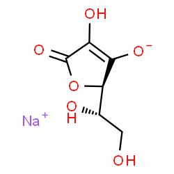
抗坏血酸钠是一种有机钠盐,由抗坏血酸3-羟基上的质子被钠离子取代而形成。它可用作食品抗氧化剂、面粉处理剂、辅酶、植物代谢物、人体代谢物、大型跳蚤代谢物和还原剂。它是一种有机钠盐,也是维生素C。它含有L-抗坏血酸。 一种与葡萄糖结合的六碳化合物。它天然存在于柑橘类水果和许多蔬菜中。抗坏血酸是人体必需的营养素,对维持结缔组织和骨骼至关重要。其生物活性形式——维生素C——在多种代谢途径中作为还原剂和辅酶发挥作用。维生素C被认为是一种抗氧化剂。 另见:抗坏血酸(含活性成分)……查看更多…… 作用机制 抗坏血酸的作用机制是清除超氧自由基。 ……抗坏血酸钠以剂量和时间依赖的方式降低黑色素瘤细胞对铁的吸收,这表明细胞内铁水平可能是抗坏血酸钠诱导细胞凋亡的关键因素。事实上,铁螯合剂去铁胺(DFO)增强了抗坏血酸钠诱导的细胞凋亡,而铁供体柠檬酸铁铵(FAC)则抑制了这一过程。此外,添加转铁蛋白可阻断抗坏血酸钠对细胞内铁水平的抑制作用,提示抗坏血酸钠可能通过转铁蛋白受体 (TfR) 依赖性途径调节铁的摄取。暴露于抗坏血酸钠的细胞表现出转铁蛋白受体 (TfR) 表达下调,且这种下调先于抗坏血酸钠诱导的细胞凋亡。总之,抗坏血酸钠介导的细胞凋亡似乎是由 TfR 表达降低启动的,导致铁摄取减少,进而诱导细胞凋亡…… 人体利用两种钠-抗坏血酸共转运蛋白(hSVCT1 和 hSVCT2)来运输膳食必需微量营养素抗坏血酸,即维生素 C 的还原活性形式。尽管人肝脏在调节和维持维生素 C 稳态方面发挥着至关重要的作用,但该器官中维生素 C 转运的生理机制以及 hSVCT 系统的调控机制仍不甚明了。因此,本研究利用人肝细胞系(HepG2)验证了部分原代人肝细胞研究结果,并确定抗坏血酸的初始摄取速率与Na⁺梯度相关,且具有pH依赖性,并在低微摩尔和高微摩尔浓度下均达到饱和。此外,HepG2细胞和天然人肝组织中hSVCT2蛋白和mRNA的表达水平均较高,且克隆的hSVCT2启动子在HepG2细胞中表现出更强的活性。使用短干扰RNA(siRNA)的结果表明,在HepG2细胞中,降低hSVCT2 mRNA水平比降低hSVCT1 mRNA水平更能有效地降低抗坏血酸的总摄取量。细胞内PKC调控通路的激活导致抗坏血酸摄取下调,但这种下调并非由hSVCT1或hSVCT2中单个预测的PKC特异性氨基酸磷酸化位点介导。然而,PKC激活导致hSVCT1内化,但不导致hSVCT2内化。对其他细胞内抗坏血酸摄取调节通路的研究表明,PKA、PTK和Ca(2+)/钙调蛋白也可能参与调节,但一氧化氮依赖性通路不参与…… 治疗用途 抗氧化剂;自由基清除剂 抗坏血酸以及抗坏血酸钙和抗坏血酸钠在制药和食品工业中用作抗氧化剂。 20例急性哮喘发作患者中,16例在静脉注射6克抗坏血酸钠后迅速康复。25例哮喘患者中,18例长期口服抗坏血酸钠(0.6-1克/天,持续60天)后哮喘症状得到控制。8例前房积血患者接受了静脉注射甘油联合抗坏血酸钠治疗。结果显示,甘油和抗坏血酸钠联合使用可在 12-24 小时内减少眼部出血。有关抗坏血酸钠(6 种类型)治疗用途的更完整数据,请访问 HSDB 记录页面。药物警告:每克抗坏血酸钠约含 5 毫当量钠;低钠饮食患者使用此药时应考虑这一点。 背景:抗坏血酸(维生素 C)是一种内源性氧化还原剂,在神经元中浓度很高(细胞外 100-500 μM,细胞内高达 10 mM)。它既可以作为抗氧化剂,也可以作为促氧化剂。[3] - 作用机制 – 金属催化氧化 (MCO):抗坏血酸通过启动金属催化氧化 (MCO) 来抑制 Cav3.2 通道。该过程涉及抗坏血酸还原痕量过渡金属(例如 Cu²⁺ 或 Fe³⁺),进而生成活性氧(ROS),包括过氧化氢和羟基自由基。这些 ROS 特异性地氧化 Cav3.2 通道 I 结构域中的关键组氨酸残基 (H191),从而抑制通道活性。由于金属结合于通道的金属结合位点,因此该反应具有位点特异性。[3] - 治疗/潜在意义:抗坏血酸对 Cav3.2 通道的选择性抑制可能在与氧化还原活性过渡金属水平升高相关的生理或病理条件下具有功能意义,例如神经元损伤、癫痫、阿尔茨海默病、门克斯病和威尔逊病。突触末端也可能释放高浓度的 Cu²⁺ (200-400 μM)。抗坏血酸可能通过这种机制作为内源性神经调节剂调节神经元兴奋性。[3] - 药理学工具:抗坏血酸是目前已知的能够区分T型通道亚型(Cav3.2、Cav3.1和Cav3.3)的选择性最高的试剂。它可能是一种有用的药理学工具,用于确定Cav3.2电流对总钙电流和细胞兴奋性的贡献。[3] |
| 分子式 |
C6H7NAO6
|
|---|---|
| 分子量 |
198.11
|
| 精确质量 |
198.014
|
| 元素分析 |
C, 36.38; H, 3.56; Na, 11.60; O, 48.46
|
| CAS号 |
134-03-2
|
| 相关CAS号 |
L-Ascorbic acid;50-81-7;L-Ascorbic acid (GMP Like);50-81-7
|
| PubChem CID |
23667548
|
| 外观&性状 |
Off-white to light yellow solid powder
|
| 密度 |
1.799 g/cm3
|
| 沸点 |
552.7ºC at 760 mmHg
|
| 熔点 |
220 °C (dec.)(lit.)
|
| 闪点 |
238.2ºC
|
| 蒸汽压 |
1.62E-14mmHg at 25°C
|
| 折射率 |
105.5 ° (C=10, H2O)
|
| tPSA |
110.05
|
| 氢键供体(HBD)数目 |
3
|
| 氢键受体(HBA)数目 |
6
|
| 可旋转键数目(RBC) |
2
|
| 重原子数目 |
13
|
| 分子复杂度/Complexity |
237
|
| 定义原子立体中心数目 |
2
|
| SMILES |
[Na+].O1C(C(=C([C@@]1([H])[C@]([H])(C([H])([H])O[H])O[H])[O-])O[H])=O
|
| InChi Key |
PPASLZSBLFJQEF-RXSVEWSESA-M
|
| InChi Code |
InChI=1S/C6H8O6.Na/c7-1-2(8)5-3(9)4(10)6(11)12-5;/h2,5,7-10H,1H2;/q;+1/p-1/t2-,5+;/m0./s1
|
| 化学名 |
sodium;(2R)-2-[(1S)-1,2-dihydroxyethyl]-4-hydroxy-5-oxo-2H-furan-3-olate
|
| 别名 |
Ascorbate; Vitamin C sodium; SODIUM ASCORBATE; 134-03-2; L-Ascorbic acid sodium salt; Sodium L-ascorbate; Vitamin C sodium; Sodium Ascorbate
|
| HS Tariff Code |
2934.99.9001
|
| 存储方式 |
Powder -20°C 3 years 4°C 2 years In solvent -80°C 6 months -20°C 1 month 注意: 请将本产品存放在密封且受保护的环境中(例如氮气保护),避免吸湿/受潮和光照。 |
| 运输条件 |
Room temperature (This product is stable at ambient temperature for a few days during ordinary shipping and time spent in Customs)
|
| 溶解度 (体外实验) |
H2O : ~100 mg/mL (~504.77 mM)
DMSO : ~1 mg/mL (~5.05 mM) |
|---|---|
| 溶解度 (体内实验) |
配方 1 中的溶解度: 50 mg/mL (252.39 mM) in PBS (这些助溶剂从左到右依次添加,逐一添加), 澄清溶液; 超声助溶。
请根据您的实验动物和给药方式选择适当的溶解配方/方案: 1、请先配制澄清的储备液(如:用DMSO配置50 或 100 mg/mL母液(储备液)); 2、取适量母液,按从左到右的顺序依次添加助溶剂,澄清后再加入下一助溶剂。以 下列配方为例说明 (注意此配方只用于说明,并不一定代表此产品 的实际溶解配方): 10% DMSO → 40% PEG300 → 5% Tween-80 → 45% ddH2O (或 saline); 假设最终工作液的体积为 1 mL, 浓度为5 mg/mL: 取 100 μL 50 mg/mL 的澄清 DMSO 储备液加到 400 μL PEG300 中,混合均匀/澄清;向上述体系中加入50 μL Tween-80,混合均匀/澄清;然后继续加入450 μL ddH2O (或 saline)定容至 1 mL; 3、溶剂前显示的百分比是指该溶剂在最终溶液/工作液中的体积所占比例; 4、 如产品在配制过程中出现沉淀/析出,可通过加热(≤50℃)或超声的方式助溶; 5、为保证最佳实验结果,工作液请现配现用! 6、如不确定怎么将母液配置成体内动物实验的工作液,请查看说明书或联系我们; 7、 以上所有助溶剂都可在 Invivochem.cn网站购买。 |
| 制备储备液 | 1 mg | 5 mg | 10 mg | |
| 1 mM | 5.0477 mL | 25.2385 mL | 50.4770 mL | |
| 5 mM | 1.0095 mL | 5.0477 mL | 10.0954 mL | |
| 10 mM | 0.5048 mL | 2.5239 mL | 5.0477 mL |
1、根据实验需要选择合适的溶剂配制储备液 (母液):对于大多数产品,InvivoChem推荐用DMSO配置母液 (比如:5、10、20mM或者10、20、50 mg/mL浓度),个别水溶性高的产品可直接溶于水。产品在DMSO 、水或其他溶剂中的具体溶解度详见上”溶解度 (体外)”部分;
2、如果您找不到您想要的溶解度信息,或者很难将产品溶解在溶液中,请联系我们;
3、建议使用下列计算器进行相关计算(摩尔浓度计算器、稀释计算器、分子量计算器、重组计算器等);
4、母液配好之后,将其分装到常规用量,并储存在-20°C或-80°C,尽量减少反复冻融循环。
计算结果:
工作液浓度: mg/mL;
DMSO母液配制方法: mg 药物溶于 μL DMSO溶液(母液浓度 mg/mL)。如该浓度超过该批次药物DMSO溶解度,请首先与我们联系。
体内配方配制方法:取 μL DMSO母液,加入 μL PEG300,混匀澄清后加入μL Tween 80,混匀澄清后加入 μL ddH2O,混匀澄清。
(1) 请确保溶液澄清之后,再加入下一种溶剂 (助溶剂) 。可利用涡旋、超声或水浴加热等方法助溶;
(2) 一定要按顺序加入溶剂 (助溶剂) 。
| NCT Number | Recruitment | interventions | Conditions | Sponsor/Collaborators | Start Date | Phases |
| NCT03508726 | COMPLETEDWITH RESULTS | Drug: Ascorbate | Soft Tissue Sarcoma | Mohammed Milhem | 2019-06-27 | Phase 1 Phase 2 |
| NCT04877587 | WITHDRAWN | Drug: Ascorbate Drug: Gemcitabine |
Bone Sarcoma Metastatic Bone Sarcoma Metastatic Soft-tissue Sarcoma Soft Tissue Sarcoma |
David Dickens | 2023-01 | Early Phase 1 |
| NCT02420314 | COMPLETEDWITH RESULTS | Drug: Paclitaxel Drug: Carboplatin Drug: Ascorbic Acid |
Carcinoma, Non-Small-Cell Lung | Joseph J. Cullen, MD, FACS | 2015-04 | Phase 2 |
| NCT06433791 | NOT YET RECRUITING | Drug: Ascorbate-Meglumine | Safety | LadeRx LLC | 2024-06-17 | Phase 1 |
| NCT04634227 | RECRUITING | Drug: Ascorbate | Bone Sarcoma Metastatic Bone Tumor Sarcoma Soft Tissue Sarcoma Unresectable Soft Tissue Sarcoma |
Mohammed Milhem, MBBS | 2020-11-24 | Early Phase 1 |
|
|
|
InvivoChem的所有产品仅用于作科学研究,不面向患者销售
Copyright 2020 InvivoChem LLC | All Rights Reserved 粤ICP备20063088号-1


 COA
COA


 463611831
