| 规格 | 价格 | 库存 | 数量 |
|---|---|---|---|
| 10 mM * 1 mL in DMSO |
|
||
| 1mg |
|
||
| 2mg |
|
||
| 5mg |
|
||
| 10mg |
|
||
| 25mg |
|
||
| 50mg |
|
||
| 100mg |
|
||
| 250mg |
|
||
| Other Sizes |
|
| 靶点 |
p53 (IC50 = 0.37 μM); NF-κB (IC50 = 0.47 μM); FACT
FACT (facilitates chromatin transcription) complex [1] FACT (facilitates chromatin transcription) complex [2] |
|---|---|
| 体外研究 (In Vitro) |
CBL0137 是胰腺癌细胞系细胞凋亡的有效诱导剂,对胰腺癌干细胞和活跃增殖的肿瘤细胞有毒。使用 CBL0137 和相关分子可以抑制 NF-B 和 HSF-1 控制的细胞应激途径,同时激活 p53 [2]。 CBL0137 结合 DNA 时不会引入任何类型的化学改变,使其无基因毒性。促进染色质转录 (FACT) 复合物是一种参与转录、复制和 DNA 修复的染色质重塑复合物,由于 CBL0137 与 DNA 结合而功能失活。在 CBL0137 处理的细胞中,FACT 从核质中丢失并被捕获在染色质中,从而抑制 FACT 依赖性转录,包括 NF-kB 介导的转录。此外,FACT 的染色质捕获导致 p53 以酪蛋白激酶 2 (CK2) 依赖性方式磷酸化和激活 [3]。
CBL0137盐酸盐(CBLC137)对人纤维肉瘤HT1080细胞具有细胞毒性(具有特定IC₅₀值),对正常二倍体成纤维细胞(Wi38、MEF)的毒性较低(P < 0.001)。用2 μM CBLC137处理24小时可诱导肿瘤细胞(HT1080、RCC45、MiaPaca)细胞周期停滞,但对正常细胞(Wi38、NKE-hTERT)影响较小。它通过酪蛋白激酶2(CK2)促进p53 Ser³⁹²磷酸化来激活p53,并通过将FACT捕获在染色质中抑制NF-κB依赖性转录,且不会诱导可检测到的DNA损伤(彗星实验和H2AX磷酸化实验证实无DNA断裂)。此外,它还会降低FACT亚基(SSRP1、SPT16)的可溶性部分,阻断NF-κB的核质穿梭和DNA结合 [1] CBL0137盐酸盐(CBLC137)是胰腺癌细胞系(MiaPaCa-2、PANC-1、BxPC-3)的强效凋亡诱导剂,用2 μM处理4-24小时后,可检测到半胱天冬酶3、7、8、9和PARP1的切割。它能抑制胰腺癌细胞和耐药癌干细胞(CSCs)的活力,降低CSC表面标志物(CD24ᵂⁱ CD44ᵂⁱ)的比例,阻止吉西他滨诱导的“侧群”富集。在集落形成实验(MiaPaCa-2、PANC-1)中与吉西他滨表现出协同作用,并能抑制吉西他滨诱导的转录反应,包括NF-κB靶基因表达和核糖核苷酸还原酶(RNR)的RRM1/RRM2亚基 [2] |
| 体内研究 (In Vivo) |
在小鼠中,CBL0137 可有效对抗多种胰腺导管腺癌 (PDA) 模型,包括原位吉西他滨耐药 PANC-1 模型和患者来源的异种移植物,其中 CBL0137 的抗肿瘤作用与 FACT 的过度表达相关[1]。根据其提出的作用机制,CBL0137靶向胶质母细胞瘤(GBM),穿透血脑屏障,并且在TMZ反应性和耐药性原位模型中均有效。这种药物能够穿过血脑屏障,尤其是静脉注射时,其治疗中枢神经系统肿瘤的潜力令人鼓舞。在原位模型中,静脉注射药物比口服药物具有更高的生物利用度,因为它们在肿瘤组织中积累更多。 CBL0137 在脑组织中的正常积累不会导致可检测到的神经毒性[3]。
CBL0137盐酸盐(CBLC137)在多种人类肿瘤异种移植小鼠模型中表现出抗肿瘤活性。对于肾细胞癌Caki-1、结直肠癌DLD-1、黑色素瘤Mel-7和胰腺导管腺癌(PDA),CBLC137处理与溶媒组相比显著降低肿瘤体积(P < 0.005,方差分析),每组5-10只小鼠。在患有自发性乳腺肿瘤的MMTV-neu转基因小鼠中,口服灌胃100 mg/kg CBLC137后24小时,肿瘤中SSRP1和SPT16的可溶性部分减少(P < 0.05) [1] CBL0137盐酸盐(CBLC137)在胰腺癌的原位和患者来源异种移植(PDX)模型中显示出疗效。在原位吉西他滨耐药PANC-1模型(每组6-7只)中,每周静脉注射90 mg/kg CBLC137,持续4周,可减少肿瘤体积,与吉西他滨(40 mg/kg腹腔注射,每4天一次)联合使用可增强抗肿瘤效果。在PDX模型(PDX#13756、#13590;每组5-10只)中,50-90 mg/kg CBLC137静脉注射每周一次(单独或与20 mg/kg吉西他滨腹腔注射每4天一次联合使用),持续4周,可抑制肿瘤生长,其抗肿瘤效果与FACT过表达相关 [2] |
| 酶活实验 |
将 CBL0137 盐酸盐应用于 MiaPaca2 和 BxPC-3 细胞 4 或 24 小时。蛋白酶和磷酸酶抑制剂存在于 1× 细胞培养裂解试剂中,用于收获细胞。在 SDS-PAGE 凝胶上分离后,将 5 至 20 μg 裂解物转移到 PVDF 膜上。靶向 SSRP1、SPT16、RRM1 和 RRM2 蛋白的抗体用于探测印迹。上样对照是 GAPDH。 ECL 试剂盒用于可视化蛋白质[1]。
从经CBL0137盐酸盐(CBLC137)、奎纳克林或紫外线处理的HT1080细胞裂解物中免疫沉淀CK2β,进行CK2激酶活性测定。使用对应于p53 C末端(311-393位氨基酸)的肽底物,检测药物诱导的CK2介导的磷酸化 [1] |
| 细胞实验 |
将细胞重悬于无血清 Dulbecco's Modified Eagle Medium (DMEM) 中,并暴露于不同浓度的 CBL0137 盐酸盐中 1 小时。然后,将来自每种处理条件的 105 个细胞接种到 6 孔板的 3 个孔中,加入 2 mL 无血清 DMEM/F12 培养基,补充有 0.4% BSA、0.2×B27、10 ng/mL 重组 EGF,并含有 0.25 %琼脂糖。在具有常规含 FBS 培养基的 6 孔板的三个孔中,将来自每种处理条件的 103 个细胞铺板。平板接种后七至十五天,在倒置显微镜下对菌落进行计数。
细胞毒性实验:将肿瘤细胞(HT1080、RCC45、MiaPaca)和正常细胞(Wi38、NKE-hTERT)用不同浓度的CBL0137盐酸盐(CBLC137)处理24-72小时,通过Cell Titer Blue实验或亚甲基蓝染色检测细胞活力。细胞周期分析:用2 μM CBLC137处理细胞24小时后,用碘化丙啶染色,通过流式细胞术(FACS)分析。蛋白质印迹法:用CBLC137(0.8-2 μM)处理HT1080细胞8-16小时,检测p53 Ser³⁹²磷酸化、FACT亚基表达以及半胱天冬酶/PARP1切割。RT-PCR/qPCR:用1-2 μM CBLC137处理HT1080或MiaPaCa-2细胞2-24小时,定量NF-κB靶基因(IL-8、TNF、IκBα)和RNR亚基(RRM1、RRM2)的mRNA表达。染色质免疫沉淀(ChIP):用1-2 μM CBLC137处理HT1080细胞2小时(有/无TNF刺激),分析SSRP1、p65和RNA聚合酶II与NF-κB依赖性基因启动子(IL-8、TNF)的结合情况。CSC相关实验:用3 μM CBLC137处理PANC-1细胞1小时,用Hoechst 33342或CSC表面标志物(CD24、CD44)染色,流式细胞术分析;药物处理后计数2D/3D培养基中的集落形成情况 [1][2] |
| 动物实验 |
使用氯胺酮/赛拉嗪对10周龄雌性无胸腺裸鼠(每组8只)进行深度麻醉。每只小鼠经剖腹手术在其胰尾接种2×10⁶个PANC-1细胞。接种两周后,通过超声检测到肿瘤,开始治疗。治疗方案如下:1)对照组,灌胃无菌水和100 mg/kg卡匹索;2)将100 mg/mL卡匹索与50~90 mg/kg CBL0137盐酸盐稀释,每周一次经尾静脉注射;3)口服10~20 mg/kg CBL0137盐酸盐,连续用药五天,停药两天。使用数字游标卡尺测量肿瘤大小。公式 LW²/2 用于确定肿瘤体积,其中 L 为最长边,W 为垂直于 W 的边。小鼠在治疗开始后至少监测 90 天,或直至每只小鼠至少有一个肿瘤体积达到 1000 mm³,以先发生者为准。
异种移植瘤模型:将人肿瘤细胞(Caki-1、DLD-1、Mel-7、PDA、PANC-1)或 PDX 组织接种到裸鼠或 SCID 小鼠体内。当肿瘤达到指定体积时,每周一次静脉注射 50-90 mg/kg 的 CBL0137 HCl (CBLC137),单独使用或与吉西他滨(20-40 mg/kg,腹腔注射,每 4 天一次)联合使用,持续 4 周。定期测量肿瘤体积,并在治疗后 1 周处死小鼠,收集肿瘤组织进行分析。自发性肿瘤模型:将MMTV-neu转基因小鼠(可触及乳腺肿瘤)经口灌胃给予100 mg/kg CBLC137,24小时后收集肿瘤裂解液,通过Western blot检测可溶性FACT亚基[1][2] |
| 毒性/毒理 (Toxicokinetics/TK) |
CBL0137 HCl (CBLC137) 对肿瘤细胞表现出选择性毒性,其对肿瘤细胞(HT1080、C8)的 IC₅₀ 值低于对正常二倍体成纤维细胞(Wi38、MEF)的 IC₅₀ 值。彗星试验(无尾矩增加)和组蛋白 H2AX 磷酸化缺失证实,CBLC137 不会在 HeLa 或 HT1080 细胞中诱导可检测的 DNA 损伤 [1]
|
| 参考文献 | |
| 其他信息 |
有效根除癌症需要针对多个靶点进行治疗。p53 和核因子 κB (NF-κB) 通路在几乎所有肿瘤中均存在异常调控,因此它们分别成为治疗激活和抑制的理想靶点。我们分离并优化了小分子化合物 curaxins 的结构,该化合物能够同时激活 p53 并抑制 NF-κB,且不引起可检测的基因毒性。Curaxins 对所有测试的小鼠体内人源肿瘤异种移植模型均表现出抗癌活性。本文报道,curaxins 对 p53 和 NF-κB 的影响及其对癌细胞的毒性均源于 FACT(促进染色质转录)复合物的“染色质捕获”。这种 FACT 复合物的不可及性导致酪蛋白激酶 2 对 p53 Ser(392) 的磷酸化,并抑制 NF-κB 依赖性转录,而 NF-κB 依赖性转录在延伸阶段需要 FACT 复合物的活性。这些结果表明,FACT 是一种潜在的抗癌靶点,能够在不诱导 DNA 损伤的情况下,同时调节多种在癌症中经常失调的信号通路。Curaxin 类化合物具有开发成有效且安全的抗癌药物的潜力。[1]
由于缺乏有效的治疗方法,胰腺导管腺癌 (PDA) 仍然是最致命的癌症之一。Curaxin 是一类具有抗癌活性的小分子化合物,已在不同的小鼠癌症模型中得到证实。先导化合物 CBL0137 近期已进入 I 期临床试验。Curaxin 通过抑制染色质重塑复合物 FACT,调节参与 PDA 发病机制的多个重要信号通路。FACT 在多种肿瘤中过表达,其中在 PDA 中的过表达率最高 (59%)。本研究在体外和体内小鼠模型中,测试了 CBL0137 单独使用或与目前标准疗法吉西他滨联合使用对不同 PDA 模型的疗效。研究发现,CBL0137 单独使用即可有效诱导胰腺癌细胞系凋亡,并且不仅对增殖的肿瘤细胞具有毒性,对胰腺癌干细胞也具有毒性。在小鼠模型中,CBL0137 对多种胰腺导管腺癌 (PDA) 模型有效,包括原位吉西他滨耐药的 PANC-1 模型和患者来源的异种移植瘤模型,其中 CBL0137 的抗肿瘤作用与 FACT 的过表达相关。此外,我们观察到 CBL0137 与吉西他滨存在协同作用,这可能是由于 CBL0137 能够抑制吉西他滨诱导的多种转录程序,包括 NF-κB 反应和核糖核苷酸还原酶的表达,而核糖核苷酸还原酶是吉西他滨在细胞中的靶点之一。这些数据表明,应在胰腺导管腺癌 (PDA) 患者的 II 期临床试验中测试 CBL0137 单药治疗以及与吉西他滨联合治疗的疗效。[2] 由于缺乏有效的治疗方法,胰腺导管腺癌 (PDA) 仍然是最致命的癌症之一。Curaxin 是一类具有抗癌活性的小分子化合物,已在不同的小鼠癌症模型中得到证实。Curaxin 的先导化合物 CBL0137 近期已进入 I 期临床试验。Curaxin 通过抑制染色质重塑复合物 FACT 来调节参与 PDA 发病机制的多个重要信号通路。FACT 在多种肿瘤中过表达,其中在 PDA 中的过表达率最高 (59%)。本研究在体外和小鼠模型中测试了 CBL0137 单药治疗以及与当前标准疗法吉西他滨联合治疗对不同 PDA 模型的疗效。研究发现,CBL0137 单独使用即可有效诱导胰腺癌细胞系凋亡,并且不仅对增殖的肿瘤细胞具有毒性,对胰腺癌干细胞也具有毒性。在小鼠模型中,CBL0137 对多种胰腺导管腺癌 (PDA) 模型有效,包括原位吉西他滨耐药的 PANC-1 模型和患者来源的异种移植瘤模型,其中 CBL0137 的抗肿瘤作用与 FACT 的过表达相关。此外,我们观察到 CBL0137 与吉西他滨存在协同作用,这可能是由于 CBL0137 能够抑制吉西他滨诱导的多种转录程序,包括 NF-κB 反应和核糖核苷酸还原酶的表达,而核糖核苷酸还原酶是吉西他滨在细胞中的靶点之一。这些数据表明,应在胰腺导管腺癌(PDA)患者的 II 期临床试验中测试 CBL0137 单药治疗以及与吉西他滨联合治疗的疗效。[3] CBL0137 HCl (CBLC137) 是一种先导化合物 curaxin,这是一类靶向 FACT 复合物的小分子抗癌药物。[1][2] 其作用机制涉及 FACT 的“染色质捕获”,从而激活 p53(通过 CK2 介导的 Ser³⁹² 磷酸化)并抑制 NF-κB(阻断转录延伸),且不引起基因毒性。[1] FACT 在多种肿瘤中过表达(胰腺导管腺癌中过表达率为 59%),CBL0137 的抗肿瘤作用与 FACT 的表达水平相关。[2] 它能清除耐药性癌干细胞,并通过抑制吉西他滨诱导的 FACT 表达增强吉西他滨的疗效。 NF-κB 反应和 RNR 表达 [2] CBL0137 已进入 I 期临床试验,并拟在胰腺癌患者中开展 II 期临床试验(单药或联合吉西他滨)[2] |
| 分子式 |
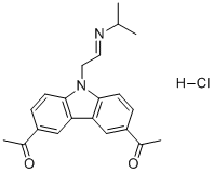
C21H24N2O2-HCL
|
|
|---|---|---|
| 分子量 |
372.89
|
|
| 精确质量 |
372.16
|
|
| 元素分析 |
C, 67.64; H, 6.76; Cl, 9.51; N, 7.51; O, 8.58
|
|
| CAS号 |
1197397-89-9
|
|
| 相关CAS号 |
CBL0137;1197996-80-7
|
|
| PubChem CID |
44519123
|
|
| 外观&性状 |
Off-white to yellow solid powder
|
|
| LogP |
5.39
|
|
| tPSA |
51.1
|
|
| 氢键供体(HBD)数目 |
2
|
|
| 氢键受体(HBA)数目 |
3
|
|
| 可旋转键数目(RBC) |
6
|
|
| 重原子数目 |
26
|
|
| 分子复杂度/Complexity |
466
|
|
| 定义原子立体中心数目 |
0
|
|
| SMILES |
CC(C1C=C2C(N(CCNC(C)C)C3=CC=C(C=C32)C(C)=O)=CC=1)=O.Cl
|
|
| InChi Key |
IXRKBBVMDMKAEB-UHFFFAOYSA-N
|
|
| InChi Code |
InChI=1S/C21H24N2O2.ClH/c1-13(2)22-9-10-23-20-7-5-16(14(3)24)11-18(20)19-12-17(15(4)25)6-8-21(19)23;/h5-8,11-13,22H,9-10H2,1-4H3;1H
|
|
| 化学名 |
1-[6-acetyl-9-[2-(propan-2-ylamino)ethyl]carbazol-3-yl]ethanone;hydrochloride
|
|
| 别名 |
|
|
| HS Tariff Code |
2934.99.9001
|
|
| 存储方式 |
Powder -20°C 3 years 4°C 2 years In solvent -80°C 6 months -20°C 1 month 注意: 请将本产品存放在密封且受保护的环境中,避免吸湿/受潮。 |
|
| 运输条件 |
Room temperature (This product is stable at ambient temperature for a few days during ordinary shipping and time spent in Customs)
|
| 溶解度 (体外实验) |
|
|||
|---|---|---|---|---|
| 溶解度 (体内实验) |
配方 1 中的溶解度: ≥ 2.5 mg/mL (6.70 mM) (饱和度未知) in 10% DMSO + 40% PEG300 + 5% Tween80 + 45% Saline (这些助溶剂从左到右依次添加,逐一添加), 澄清溶液。
例如,若需制备1 mL的工作液,可将100 μL 25.0 mg/mL澄清DMSO储备液加入到400 μL PEG300中,混匀;然后向上述溶液中加入50 μL Tween-80,混匀;加入450 μL生理盐水定容至1 mL。 *生理盐水的制备:将 0.9 g 氯化钠溶解在 100 mL ddH₂O中,得到澄清溶液。 配方 2 中的溶解度: ≥ 2.5 mg/mL (6.70 mM) (饱和度未知) in 10% DMSO + 90% (20% SBE-β-CD in Saline) (这些助溶剂从左到右依次添加,逐一添加), 澄清溶液。 例如,若需制备1 mL的工作液,可将 100 μL 25.0 mg/mL澄清DMSO储备液加入900 μL 20% SBE-β-CD生理盐水溶液中,混匀。 *20% SBE-β-CD 生理盐水溶液的制备(4°C,1 周):将 2 g SBE-β-CD 溶解于 10 mL 生理盐水中,得到澄清溶液。 View More
配方 3 中的溶解度: 5%DMSO+40%PEG300+5%Tween80+50%ddH2O: 0.75mg/ml 1、请先配制澄清的储备液(如:用DMSO配置50 或 100 mg/mL母液(储备液)); 2、取适量母液,按从左到右的顺序依次添加助溶剂,澄清后再加入下一助溶剂。以 下列配方为例说明 (注意此配方只用于说明,并不一定代表此产品 的实际溶解配方): 10% DMSO → 40% PEG300 → 5% Tween-80 → 45% ddH2O (或 saline); 假设最终工作液的体积为 1 mL, 浓度为5 mg/mL: 取 100 μL 50 mg/mL 的澄清 DMSO 储备液加到 400 μL PEG300 中,混合均匀/澄清;向上述体系中加入50 μL Tween-80,混合均匀/澄清;然后继续加入450 μL ddH2O (或 saline)定容至 1 mL; 3、溶剂前显示的百分比是指该溶剂在最终溶液/工作液中的体积所占比例; 4、 如产品在配制过程中出现沉淀/析出,可通过加热(≤50℃)或超声的方式助溶; 5、为保证最佳实验结果,工作液请现配现用! 6、如不确定怎么将母液配置成体内动物实验的工作液,请查看说明书或联系我们; 7、 以上所有助溶剂都可在 Invivochem.cn网站购买。 |
| 制备储备液 | 1 mg | 5 mg | 10 mg | |
| 1 mM | 2.6818 mL | 13.4088 mL | 26.8176 mL | |
| 5 mM | 0.5364 mL | 2.6818 mL | 5.3635 mL | |
| 10 mM | 0.2682 mL | 1.3409 mL | 2.6818 mL |
1、根据实验需要选择合适的溶剂配制储备液 (母液):对于大多数产品,InvivoChem推荐用DMSO配置母液 (比如:5、10、20mM或者10、20、50 mg/mL浓度),个别水溶性高的产品可直接溶于水。产品在DMSO 、水或其他溶剂中的具体溶解度详见上”溶解度 (体外)”部分;
2、如果您找不到您想要的溶解度信息,或者很难将产品溶解在溶液中,请联系我们;
3、建议使用下列计算器进行相关计算(摩尔浓度计算器、稀释计算器、分子量计算器、重组计算器等);
4、母液配好之后,将其分装到常规用量,并储存在-20°C或-80°C,尽量减少反复冻融循环。
计算结果:
工作液浓度: mg/mL;
DMSO母液配制方法: mg 药物溶于 μL DMSO溶液(母液浓度 mg/mL)。如该浓度超过该批次药物DMSO溶解度,请首先与我们联系。
体内配方配制方法:取 μL DMSO母液,加入 μL PEG300,混匀澄清后加入μL Tween 80,混匀澄清后加入 μL ddH2O,混匀澄清。
(1) 请确保溶液澄清之后,再加入下一种溶剂 (助溶剂) 。可利用涡旋、超声或水浴加热等方法助溶;
(2) 一定要按顺序加入溶剂 (助溶剂) 。
CBL0137 and gemcitabine toxicity to pancreatic ductal adenocarcinoma cell lines.Oncotarget.2014 Nov 30;5(22):11038-53. |
|---|
Effect of CBL0137 and gemcitabine on orthotopic PANC1 pancreatic tumor growth in nude mice.Oncotarget.2014 Nov 30;5(22):11038-53. |
Morphology and expression of FACT subunits (SSRP1, SPT16) and proliferation marker Ki67 in PDX samples of pancreatic ductal adenocarcinoma (PDA) used in the study.Oncotarget.2014 Nov 30;5(22):11038-53. |
Effect of CBL0137 and gemcitabine on patient derived PDA xenograft models.Oncotarget.2014 Nov 30;5(22):11038-53. |
|---|
CBL0137 inhibit gemcitabine induced transcriptional responses.Oncotarget.2014 Nov 30;5(22):11038-53. |
CBL0137 is toxic for cancer stem cells (CSC).Oncotarget.2014 Nov 30;5(22):11038-53. |
 PROTAC LZK-IN-1
 Seldegamadlin (KT-253)
 MD-265
 p53 Activator 13
InvivoChem的所有产品仅用于作科学研究,不面向患者销售
Copyright 2020 InvivoChem LLC | All Rights Reserved 粤ICP备20063088号-1


 COA
COA


 463611831
