| 规格 | 价格 | 库存 | 数量 |
|---|---|---|---|
| 5mg |
|
||
| 10mg |
|
||
| 25mg |
|
||
| 50mg |
|
||
| 100mg |
|
||
| 250mg |
|
||
| 500mg |
|
||
| 1g |
|
||
| Other Sizes |
|
| 靶点 |
PKC:660 nM (IC50); PKA:0.17 mM (IC50); TPK:0.1 mM (IC50)
Chelerythrine displaces the BH3-containing protein Bax from BclXL and inhibits the BclXL-Bak BH3 peptide binding with an IC50 of 1.5 μM. Loaded cells treated with chelerythrine underwent cell engraftment, which was characterized by mitochondrial involvement [1]. Chelidonine treatment activates and inhibits LPS-induced TNF-α levels and LPS-induced mouse peritoneal giant cells by blocking p38 mitogen protein activator (MAPK) and extracellular signal regulatory protein activator 1 and 2 (ERK1/2). Furthermore, the control of intermediate mediator expression by p38 MAPK and ERK1/2 may account for the effects of chelerythrine on the generation of NO and the cytokine TNF-α [2]. With an LD50 value of 3.46 μM, erythrine is cytotoxic to human monocyte leukocytes. Cells exposed to sanguinarine and chelerythrine dramatically decreased the expression of CCL-2 by 3.5 and 1.9 times, respectively, two hours after LPS stimulation [3]. In a dose-dependent way, chelidonine chloride markedly increased ERK1/2 phosphorylation. Furthermore, p38 phosphorylation can be inhibited by chelerythrine chloride [4]. |
|---|---|
| 体外研究 (In Vitro) |
白屈菜红碱取代 BclXL 中含有 BH3 的蛋白 Bax,并抑制 BclXL-Bak BH3 肽结合,IC50 为 1.5 μM。用白屈菜红碱处理的负载细胞进行细胞植入,其特征是线粒体参与[1]。 Chelidonine 治疗通过阻断 p38 丝裂原蛋白激活剂 (MAPK) 和细胞外信号调节蛋白激活剂 1 和 2 (ERK1/2) 来激活和抑制 LPS 诱导的 TNF-α 水平以及 LPS 诱导的小鼠腹膜巨细胞。此外,p38 MAPK 和 ERK1/2 对中间介质表达的控制可能解释了白屈菜赤碱对 NO 和细胞因子 TNF-α 生成的影响 [2]。赤藓碱的 LD50 值为 3.46 μM,对人单核白细胞具有细胞毒性。 LPS 刺激后两小时,暴露于血根碱和白屈菜红碱的细胞 CCL-2 的表达分别显着降低 3.5 倍和 1.9 倍 [3]。以剂量依赖性方式,白屈菜碱氯化物显着增加 ERK1/2 磷酸化。此外,p38 磷酸化可以被氯化白屈菜红碱抑制[4]。
白屈菜红碱 (Chelerythrine, CHE) 以浓度依赖性方式(浓度为10⁻⁵, 10⁻⁴, 10⁻³, 10⁻², 10⁻¹ µg/ml)显著抑制脂多糖 (LPS) 诱导的小鼠腹腔巨噬细胞一氧化氮 (NO) 的产生。其抑制效果与特异性p38 MAPK抑制剂SB203580 (10 µM) 和ERK1/2抑制剂PD98059 (20 µM) 相当。[1] 白屈菜红碱 (Chelerythrine, CHE) 以浓度依赖性方式(浓度为10⁻⁵, 10⁻⁴, 10⁻³, 10⁻², 10⁻¹ µg/ml)随时间(4, 6, 12, 24小时)显著降低脂多糖 (LPS) 诱导的小鼠腹腔巨噬细胞肿瘤坏死因子-α (TNF-α) 的产生。其抑制效果与特异性p38 MAPK抑制剂SB203580 (10 µM) 和ERK1/2抑制剂PD98059 (20 µM) 相当。[1] 白屈菜红碱 (Chelerythrine, CHE) 以浓度依赖性方式(浓度为10⁻⁴, 10⁻², 10⁻¹ µg/ml)显著抑制脂多糖 (LPS) 诱导的小鼠腹腔巨噬细胞中ERK1/2和p38 MAPK的磷酸化,Western blot分析证实了这一点。其抑制效果与特异性抑制剂SB203580和PD98059相当。[1] 根据MTT法测定,白屈菜红碱 (Chelerythrine, CHE) 在1和10 µg/ml浓度下处理24小时对小鼠腹腔巨噬细胞表现出细胞毒性效应。浓度高达10⁻¹ µg/ml时未显著影响细胞活力。[1] |
| 体内研究 (In Vivo) |
当接触 LPS 时,白屈菜红碱可抑制血清中一氧化氮 (NO) 和肿瘤坏死因子-α (TNF-α) 的产生,从而在体内实验诱导的小鼠内毒素休克模型中表现出显着的抗炎作用 [2]。当腹膜内给药时,氯化白屈菜红碱(5 mg/kg/天)会导致肾细胞癌细胞凋亡,但不会对小鼠造成相当大的伤害。 P53 以剂量依赖性方式响应氯化胆碱治疗而积累[4]。
白屈菜红碱 (Chelerythrine, CHE) 预处理(腹腔注射,剂量为1、5或10 mg/kg,于LPS注射前24小时和1小时给药)以剂量依赖性方式显著提高了LPS诱导的内毒素休克模型小鼠的存活率。10 mg/kg剂量提供完全保护(8/8小鼠存活),5 mg/kg提供75%保护(6/8存活),1 mg/kg提供37.5%保护(3/8存活),观察时长为72小时。[1] 白屈菜红碱 (Chelerythrine, CHE) 预处理(腹腔注射,剂量为1、5或10 mg/kg)以剂量依赖性方式显著抑制了LPS注射后3小时和6小时血清一氧化氮 (NO) 水平的升高。[1] 白屈菜红碱 (Chelerythrine, CHE) 预处理(腹腔注射,剂量为1、5或10 mg/kg)以剂量依赖性方式显著降低了LPS注射后1、3和6小时血清肿瘤坏死因子-α (TNF-α) 水平的升高。[1] |
| 酶活实验 |
抗凋亡Bcl-2家族成员的小分子抑制剂的鉴定开辟了新的治疗机会,而天然产物的化学结构和生物活性的巨大多样性尚未被系统地利用。在这里,我们报告通过高通量筛选从天然产物中提取的107,423个提取物,鉴定出chelerythrine是BclXL-Bak Bcl-2同源3 (BH3)结构域结合的抑制剂。Chelerythrine抑制BclXL- bak BH3肽结合的IC50为1.5 μ m,并取代BclXL中含有BH3的蛋白Bax。用chelerythrine处理的哺乳动物细胞发生凋亡,其特征表明与线粒体途径有关。在完整细胞中,星孢素、H7、依托opo苷和车车红碱释放线粒体中的细胞色素c,而只有车车红碱释放离体线粒体中的细胞色素c。此外,bclxl过表达的细胞对本研究中使用的凋亡刺激完全耐药,但对车车菊碱仍然敏感。虽然chelerythrine被广泛认为是一种蛋白激酶C抑制剂,但其介导细胞凋亡的机制仍然存在争议。我们的数据表明,赤藓碱通过直接靶向Bcl-2家族蛋白的机制触发细胞凋亡[5]。
In Vitro Binding Analysis-Labeled Bax是利用Promega的TnT t7偶联网状细胞裂解液系统,通过体外转录/翻译pXJHA-Bax制备的。GST结合实验按照之前的描述进行,只是在加入[35S]Bax之前,在GSTBclXLΔ19中孵育浓度增加的车车菊氨酸30分钟。[5] |
| 细胞实验 |
MTT法测定细胞活力。将100µL培养基中的细胞(2×10³ HEK-293细胞/孔和3×103 SW-839细胞/孔)接种到96孔板中,孵育12小时。然后,将每孔中的培养基替换为含有不同浓度氯化Chelerythrine的培养基,在37°C下再孵育24和48小时。随后,每孔中加入20µL MTT (5 mg/mL)。在37℃下孵育4小时后,去除上清,每孔加入100µL DMSO。使用iMark™微孔板吸光度仪测定吸光度值(在540nm处读取)[1]。
先前的研究表明,苯并[c]菲啶生物碱白屈菜红碱氯化物(CC)对各种肿瘤具有抑制作用。然而,CC的抗癌活性及其潜在机制尚未在癌症肾细胞中阐明。本研究观察了CC对肾癌症细胞生长抑制和凋亡的影响。流式细胞术和3-(4,5-二甲基噻唑-2-基)-2,5-二苯基四唑鎓溴化物测定显示,CC以时间和剂量依赖的方式显著抑制HEK-293和人癌症SW-839细胞的生长。在裸小鼠中进行的异种移植物小鼠模型显示,CC治疗后肿瘤生长减少。此外,本研究表明,CC显著降低了细胞外信号调节激酶(ERK)和Akt的磷酸化,同时上调了p53、B细胞淋巴瘤2(Bcl-2)相关X蛋白、切割的半胱氨酸天冬氨酸蛋白酶-3和切割的聚二磷酸腺苷核糖聚合酶(PARP),下调了Bcl-2、半胱氨酸天冬氨酰蛋白酶-3和PARP。此外,使用PD98059,一种特异性促有丝分裂原活化的蛋白激酶抑制剂,增强了CC的促凋亡作用,这表明CC可能部分通过抑制ERK活性诱导癌症细胞凋亡。总体而言,本研究的结果表明,CC可能被开发为癌症患者的潜在抗癌治疗[3]。 细胞活力测定 (MTT法): 从注射了硫基乙酸盐的小鼠体内分离腹腔巨噬细胞。将细胞接种于96孔板中,并用不同浓度的白屈菜红碱 (Chelerythrine, CHE)(10⁻⁵ 至 10 µg/ml)单独或与LPS (10 µg/ml) 联合处理24小时。孵育后,向每孔加入MTT试剂。再孵育4小时后,使用DMSO溶解生成的甲瓒结晶。在490 nm波长下测量吸光度以评估细胞活力。[1] 一氧化氮 (NO) 测定: 将腹腔巨噬细胞接种于24孔板中,并分别用不同浓度的白屈菜红碱 (Chelerythrine, CHE)(10⁻⁵ 至 10⁻¹ µg/ml)、SB203580 (10 µM) 或PD98059 (20 µM) 预处理24小时,然后暴露于LPS (10 µg/ml) 12小时。收集细胞培养上清液并与Griess试剂混合。在室温避光条件下孵育后,在540 nm波长下测量吸光度。使用亚硝酸钠标准曲线确定亚硝酸盐浓度。[1] 肿瘤坏死因子-α (TNF-α) ELISA: 腹腔巨噬细胞用不同浓度的白屈菜红碱 (Chelerythrine, CHE)(10⁻⁵ 至 10⁻¹ µg/ml)预处理24小时,然后暴露于LPS (10 µg/ml) 4、6、12或24小时。收集细胞培养上清液。使用商业ELISA试剂盒按照制造商说明测量TNF-α水平。[1] Western Blot分析: 腹腔巨噬细胞用不同浓度的白屈菜红碱 (Chelerythrine, CHE)(10⁻⁴, 10⁻², 10⁻¹ µg/ml)、SB203580 (10 µM) 或PD98059 (20 µM) 预处理24小时,然后暴露于LPS (10 µg/ml) 30分钟。使用裂解液提取总细胞蛋白。测定蛋白质浓度。等量蛋白质通过SDS-PAGE分离并转印至膜上。封闭膜后,与针对磷酸化p38、磷酸化ERK1/2和GAPDH的一抗在4°C孵育过夜。洗涤后,膜与辣根过氧化物酶标记的二抗孵育。使用增强化学发光系统显影蛋白条带。[1] |
| 动物实验 |
溶于PBS;5 mg/kg;腹腔注射。携带SQ-20B异种移植瘤的小鼠:将5×10⁶个SW-839细胞与Matrigel®混合,皮下注射到14只5周龄雄性BALB/c裸鼠的侧腹部。小鼠饲养于18×30 cm的笼子中,每笼3只,温度为22°C,光照/黑暗周期为12小时。食物和水自由摄取。小鼠随机分为两组(n=7)。如前所述,小鼠腹腔注射氯化螯合红碱,剂量为5 mg/kg/天,持续5周,首次注射氯化螯合红碱在SW-839细胞注射后24小时进行。对照组小鼠注射等体积的含1% DMSO的PBS。每周测量一次小鼠肿瘤的体积和重量。所有小鼠在接种癌细胞后36天处死,并切除肿瘤。[1]
急性毒性研究:健康雄性小鼠腹腔注射白屈菜红碱(CHE),初始剂量为1 mg/kg。观察小鼠的毒性症状、一般行为和死亡率,持续72小时。每日测量体重。如果未观察到毒性或死亡,则使用更高剂量(5、10和15 mg/kg)重复上述步骤。在本研究中,10 mg/kg的剂量未产生任何毒性症状。[1] LPS诱导的内毒素休克模型:将雄性小鼠分组。各治疗组分别在腹腔注射LPS(100 µg/kg)前24小时和1小时腹腔注射不同剂量(1、5或10 mg/kg)的白屈菜红碱 (CHE)。对照组注射溶剂,LPS组仅注射LPS。分别于LPS注射后0、1、3和6小时经静脉窦采集血样。离心分离血清,并储存于-80°C,用于TNF-α和NO水平的分析。监测小鼠存活72小时。[1] 腹腔巨噬细胞分离用于细胞分析:雌性小鼠腹腔注射1%硫代乙醇酸钠溶液。六天后,将小鼠安乐死,并用磷酸盐缓冲液(PBS)灌洗腹腔,收集腹腔巨噬细胞。离心收集细胞,洗涤后,在添加了胎牛血清和抗生素的RPMI-1640培养基中培养。[1] |
| 毒性/毒理 (Toxicokinetics/TK) |
毒性概述
白屈菜红碱是一种强效、选择性强且可透过细胞膜的蛋白激酶C (PKC) 抑制剂。它也是植物刺花椒(Zanthoxylum clava-herculis)中的主要活性天然产物,对金黄色葡萄球菌具有抗菌活性。(维基百科) 白屈菜红碱是A组和B组PKC亚型的选择性抑制剂,具有抗肿瘤活性。氯化白屈菜红碱抑制PKC可通过激活中性鞘磷脂酶、神经酰胺积累和鞘磷脂耗竭诱导细胞凋亡。白屈菜红碱对PKC的选择性至少比其他激酶高100倍。白屈菜红碱与PKC的保守催化位点竞争,似乎是A组和B组激酶的强效特异性抑制剂。体外实验表明,白屈菜红碱对九种人类肿瘤细胞系均具有细胞毒活性。体外实验中,经白屈菜红碱处理后,放射抗性和化疗抗性的头颈部鳞状细胞癌(HNSCC)细胞系迅速发生凋亡。用白屈菜红碱治疗荷瘤SQ-20B HNSCC细胞的裸鼠可显著延缓肿瘤生长。此外,白屈菜红碱治疗的毒性极低。 小鼠皮下注射LD50为95 mg/kg,Planta Medica., 43(161), 1981 [PMID:7312984] 小鼠静脉注射LD50为18500 μg/kg,Planta Medica., 43(161), 1981 [PMID:7312984] 在小鼠急性毒性研究中,腹腔注射10 mg/kg剂量的白屈菜红碱(CHE),在72小时的观察期内,未观察到小鼠一般外观或毒性症状的任何变化。[1] |
| 参考文献 |
|
| 其他信息 |
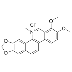
氯化白屈菜碱是一种苯并菲啶生物碱。
季铵苯并[c]生物碱白屈菜碱(CHE)是一种传统中药,已被用于治疗多种炎症性疾病。为了深入了解CHE的抗炎作用及其分子机制,我们利用实验诱导的小鼠内毒素休克模型和脂多糖(LPS)诱导的小鼠腹腔巨噬细胞模型,研究了CHE的抗炎功能。结果表明,CHE在体内实验诱导的小鼠内毒素休克模型中表现出显著的抗炎作用,其机制是通过抑制LPS诱导的血清肿瘤坏死因子-α(TNF-α)水平和一氧化氮(NO)的产生。此外,我们的数据表明,CHE 处理通过选择性抑制 p38 丝裂原活化蛋白激酶 (MAPK) 和细胞外信号调节激酶 1 和 2 (ERK1/2) 的激活,抑制 LPS 诱导的小鼠腹腔巨噬细胞中 TNF-α 水平和 NO 的产生。此外,CHE 对 NO 和细胞因子 TNF-α 产生的影响可能与 p38 MAPK 和 ERK1/2 在炎症介质表达调控中的作用有关。[1] 季铵苯并[c]菲啶生物碱血根碱和白屈菜红碱因其广泛的药用特性而被用于民间医学。它们的主要作用之一是抗炎活性,但其具体机制尚未完全阐明。本研究着重探讨了这些生物碱调节促炎细胞因子肿瘤坏死因子α (TNF-α)、单核细胞趋化蛋白1 (MCP-1,又称CCL-2)、白细胞介素 (IL)-6、IL-1β以及抗炎细胞因子IL-1受体拮抗剂 (IL-1RA) 和IL-10基因表达的能力。并将这些生物碱的作用与传统药物泼尼松进行了比较。人单核细胞来源的巨噬细胞预先用生物碱或泼尼松处理,然后用脂多糖诱导炎症反应。检测了上述细胞因子转录水平的基因表达变化。本研究发现,主要受影响的促炎细胞因子是CCL-2和IL-6。 LPS刺激2小时后,经血根碱和白屈菜红碱处理的细胞中CCL-2表达显著降低,分别降低了3.5倍(p<0.001)和1.9倍(p<0.01);而经泼尼松处理的细胞中CCL-2表达降低5.3倍(p<0.001)。LPS诱导8小时后,两种生物碱均显著降低了CCL-2的表达。血根碱处理的细胞中CCL-2表达降低更为显著,比溶剂对照组降低了4.3倍(p<0.001)。LPS刺激2小时后,与溶剂对照组相比,血根碱处理的细胞中IL-6 mRNA水平降低了3.9倍(p<0.001)。白屈菜红碱处理的细胞中IL-6 mRNA水平降低了1.6倍(p<0.001)。与白屈菜红碱相比,血根碱更能降低CCL-2和IL-6的基因表达,其作用与泼尼松非常相似。LPS刺激4小时后,经血根碱预处理的细胞中IL-1RA的表达显著高于未用血根碱处理的细胞(高出1.7倍,p<0.001)。我们的研究结果有助于阐明这些生物碱在炎症过程中的可能作用机制。[2] 白屈菜红碱 (CHE)是一种季铵苯并[c]菲啶生物碱,存在于罂粟科和芸香科植物中,例如白屈菜和博落回。[1] 白屈菜红碱 (CHE)传统上用于治疗各种炎症性疾病,并具有广泛的生物活性,包括抗菌和抗炎作用。 [1] 在LPS激活的巨噬细胞中,白屈菜红碱(CHE)的抗炎机制涉及MAPK信号通路的下调(特别是抑制p38和ERK1/2的磷酸化),从而减少TNF-α和NO等促炎介质的产生。[1] 该研究表明,白屈菜红碱(CHE)通过调节巨噬细胞活化,具有治疗炎症介导疾病(如内毒素休克)的治疗潜力。[1] |
| 分子式 |
C21H18NO4.HCL
|
|
|---|---|---|
| 分子量 |
384.83
|
|
| 精确质量 |
383.092
|
|
| 元素分析 |
C, 65.71; H, 4.73; Cl, 9.24; N, 3.65; O, 16.67
|
|
| CAS号 |
3895-92-9
|
|
| 相关CAS号 |
Chelerythrine;34316-15-9; 478-03-5 (OH-); 34316-15-9; 3895-92-9 (chloride)
|
|
| PubChem CID |
72311
|
|
| 外观&性状 |
Typically exists as Yellow to orange solids at room temperature
|
|
| 密度 |
1.36g/cm3
|
|
| 沸点 |
711.4ºC at 760 mmHg
|
|
| 熔点 |
195-205ºC
|
|
| 闪点 |
219.3ºC
|
|
| 折射率 |
1.681
|
|
| LogP |
0.72
|
|
| tPSA |
40.8
|
|
| 氢键供体(HBD)数目 |
0
|
|
| 氢键受体(HBA)数目 |
5
|
|
| 可旋转键数目(RBC) |
2
|
|
| 重原子数目 |
27
|
|
| 分子复杂度/Complexity |
516
|
|
| 定义原子立体中心数目 |
0
|
|
| SMILES |
[Cl-].O1C([H])([H])OC2=C1C([H])=C1C(=C2[H])C([H])=C([H])C2=C3C([H])=C([H])C(=C(C3=C([H])[N+](C([H])([H])[H])=C21)OC([H])([H])[H])OC([H])([H])[H]
|
|
| InChi Key |
WEEFNMFMNMASJY-UHFFFAOYSA-M
|
|
| InChi Code |
InChI=1S/C21H18NO4.ClH/c1-22-10-16-13(6-7-17(23-2)21(16)24-3)14-5-4-12-8-18-19(26-11-25-18)9-15(12)20(14)22;/h4-10H,11H2,1-3H3;1H/q+1;/p-1
|
|
| 化学名 |
|
|
| 别名 |
|
|
| HS Tariff Code |
2934.99.9001
|
|
| 存储方式 |
Powder -20°C 3 years 4°C 2 years In solvent -80°C 6 months -20°C 1 month 注意: 请将本产品存放在密封且受保护的环境中,避免吸湿/受潮。 |
|
| 运输条件 |
Room temperature (This product is stable at ambient temperature for a few days during ordinary shipping and time spent in Customs)
|
| 溶解度 (体外实验) |
|
|||
|---|---|---|---|---|
| 溶解度 (体内实验) |
配方 1 中的溶解度: ≥ 0.44 mg/mL (1.15 mM) (饱和度未知) in 10% DMSO + 90% (20% SBE-β-CD in Saline) (这些助溶剂从左到右依次添加,逐一添加), 澄清溶液。
例如,若需制备1 mL的工作液,可将100 μL 4.4mg/mL澄清的DMSO储备液加入到900μL 20%SBE-β-CD生理盐水中,混匀。 *20% SBE-β-CD 生理盐水溶液的制备(4°C,1 周):将 2 g SBE-β-CD 溶解于 10 mL 生理盐水中,得到澄清溶液。 配方 2 中的溶解度: ≥ 0.44 mg/mL (1.15 mM) (饱和度未知) in 10% DMSO + 90% Corn Oil (这些助溶剂从左到右依次添加,逐一添加), 澄清溶液。 例如,若需制备1 mL的工作液,可将 100 μL 4.4 mg/mL 澄清 DMSO 储备液加入900 μL 玉米油中,混合均匀。 View More
配方 3 中的溶解度: ≥ 0.43 mg/mL (1.12 mM) (饱和度未知) in 10% DMSO + 40% PEG300 + 5% Tween80 + 45% Saline (这些助溶剂从左到右依次添加,逐一添加), 澄清溶液。 1、请先配制澄清的储备液(如:用DMSO配置50 或 100 mg/mL母液(储备液)); 2、取适量母液,按从左到右的顺序依次添加助溶剂,澄清后再加入下一助溶剂。以 下列配方为例说明 (注意此配方只用于说明,并不一定代表此产品 的实际溶解配方): 10% DMSO → 40% PEG300 → 5% Tween-80 → 45% ddH2O (或 saline); 假设最终工作液的体积为 1 mL, 浓度为5 mg/mL: 取 100 μL 50 mg/mL 的澄清 DMSO 储备液加到 400 μL PEG300 中,混合均匀/澄清;向上述体系中加入50 μL Tween-80,混合均匀/澄清;然后继续加入450 μL ddH2O (或 saline)定容至 1 mL; 3、溶剂前显示的百分比是指该溶剂在最终溶液/工作液中的体积所占比例; 4、 如产品在配制过程中出现沉淀/析出,可通过加热(≤50℃)或超声的方式助溶; 5、为保证最佳实验结果,工作液请现配现用! 6、如不确定怎么将母液配置成体内动物实验的工作液,请查看说明书或联系我们; 7、 以上所有助溶剂都可在 Invivochem.cn网站购买。 |
| 制备储备液 | 1 mg | 5 mg | 10 mg | |
| 1 mM | 2.5986 mL | 12.9928 mL | 25.9855 mL | |
| 5 mM | 0.5197 mL | 2.5986 mL | 5.1971 mL | |
| 10 mM | 0.2599 mL | 1.2993 mL | 2.5986 mL |
1、根据实验需要选择合适的溶剂配制储备液 (母液):对于大多数产品,InvivoChem推荐用DMSO配置母液 (比如:5、10、20mM或者10、20、50 mg/mL浓度),个别水溶性高的产品可直接溶于水。产品在DMSO 、水或其他溶剂中的具体溶解度详见上”溶解度 (体外)”部分;
2、如果您找不到您想要的溶解度信息,或者很难将产品溶解在溶液中,请联系我们;
3、建议使用下列计算器进行相关计算(摩尔浓度计算器、稀释计算器、分子量计算器、重组计算器等);
4、母液配好之后,将其分装到常规用量,并储存在-20°C或-80°C,尽量减少反复冻融循环。
计算结果:
工作液浓度: mg/mL;
DMSO母液配制方法: mg 药物溶于 μL DMSO溶液(母液浓度 mg/mL)。如该浓度超过该批次药物DMSO溶解度,请首先与我们联系。
体内配方配制方法:取 μL DMSO母液,加入 μL PEG300,混匀澄清后加入μL Tween 80,混匀澄清后加入 μL ddH2O,混匀澄清。
(1) 请确保溶液澄清之后,再加入下一种溶剂 (助溶剂) 。可利用涡旋、超声或水浴加热等方法助溶;
(2) 一定要按顺序加入溶剂 (助溶剂) 。
InvivoChem的所有产品仅用于作科学研究,不面向患者销售
Copyright 2020 InvivoChem LLC | All Rights Reserved 粤ICP备20063088号-1


 COA
COA
 463611831
