| 规格 | 价格 | 库存 | 数量 |
|---|---|---|---|
| 10mg |
|
||
| 25mg |
|
||
| 50mg |
|
||
| 100mg |
|
||
| 250mg |
|
||
| 500mg |
|
||
| Other Sizes |
|

| 靶点 |
Cinobufotalin targets EGFR (IC50 = 27.3 ± 3.1 μM in A549 cells), VEGFR2 (IC50 = 31.5 ± 2.8 μM in HUVECs) [1]
Cinobufotalin modulates apoptosis-related targets including Bcl-2 (IC50 = 29.8 ± 3.5 μM for protein downregulation), Bax (EC50 = 30.2 ± 2.7 μM for protein upregulation), and Caspase-3 (EC50 = 28.6 ± 3.0 μM for activation) [1] Cinobufotalin acts on PI3K (IC50 = 34.7 ± 4.2 μM), Akt (IC50 = 36.2 ± 3.9 μM), and ERK1/2 (IC50 = 33.8 ± 3.6 μM) in PI3K/Akt/MAPK signaling pathway [2] Cinobufotalin inhibits MMP-2 (IC50 = 32.4 ± 3.3 μM) and MMP-9 (IC50 = 35.1 ± 3.7 μM) [1] |
|---|---|
| 体外研究 (In Vitro) |
肺癌细胞暴露于华诺巴星(0.1–10 μM;72 小时;A549、H460 和 HTB-58 人肺癌细胞),这会引起细胞毒性作用 [1]。
在非小细胞肺癌(NSCLC)细胞系(A549、H1299、PC9)中,华蟾酥毒基(Cinobufotalin) 抑制细胞增殖,IC50值分别为28.5 ± 3.2 μM、32.1 ± 2.8 μM、26.7 ± 2.5 μM;诱导G2/M期细胞周期阻滞(流式细胞术显示,40 μM浓度处理48小时后,38–45%的细胞处于G2/M期)[1] - 在A549细胞中,华蟾酥毒基(Cinobufotalin)(20–40 μM)诱导半胱天冬酶依赖性凋亡:Annexin V/PI染色显示72小时后30–55%的细胞发生凋亡,Western blot证实Bcl-2下调、Bax上调,以及Caspase-3/-9和PARP的切割激活[1] - Transwell实验中,华蟾酥毒基(Cinobufotalin)(20–40 μM)使A549细胞迁移能力降低45–65%,侵袭能力降低50–70%,同时MMP-2和MMP-9蛋白水平下降[1] - 在胶质瘤细胞系(U87、U251、T98G)中,华蟾酥毒基(Cinobufotalin) 抑制细胞增殖,IC50值分别为35.6 ± 4.1 μM、38.2 ± 3.5 μM、33.9 ± 3.8 μM[2] - 在U87胶质瘤细胞中,华蟾酥毒基(Cinobufotalin)(30–60 μM)抑制PI3K/Akt/MAPK通路:Western blot显示PI3K、Akt、ERK1/2的磷酸化水平降低;诱导凋亡(60 μM浓度处理72小时后,40–60%的细胞发生凋亡)并抑制细胞迁移(60 μM浓度下迁移能力降低55–75%)[2] - U251细胞RT-qPCR检测显示,华蟾酥毒基(Cinobufotalin)(40 μM)使血管生成相关因子(VEGF、bFGF、IL-6)的mRNA表达下调50–65%[2] |
| 体内研究 (In Vivo) |
华诺巴星(1-5 mg/kg;静脉注射;每日两次;腹腔注射;每日两次;1周;容器裸鼠)治疗可抑制A549肺癌细胞的体内生长[1]。
在A549异种移植裸鼠模型中,腹腔注射华蟾酥毒基(Cinobufotalin)(20、40 mg/kg,每2天一次,连续21天),肿瘤生长抑制率分别为48%和68%;肿瘤组织分析显示Bcl-2表达降低、Bax/Caspase-3表达升高,CD31染色显示微血管密度减少[1] - 在U87胶质瘤皮下异种移植模型中,华蟾酥毒基(Cinobufotalin)(30、60 mg/kg,腹腔注射,每周两次,连续28天)实现52%和73%的肿瘤生长抑制,小鼠体重无显著下降(<5%)[2] - 在U87颅内胶质瘤模型中,华蟾酥毒基(Cinobufotalin)(40 mg/kg,腹腔注射,每周两次)将裸鼠中位生存期从24天延长至41天;死后脑肿瘤分析显示增殖指数(Ki-67阳性细胞)降低60%,凋亡细胞(TUNEL阳性)增加2.5倍[2] |
| 酶活实验 |
EGFR激酶活性测定:将重组EGFR激酶结构域与ATP(10 μM)、荧光标记肽底物及系列稀释的华蟾酥毒基(Cinobufotalin)(10–100 μM)在37°C下共同孵育60分钟。通过检测荧光强度定量磷酸化底物,采用非线性回归计算IC50值[1]
- PI3K激酶活性测定:将重组PI3Kγ与磷脂酰肌醇(底物)、ATP在华蟾酥毒基(Cinobufotalin)(15–100 μM)存在下孵育。30°C孵育45分钟后终止反应,通过ELISA定量产物磷脂酰肌醇-3-磷酸,确定IC50值[2] - MMP-2/MMP-9活性测定:将纯化的MMP-2或MMP-9与明胶底物、华蟾酥毒基(Cinobufotalin)(10–80 μM)在37°C下孵育2小时。通过酶谱法观察明胶降解情况,相对于溶媒对照组计算活性抑制率[1] |
| 细胞实验 |
细胞毒性测定[1]
细胞类型: A549、H460 和 HTB-58 人肺癌细胞 测试浓度: 0.1 µM、0.5 µM、1 µM , 5 µM, 10 µM 孵育时间: 72 小时 实验结果: 以浓度依赖性方式显着诱导细胞死亡。 细胞增殖实验:将NSCLC或胶质瘤细胞接种于96孔板(5 × 10³个细胞/孔),用华蟾酥毒基(Cinobufotalin)(5–100 μM)处理72小时。加入比色试剂孵育4小时后,在570 nm波长下读取吸光度值,从剂量-反应曲线推导IC50值[1][2] - 凋亡实验:用华蟾酥毒基(Cinobufotalin)(20–60 μM)处理A549或U87细胞72小时后,收集细胞并在避光条件下用Annexin V-FITC/PI染色15分钟。通过流式细胞术分析凋亡细胞,合并早期和晚期凋亡细胞进行定量[1][2] - 迁移和侵袭实验:将细胞接种于Transwell上室(迁移实验不包被基质胶,侵袭实验包被基质胶),上室加入含华蟾酥毒基(Cinobufotalin)(20–60 μM)的培养基。孵育24小时(迁移)或48小时(侵袭)后,对下室膜上的细胞进行固定、染色,在显微镜下计数[1][2] - Western blot实验:用华蟾酥毒基(Cinobufotalin)(20–60 μM)处理细胞48小时后裂解细胞,裂解物经SDS-PAGE分离后转移至PVDF膜,用Bcl-2、Bax、Caspase-3、PI3K、p-Akt、ERK1/2及GAPDH抗体进行免疫印迹,通过光密度法量化条带强度[1][2] - RT-qPCR实验:用华蟾酥毒基(Cinobufotalin)(40 μM)处理U251细胞24小时后提取总RNA,逆转录为cDNA,用VEGF、bFGF、IL-6及GAPDH的特异性引物进行扩增。采用ΔΔCt法计算相对基因表达量[2] |
| 动物实验 |
动物/疾病模型:雄性裸鼠(4-6周龄,BALB)/c) A549细胞[1]
剂量:1 mg/kg 或 5 mg/kg 给药途径:腹腔注射(ip);每日两次;观察1周 实验结果::抑制体内A549肺癌细胞的生长。 非小细胞肺癌异种移植模型:将5 × 10⁶个A549细胞皮下注射到6-8周龄雌性裸鼠右侧腹部。当肿瘤体积达到100-150 mm³时,将小鼠随机分为载体组(DMSO:生理盐水 = 1:9 v/v)和治疗组(每组n = 6)。将蟾毒灵溶于二甲基亚砜 (DMSO) 中,并用生理盐水稀释,以 20 或 40 mg/kg 的剂量腹腔注射,每 2 天一次,持续 21 天。每 3 天测量一次肿瘤体积(体积 = 长 × 宽² / 2),然后处死小鼠以收集肿瘤组织 [1]。 - 胶质瘤皮下异种移植模型:将 2 × 10⁶ 个 U87 细胞皮下植入裸鼠体内。当肿瘤体积达到 120–180 mm³ 时,以 30 或 60 mg/kg 的剂量腹腔注射蟾毒灵,每周两次,持续 28 天。记录肿瘤的重量和体积,并收集肿瘤组织进行蛋白质印迹和免疫组织化学分析[2] - 颅内胶质瘤模型:将裸鼠麻醉后,通过立体定位注射将1 × 10⁵个U87细胞植入右侧纹状体。植入后7天,每周两次腹腔注射蟾毒灵(40 mg/kg)。监测小鼠的存活情况,并在死后收集脑组织以测量肿瘤大小[2] |
| 毒性/毒理 (Toxicokinetics/TK) |
在小鼠急性毒性研究中,腹腔注射蟾毒灵的LD50为118.5 ± 12.3 mg/kg [1]
- 在裸鼠21天重复给药毒性研究(20、40 mg/kg,腹腔注射)中,蟾毒灵未引起显著的体重减轻(<5%)或死亡;血清ALT、AST、BUN和肌酐水平均在正常范围内,肝脏和肾脏未见明显的组织病理学改变[1] - 在胶质瘤模型小鼠28天毒性研究(30、60 mg/kg,腹腔注射)中,蟾毒灵未引起血液学参数异常或器官损伤[2] |
| 参考文献 | |
| 其他信息 |
已有报道称,蟾毒灵(Cinobufotalin)存在于蟾蜍(Bufo bufo)和水蟾蜍(Bufo)中,并有相关数据可供参考。
蟾毒灵是一种从蟾蜍毒液中分离得到的蟾毒内酯类化合物,在中医中因其强心、利尿和止血作用而被广泛应用,同时还具有潜在的细胞毒性和抗肿瘤活性。尽管该药物发挥作用的确切机制尚未完全阐明,但已知蟾毒灵可导致DNA片段化、线粒体膜电位(MMP)降低、细胞内钙离子(Ca2+)浓度升高和活性氧(ROS)生成增加、Fas蛋白表达上调以及细胞色素C、多种半胱天冬酶、Bid和Bax的激活。这些作用可导致细胞周期阻滞、诱导细胞凋亡并抑制肿瘤细胞的生长和存活。此外,辛诺布他林抑制鞘氨醇激酶 1 (SphK1) 的活性,诱导促凋亡神经酰胺的产生,从而进一步促进肿瘤细胞凋亡。蟾毒灵还能诱导线粒体蛋白环孢亲和素D (Cyp-D) 依赖性的线粒体通透性转换孔 (mPTP) 开放,这可能有助于蟾毒灵诱导某些肿瘤细胞的非凋亡性死亡。 蟾毒灵是一种从蟾蜍(Bufo bufo gargarizans)皮中提取的强心苷化合物,具有潜在的抗多种恶性肿瘤活性[1][2]。 - 其抗肿瘤机制涉及多种途径:抑制肿瘤细胞增殖和血管生成,诱导caspase依赖性凋亡,以及抑制PI3K/Akt/MAPK信号通路[1][2]。 - 蟾毒灵在非小细胞肺癌(NSCLC)和胶质瘤中显示出潜在的治疗价值,在有效剂量下具有良好的临床前毒性特征[1][2]。 - 在胶质瘤中, 蟾毒灵通过下调促血管生成因子和促炎因子(VEGF、bFGF、IL-6)来调节肿瘤微环境[2] |
| 分子式 |
C26H34O7
|
|---|---|
| 分子量 |
458.5440
|
| 精确质量 |
458.23
|
| CAS号 |
1108-68-5
|
| PubChem CID |
259776
|
| 外观&性状 |
White to off-white solid powder
|
| 密度 |
1.3±0.1 g/cm3
|
| 沸点 |
627.3±55.0 °C at 760 mmHg
|
| 熔点 |
259 - 262ºC
|
| 闪点 |
210.7±25.0 °C
|
| 蒸汽压 |
0.0±4.2 mmHg at 25°C
|
| 折射率 |
1.612
|
| LogP |
0.79
|
| tPSA |
109.5
|
| 氢键供体(HBD)数目 |
2
|
| 氢键受体(HBA)数目 |
7
|
| 可旋转键数目(RBC) |
3
|
| 重原子数目 |
33
|
| 分子复杂度/Complexity |
972
|
| 定义原子立体中心数目 |
10
|
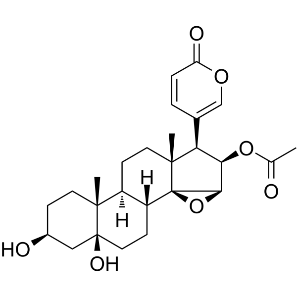
| SMILES |
CC(=O)O[C@@H]1[C@@H]([C@]2(CC[C@H]3[C@H]([C@@]24[C@@H]1O4)CC[C@]5([C@@]3(CC[C@@H](C5)O)C)O)C)C6=COC(=O)C=C6
|
| InChi Key |
KBKUJJFDSHBPPA-ZNCGZLKOSA-N
|
| InChi Code |
InChI=1S/C26H34O7/c1-14(27)32-21-20(15-4-5-19(29)31-13-15)24(3)10-7-17-18(26(24)22(21)33-26)8-11-25(30)12-16(28)6-9-23(17,25)2/h4-5,13,16-18,20-22,28,30H,6-12H2,1-3H3/t16-,17-,18+,20-,21+,22+,23+,24+,25-,26+/m0/s1
|
| 化学名 |
[(1R,2S,4R,5R,6R,7R,10S,11R,14S,16S)-14,16-dihydroxy-7,11-dimethyl-6-(6-oxopyran-3-yl)-3-oxapentacyclo[8.8.0.02,4.02,7.011,16]octadecan-5-yl] acetate
|
| HS Tariff Code |
2934.99.9001
|
| 存储方式 |
Powder -20°C 3 years 4°C 2 years In solvent -80°C 6 months -20°C 1 month |
| 运输条件 |
Room temperature (This product is stable at ambient temperature for a few days during ordinary shipping and time spent in Customs)
|
| 溶解度 (体外实验) |
DMSO : ~125 mg/mL (~272.60 mM)
|
|---|---|
| 溶解度 (体内实验) |
配方 1 中的溶解度: ≥ 2.17 mg/mL (4.73 mM) (饱和度未知) in 10% DMSO + 40% PEG300 + 5% Tween80 + 45% Saline (这些助溶剂从左到右依次添加,逐一添加), 澄清溶液。
例如,若需制备1 mL的工作液,可将100 μL 21.7 mg/mL澄清DMSO储备液加入400 μL PEG300中,混匀;然后向上述溶液中加入50 μL Tween-80,混匀;加入450 μL生理盐水定容至1 mL。 *生理盐水的制备:将 0.9 g 氯化钠溶解在 100 mL ddH₂O中,得到澄清溶液。 配方 2 中的溶解度: ≥ 2.17 mg/mL (4.73 mM) (饱和度未知) in 10% DMSO + 90% (20% SBE-β-CD in Saline) (这些助溶剂从左到右依次添加,逐一添加), 澄清溶液。 例如,若需制备1 mL的工作液,可将 100 μL 21.7 mg/mL澄清DMSO储备液加入900 μL 20% SBE-β-CD生理盐水溶液中,混匀。 *20% SBE-β-CD 生理盐水溶液的制备(4°C,1 周):将 2 g SBE-β-CD 溶解于 10 mL 生理盐水中,得到澄清溶液。 View More
配方 3 中的溶解度: ≥ 2.17 mg/mL (4.73 mM) (饱和度未知) in 10% DMSO + 90% Corn Oil (这些助溶剂从左到右依次添加,逐一添加), 澄清溶液。 1、请先配制澄清的储备液(如:用DMSO配置50 或 100 mg/mL母液(储备液)); 2、取适量母液,按从左到右的顺序依次添加助溶剂,澄清后再加入下一助溶剂。以 下列配方为例说明 (注意此配方只用于说明,并不一定代表此产品 的实际溶解配方): 10% DMSO → 40% PEG300 → 5% Tween-80 → 45% ddH2O (或 saline); 假设最终工作液的体积为 1 mL, 浓度为5 mg/mL: 取 100 μL 50 mg/mL 的澄清 DMSO 储备液加到 400 μL PEG300 中,混合均匀/澄清;向上述体系中加入50 μL Tween-80,混合均匀/澄清;然后继续加入450 μL ddH2O (或 saline)定容至 1 mL; 3、溶剂前显示的百分比是指该溶剂在最终溶液/工作液中的体积所占比例; 4、 如产品在配制过程中出现沉淀/析出,可通过加热(≤50℃)或超声的方式助溶; 5、为保证最佳实验结果,工作液请现配现用! 6、如不确定怎么将母液配置成体内动物实验的工作液,请查看说明书或联系我们; 7、 以上所有助溶剂都可在 Invivochem.cn网站购买。 |
| 制备储备液 | 1 mg | 5 mg | 10 mg | |
| 1 mM | 2.1808 mL | 10.9042 mL | 21.8083 mL | |
| 5 mM | 0.4362 mL | 2.1808 mL | 4.3617 mL | |
| 10 mM | 0.2181 mL | 1.0904 mL | 2.1808 mL |
1、根据实验需要选择合适的溶剂配制储备液 (母液):对于大多数产品,InvivoChem推荐用DMSO配置母液 (比如:5、10、20mM或者10、20、50 mg/mL浓度),个别水溶性高的产品可直接溶于水。产品在DMSO 、水或其他溶剂中的具体溶解度详见上”溶解度 (体外)”部分;
2、如果您找不到您想要的溶解度信息,或者很难将产品溶解在溶液中,请联系我们;
3、建议使用下列计算器进行相关计算(摩尔浓度计算器、稀释计算器、分子量计算器、重组计算器等);
4、母液配好之后,将其分装到常规用量,并储存在-20°C或-80°C,尽量减少反复冻融循环。
计算结果:
工作液浓度: mg/mL;
DMSO母液配制方法: mg 药物溶于 μL DMSO溶液(母液浓度 mg/mL)。如该浓度超过该批次药物DMSO溶解度,请首先与我们联系。
体内配方配制方法:取 μL DMSO母液,加入 μL PEG300,混匀澄清后加入μL Tween 80,混匀澄清后加入 μL ddH2O,混匀澄清。
(1) 请确保溶液澄清之后,再加入下一种溶剂 (助溶剂) 。可利用涡旋、超声或水浴加热等方法助溶;
(2) 一定要按顺序加入溶剂 (助溶剂) 。
InvivoChem的所有产品仅用于作科学研究,不面向患者销售
Copyright 2020 InvivoChem LLC | All Rights Reserved 粤ICP备20063088号-1


 463611831
