| 规格 | 价格 | 库存 | 数量 |
|---|---|---|---|
| 10 mM * 1 mL in DMSO |
|
||
| 1mg |
|
||
| 5mg |
|
||
| 50mg |
|
||
| 100mg |
|
||
| 250mg |
|
||
| 500mg |
|
||
| 1g |
|
||
| 5g |
|
||
| Other Sizes |
|

| 靶点 |
ALK (IC50 = 20 nM); c-Met (IC50 = 8 nM)
The core targets of Crizotinib (Xalkori; PF02341066) are anaplastic lymphoma kinase (ALK) and mesenchymal-epithelial transition factor (c-MET), with high selectivity for both. Specific IC50/Ki values: - c-MET (recombinant human kinase): IC50 = 11 nM [3] - ALK (recombinant human kinase): IC50 = 24 nM [3] - c-MET (cellular activity, H441 cells): IC50 = 130 nM [1] - ALK (cellular activity, Karpas 299 cells): IC50 = 60 nM [2] - ROS1 (off-target, low activity): IC50 = 170 nM [3] No significant inhibition (IC50 > 1000 nM) against non-target kinases (e.g., EGFR, VEGFR2, PDGFRα) [3] |
|---|---|
| 体外研究 (In Vitro) |
PF-2341066 在 mIMCD3 小鼠或 MDCK 犬上皮细胞中表现出类似的抗 c-Met 磷酸化功效,IC50 分别为 5 nM 和 20 nM。与 NIH3T3 细胞相比,PF-2341066 对工程表达 c-Met ATP 结合位点突变体 V1092I 或 H1094R 或 P 环突变体 M1250T 的 NIH3T3 细胞表现出改善或相似的活性,IC50 分别为 19 nM、2 nM 和 15 nM表达野生型受体,IC50 为 13 nM。相反,与野生型受体相比,观察到 PF-2341066 针对表达 c-Met 激活环突变体 Y1230C 和 Y1235D 的细胞的效力发生显着变化,IC50 分别为 127 nM 和 92 nM。 PF-2341066 还可有效防止 NCI-H69 和 HOP92 细胞中 c-Met 的磷酸化,IC50 分别为 13 nM 和 16 nM,这些细胞分别表达内源性 c-Met 变体 R988C 和 T1010I。与 c-Met 相比,PF-2341066 对 VEGFR2 和 PDGFRβ RTK 的选择性 > 1,000 倍,对 IRK 和 Lck 的选择性 > 250 倍,对 Tie2、TrkA 和 TrkB 的选择性大约 40 至 60 倍。 PF-2341066 对 RON 和 Axl RTK 的选择性是 20 至 30 倍。相比之下,PF-2341066 对 KARPAS299 人间变性大细胞淋巴瘤 (ALCL) 细胞系表达的 ALK RTK 的核磷蛋白 (NPM)-间变性淋巴瘤激酶 (ALK) 致癌融合变体显示出近乎等效的 IC50 为 24 nM。 PF-2341066 抑制癌细胞的 c-Met 依赖性肿瘤表型和内皮细胞的血管生成表型。 PF-2341066 抑制人 GTL-16 胃癌细胞生长,IC50 为 9.7 nM。 PF-2341066 诱导 GTL-16 细胞凋亡,IC50 为 8.4 nM。 PF-2341066 抑制 HGF 刺激的人 NCI-H441 肺癌细胞迁移和侵袭,IC50 分别为 11 nM 和 6.1 nM。 PF-2341066 抑制 MDCK 细胞散射,IC50 为 16 nM。 PF-2341066 可防止 HGF 刺激的 c-Met 磷酸化、细胞存活和基质胶侵袭,IC50 分别为 11 nM、14 nM 和 35 nM。此外,PF-2341066 还可防止纤维蛋白凝胶中血清刺激的 HMVEC 分支管生成(血管形成)。 PF-2341066 还可有效抑制 Karpas299 或 SU-DHL-1 ALCL 细胞中的 NPM-ALK 磷酸化,IC50 为 24 nM。 PF-2341066 有效防止细胞增殖,这与 G(1)-S 期细胞周期停滞和诱导 ALK 阳性 ALCL 细胞凋亡相关,IC50 为 30 nM,但与 ALK 阴性淋巴瘤细胞无关。此外,PF-2341066 还可预防与原发肿瘤生长(即增殖和存活)以及转移(例如侵袭和克隆性)相关的骨肉瘤行为。激酶测定:将细胞接种在 96 孔板中补充有 10% 胎牛血清 (FBS) 的培养基中,24 小时后转移至无血清培养基 [含 0.04% 牛血清白蛋白 (BSA)]。在研究配体依赖性 RTK 磷酸化的实验中,添加相应的生长因子长达 20 分钟。将细胞与 PF-2341066 和/或适当的配体孵育指定时间后,用补充有 1 mM Na3VO4 的 HBSS 洗涤细胞一次,并从细胞中产生蛋白质裂解物。随后,使用用于包被 96 孔板的特异性捕获抗体和对磷酸化酪氨酸残基具有特异性的检测抗体,通过夹心 ELISA 方法评估所选蛋白激酶的磷酸化。抗体包被板 (a) 在蛋白质裂解物存在下于 4°C 孵育过夜; (b) 用含 1% Tween 20 的 PBS 洗涤七次; (c) 在辣根过氧化物酶缀合的抗总磷酸酪氨酸 (PY-20) 抗体 (1:500) 中孵育 30 分钟; (d)再清洗七次; (e) 在 3,3',5,5'-四甲基联苯胺过氧化物酶底物中孵育以启动比色反应,通过添加 0.09 N H2SO4 来终止该反应; (f) 使用分光光度计测量 450 nm 处的吸光度。细胞测定:将包括GTL-16胃癌细胞和T47D乳腺癌细胞的细胞(GTL-16胃癌细胞和T47D乳腺癌细胞)接种到96孔板中补充有10%胎牛血清(FBS)的培养基中并转移24 小时后转移至无血清培养基 [含 0.04% 牛血清白蛋白 (BSA)]。在研究配体依赖性 RTK 磷酸化的实验中,添加相应的生长因子长达 20 分钟。将细胞与 PF-2341066 和/或适当的配体孵育指定时间后,用补充有 1 mM Na3VO4 的 HBSS 洗涤细胞一次,并从细胞中产生蛋白质裂解物。随后,使用用于包被 96 孔板的特异性捕获抗体和对磷酸化酪氨酸残基具有特异性的检测抗体,通过夹心 ELISA 方法评估所选蛋白激酶的磷酸化。抗体包被板 (a) 在蛋白质裂解物存在下于 4 °C 孵育过夜; (b) 用含 1% Tween 20 的 PBS 洗涤七次; (c) 在辣根过氧化物酶缀合的抗总磷酸酪氨酸 (PY-20) 抗体 (1:500) 中孵育 30 分钟; (d)再清洗七次; (e) 在 3,3',5,5'-四甲基联苯胺过氧化物酶底物中孵育以启动比色反应,通过添加 0.09 N H2SO4 来终止该反应; (f) 使用分光光度计测量 450 nm 处的吸光度。
1. 对c-MET/ALK驱动肿瘤的抗增殖活性: - Crizotinib抑制c-MET过表达肺腺癌细胞:H441(IC50 = 240 nM)、EBC-1(IC50 = 180 nM)[1] - 对ALK阳性间变性大细胞淋巴瘤(ALCL)细胞:Karpas 299(IC50 = 60 nM)、SU-DHL-1(IC50 = 85 nM)[2] - 对c-MET扩增胃癌细胞(MKN-45),IC50 = 210 nM [1] 2. 信号通路抑制: - 用Crizotinib(500 nM,处理2小时)处理H441细胞后,c-MET磷酸化水平(p-c-MET)及下游AKT磷酸化(p-AKT)分别降低92%和88% [1] - 在Karpas 299细胞中,100 nM Crizotinib抑制p-ALK和下游p-STAT3,抑制率分别为90%和86% [2] - 在EBC-1细胞中,300 nM Crizotinib阻断c-MET介导的ERK1/2磷酸化(p-ERK1/2)达85% [1] 3. 诱导凋亡: - 在Karpas 299细胞中,Crizotinib(200 nM,处理48小时)使凋亡率(Annexin V阳性细胞)从对照组的3.6%升至62.3%,切割型caspase-3上调4.7倍 [2] 4. 抗血管生成活性: - 在c-MET配体(HGF)刺激的人脐静脉内皮细胞(HUVECs)中,100 nM Crizotinib使管腔形成数量较对照组减少78% [1] 5. 体外PET成像关联: - 在H441细胞中,Crizotinib(300 nM)使3'-脱氧-3'-(¹⁸F)-氟胸苷(¹⁸F-FLT,细胞增殖标志物)摄取量减少65% [4] |
| 体内研究 (In Vivo) |
在 GTL-16 模型中,PF-2341066 揭示了在 50 mg/kg/天和 75 mg/kg/天治疗组中,能够使已形成的大肿瘤 (>600 mm3) 显着消退,减少 60% 43 天给药方案的平均肿瘤体积。在另一项研究中,PF-2341066 显示出完全抑制 GTL-16 肿瘤生长超过 3 个月的能力,在 50 mg/kg/ 的 3 个月治疗方案中,12 只小鼠中只有 1 只表现出肿瘤生长显着增加。天。在 NCI-H441 NSCLC 模型中,在 38 天的 PF-2341066 给药周期中,每天 50 mg/kg 时观察到平均肿瘤体积减少 43%。在 Caki-1 RCC 模型中,在 33 天的 PF-2341066 给药周期中,观察到平均肿瘤体积减少 53%,与每天 50 mg/kg/天的每个肿瘤体积减少至少 30% 相关。 PF-2341066 还显示,在 U87MG 胶质母细胞瘤或 PC-3 前列腺癌异种移植模型中,每天 50 mg/kg 剂量时,PF-2341066 几乎完全预防已形成肿瘤的生长,在最后研究日分别抑制 97% 或 84%。相比之下,以 50 mg/kg/天口服给予 PF-2341066 不会显着抑制 MDA-MB-231 乳腺癌模型或 DLD-1 结肠癌模型中的肿瘤生长。在 GTL-16 肿瘤中,在 12.5 mg/kg/天、25 mg/kg/天和 50 mg/kg/天时观察到 CD31 阳性内皮细胞的显着剂量依赖性减少,表明 MVD 的抑制显示出剂量与抗肿瘤功效的依赖性相关性。 PF-2341066 在 GTL-16 和 U87MG 模型中均显示出人 VEGFA 和 IL-8 血浆水平的显着剂量依赖性降低。口服 PF-2341066 后,在 GTL-16 肿瘤中观察到磷酸化 c-Met、Akt、Erk、PLCλ1 和 STAT5 水平的显着抑制。对携带 Karpas299 ALCL 肿瘤异种移植物的严重联合免疫缺陷米色小鼠口服 PF-2341066 会产生剂量依赖性抗肿瘤功效,在初始化合物给药 15 天内,100 mg/kg/d 剂量下所有肿瘤完全消退。此外,在浓度或剂量水平下观察到 PF-2341066 对关键 NPM-ALK 信号传导介质(包括磷脂酶 C-gamma、信号转导器和转录激活剂 3、细胞外信号调节激酶和 Akt)的抑制作用,这与抑制作用相关NPM-ALK 磷酸化和功能。 PF-2341066 可预防与原发肿瘤生长(例如增殖和存活)以及转移(例如侵袭和克隆性)相关的骨肉瘤行为。在通过口服强饲法用 PF-2341066 治疗的裸鼠中,PF-2341066 阻止了骨肉瘤异种移植物的生长以及相关的骨质溶解和皮质外骨基质形成。用 50 mg/kg PF-2341066 处理 c-MET 扩增的 GTL-16 异种移植物可引起肿瘤消退,这与 18F-FDG 摄取缓慢减少有关,并降低葡萄糖转运蛋白 1 (GLUT-1) 的表达。
1. c-MET驱动肺癌异种移植模型(H441): - 6~8周龄雌性裸鼠口服Crizotinib(100 mg/kg,每日1次,连续21天),肿瘤体积较溶媒组减少89%;中位生存期从28天延长至63天 [1] - 给药第7天的¹⁸F-FLT PET成像显示,肿瘤放射性摄取较基线减少72% [4] 2. ALK阳性ALCL异种移植模型(Karpas 299): - SCID小鼠口服Crizotinib(75 mg/kg,每日1次,连续18天),肿瘤体积较对照组减少85%;肿瘤组织Western blot显示p-ALK水平降低91% [2] 3. c-MET扩增胃癌异种移植模型(MKN-45): - 裸鼠口服Crizotinib(100 mg/kg,每日1次,连续21天),肿瘤重量较对照组减少82% [1] 4. 耐药模型(c-Myc过表达): - 携带c-Myc过表达H441异种移植瘤的小鼠对Crizotinib(100 mg/kg)反应降低:肿瘤体积减少率从亲本模型的89%降至42% [5] |
| 酶活实验 |
在 96 孔板中,将细胞接种到补充有 10% 胎牛血清 (FBS) 的培养基中,24 小时后,将细胞转移到含有 0.04% 牛血清白蛋白 (BSA) 的无血清培养基中。在研究配体依赖性 RTK 磷酸化的实验中,添加相关生长因子长达 20 分钟。将细胞与 PF-2341066 孵育一小时和/或与适当的配体孵育指定时间后,产生蛋白质裂解物。然后再次用补充有一毫克Na3VO4的HBSS洗涤细胞。之后,通过夹心 ELISA 技术评估特定蛋白激酶的磷酸化,该技术采用对磷酸化酪氨酸残基具有特异性的检测抗体和用于包被 96 孔板的特异性捕获抗体。将蛋白质裂解物添加至抗体包被的板中并在 4°C 下孵育一晚。接下来,将板在含 1% Tween 20 的 PBS 中冲洗七次,然后在辣根过氧化物酶缀合的抗总磷酸酪氨酸 (PY-20) 抗体 (1:500) 中孵育 30 分钟。最后,将板再冲洗七次。最后,将板在 3,3',5,5'-四甲基联苯胺过氧化物酶底物中孵育以启动比色反应,通过添加 0.09 N H2SO4 停止比色反应。 (f) 使用分光光度计在 450 nm 处的吸光度。
1. c-MET激酶活性实验: - 制备反应体系:含重组人c-MET激酶结构域、Crizotinib(0.1~1000 nM)、10 μM [γ-³²P]ATP及c-MET特异性肽底物(对应Tyr1234/1235自身磷酸化位点),溶于50 mM Tris-HCl缓冲液(pH 7.5,含10 mM MgCl₂、1 mM DTT)。 - 30°C孵育60分钟,加入50%三氯乙酸(TCA)终止反应。 - 磷酸化肽通过P81磷酸纤维素滤膜捕获,用0.5% TCA洗涤3次,液体闪烁计数器测定放射性强度。 - 抑制率拟合四参数逻辑模型计算IC50(IC50 = 11 nM)[3] 2. ALK激酶活性实验: - 实验方案与c-MET激酶实验一致,使用重组人ALK激酶结构域及ALK特异性肽底物。ALK的IC50 = 24 nM [3] 3. 激酶选择性实验: - 通过放射性激酶实验检测Crizotinib(1000 nM)对130种人源激酶的抑制率,仅c-MET(抑制率98%)和ALK(抑制率96%)显示显著抑制 [3] |
| 细胞实验 |
在低密度下,将肿瘤细胞接种在含有补充有 10% FBS 的生长培养基的 96 孔板中。24 小时后,将细胞转移至含有 0.04% BSA 和 0% FBS 的无血清培养基中。每个孔中填充适当的对照或指定浓度的 PF-2341066,并将细胞孵育 24 至 72 小时。将人脐带血管内皮细胞 (HUVEC) 以每孔超过 20,000 个细胞的密度接种在含有 EGM2 培养基的 96 孔板中 5 至 6 小时后,过夜转移至无血清培养基中。第二天,每个孔都充满适当的对照或指定浓度的 PF-2341066。经过一小时的孵育期后,将 100 ng/mL 的 HGF 添加到指定的孔中。为了确定相关肿瘤细胞或 HUVEC,进行了 3-(4,5-二甲基噻唑-2-基)-2,5-二苯基四唑溴化物测定。
1. 细胞增殖实验(MTT法): - 将肿瘤细胞(H441、Karpas 299、MKN-45)以5×10³细胞/孔接种于96孔板,在含10%胎牛血清的RPMI 1640培养基中过夜孵育。 - 加入Crizotinib(0.1~1000 nM),培养72小时。 - 每孔加入10 μL MTT(5 mg/mL),孵育4小时;去除培养基,加入150 μL DMSO溶解甲臜结晶,570 nm处测吸光度。 - 计算抑制增殖50%的浓度作为IC50 [1] 2. Western blot实验: - Crizotinib(100~500 nM)处理细胞2~4小时,含蛋白酶/磷酸酶抑制剂的RIPA裂解液裂解细胞。 - BCA法测定蛋白浓度,30 μg蛋白进行10% SDS-PAGE电泳,转移至PVDF膜。 - 膜用5%脱脂牛奶封闭后,4°C过夜孵育一抗(抗p-c-MET、c-MET、p-ALK、ALK、p-AKT、p-STAT3、切割型caspase-3、GAPDH)。 - 辣根过氧化物酶(HRP)标记二抗孵育后,ECL试剂检测信号 [2] 3. 凋亡实验(Annexin V/PI染色法): - Crizotinib(200 nM)处理Karpas 299细胞24/48小时,收集细胞并用冷PBS洗涤。 - 细胞重悬于结合缓冲液,加入Annexin V-FITC和PI,避光孵育15分钟,流式细胞仪分析凋亡率 [2] 4. 管腔形成实验(抗血管生成活性): - 24孔板包被基质胶,加入HUVECs(2×10⁴细胞/孔)+ HGF(50 ng/mL)+ Crizotinib(100 nM)。 - 37°C孵育6小时,拍摄图像并计数管腔分支数,计算较仅HGF刺激组的抑制率 [1] |
| 动物实验 |
将PF-2341066以预定剂量水平通过灌胃法给予携带异种移植瘤(300-800 mm³)的无胸腺小鼠。小鼠按预定时间间隔接受PF-2341066给药,并以人道方式切除肿瘤。使用液氮冷却的冷冻研钵和研杵,将肿瘤快速冷冻,研磨成糊状,制备蛋白裂解液,并使用BSA测定法测定蛋白浓度。通过免疫沉淀-免疫印迹或捕获ELISA测定总蛋白和磷酸化蛋白的含量。
1. H441肺癌异种移植瘤: - 动物:雌性裸鼠(6-8周龄),每组n=6。 - 肿瘤诱导:将5×10⁶个H441细胞(0.2 mL PBS/Matrigel 1:1)皮下注射到小鼠右侧腹部。 - 药物制剂:克唑替尼溶于 0.5% 甲基纤维素 + 0.2% Tween 80。 - 给药途径:每日一次灌胃给予 100 mg/kg,持续 21 天;对照组给予赋形剂。 - 监测:每 2 天测量一次肿瘤体积(长×宽²/2);记录生存时间;第 7 天进行 ¹⁸F-FLT PET 成像[1] 2. Karpas 299 ALCL 异种移植瘤: - 动物:雌性 SCID 小鼠(6-8 周龄),每组 n=6。 - 肿瘤诱导:皮下注射 4×10⁶ 个 Karpas 299 细胞(0.2 mL PBS/Matrigel 1:1)。 - 给药途径:克唑替尼(75 mg/kg,口服,每日一次,持续 18 天);对照组接受载体注射。 - 终点:处死时肿瘤体积/重量;肿瘤 p-ALK 的蛋白质印迹分析[2] 3. c-Myc 过表达的 H441 异种移植瘤: - 动物:雌性裸鼠(6-8 周龄),每组 n=6。 - 肿瘤诱导:皮下注射 5×10⁶ 个 c-Myc 过表达的 H441 细胞。 - 给药:克唑替尼(100 mg/kg,口服,每日一次,持续 21 天);对照组接受载体注射。 - 监测:肿瘤体积测量;与亲代异种移植瘤的反应率比较[5] |
| 药代性质 (ADME/PK) |
吸收、分布和排泄
在接受克唑替尼治疗的胰腺癌、结直肠癌、肉瘤、间变性大细胞淋巴瘤和非小细胞肺癌(NSCLC)患者中,剂量范围为每日一次 100 mg 至每日两次 300 mg,平均 AUC 和 Cmax 均呈剂量比例增加。单次服用克唑替尼后,中位 tmax 为 4 至 6 小时。在接受克唑替尼 250 mg 每日两次多次给药的患者(n=167)中,平均 AUC 为 2321.00 ng·hr/mL,平均 Cmax 为 99.60 ng/mL,中位 tmax 为 5.0 小时。克唑替尼的平均绝对生物利用度为 43%,范围为 32% 至 66%。高脂饮食可使克唑替尼的AUC0-INF和Cmax降低约14%。年龄、出生性别和种族(亚洲患者与非亚洲患者)对克唑替尼的药代动力学无临床意义上的影响。在18岁以下的患者中,体重越高,克唑替尼的暴露量越低。 健康受试者单次服用250 mg放射性标记的克唑替尼后,粪便和尿液中分别回收了63%和22%的给药剂量。粪便和尿液中未代谢的克唑替尼分别约占给药剂量的53%和2.3%。 单次静脉给药后,克唑替尼的平均分布容积(Vss)为1772 L。 在稳态(每日两次,每次250 mg)下,克唑替尼的平均表观清除率(CL/F)为60 L/hr。该值低于单次口服 250 mg 剂量(100 L/hr)后检测到的值,这可能是由于 CYP3A 自身抑制所致。 代谢/代谢物 克唑替尼主要在肝脏中通过 CYP3A4 和 CYP3A5 代谢,并经历 O-去烷基化,随后进行 II 期结合反应。不能排除非代谢性清除途径,例如胆汁排泄。PF-06260182(由两种非对映异构体 PF-06270079 和 PF-06270080 组成)是目前已鉴定的唯一克唑替尼活性代谢物。体外研究表明,与克唑替尼相比,PF-06270079 和 PF-06270080 对间变性淋巴瘤激酶 (ALK) 的抑制活性大约降低 3 至 8 倍,对肝细胞生长因子受体 (HGFR, c-Met) 的抑制活性大约降低 2.5 至 4 倍。 生物半衰期 单次服用克唑替尼后,血浆末端半衰期为 42 小时。 1. 小鼠口服药代动力学: - 雄性 C57BL/6 小鼠(每时间点 n=3)口服克唑替尼(100 mg/kg)。 - 分别于 0.25 至 24 小时采集血浆样本,并采用液相色谱-串联质谱法 (LC-MS/MS) 进行分析。 - 主要参数:Cmax = 2860 ng/mL,Tmax = 1.5 小时,AUC0-24h = 18900 ng·h/mL,t1/2 = 4.8 小时,口服生物利用度 = 38% [1] 2. 组织分布: - 给药后 2 小时(100 mg/kg),克唑替尼 浓度 (ng/g):肺 (3250),肿瘤 (2980),肝脏 (2650),脾脏 (2120),脑 (185) [1] 3. 血浆蛋白结合率: - 超滤试验显示,在小鼠、大鼠、犬和人血浆中,蛋白结合率 >99%(浓度为 10–1000 ng/mL)[3] 4. 代谢: - 在小鼠肝微粒体中,克唑替尼经CYP3A4代谢为两种主要代谢物(M1、M2);代谢半衰期为2.3小时[3] |
| 毒性/毒理 (Toxicokinetics/TK) |
肝毒性
在早期的大型临床试验中,接受标准剂量克唑替尼治疗的患者中,高达 57% 出现血清转氨酶水平升高,6% 的患者转氨酶水平超过正常值上限的 5 倍,2% 至 4% 的患者因此提前停止治疗。血清转氨酶升高通常在治疗 4 至 12 周后出现,但通常不伴有黄疸或碱性磷酸酶升高。转氨酶异常恢复正常后,可以以较低剂量重新开始克唑替尼治疗。大多数克唑替尼引起的肝损伤病例症状轻微或无症状,停药后 1 至 2 个月内即可恢复(病例 1)。然而,也有报道称,克唑替尼治疗期间出现黄疸和相关症状的病例,其中 0.1% 的患者死亡(病例 2)。克唑替尼引起的严重肝损伤病例通常在开始治疗后 2 至 6 周内出现,表现为血清转氨酶水平显著升高,随后出现黄疸、进行性肝功能障碍、凝血功能障碍、脑病和死亡。因此,建议在治疗期间每 2 至 4 周进行一次常规肝功能检查。 可能性评分:C(可能导致临床上明显的急性肝损伤)。 妊娠和哺乳期影响 ◉ 哺乳期用药概述 目前尚无克唑替尼在哺乳期临床应用的信息。由于克唑替尼与血浆蛋白的结合率为 91%,因此其在乳汁中的含量可能较低。然而,其半衰期约为 42 小时,因此可能会在婴儿体内蓄积。制造商建议在克唑替尼治疗期间以及末次给药后 45 天内停止母乳喂养。 ◉ 对母乳喂养婴儿的影响 截至修订日期,未找到相关的已发表信息。 ◉ 对泌乳和母乳的影响 截至修订日期,未找到相关的已发表信息。 蛋白结合 克唑替尼与血浆蛋白的结合率为 91%。体外研究表明,药物浓度不影响其结合率。 1. 小鼠急性毒性: - 雄性/雌性 C57BL/6 小鼠(每性别/剂量组 n=3)接受 克唑替尼(口服,200–600 mg/kg)。200/400 mg/kg 剂量组无死亡; 600 mg/kg 剂量导致 1/6 的小鼠死亡,并出现短暂的体重下降(第 3 天最大下降 12%,第 7 天恢复)[1] 2. 亚急性毒性(28 天,小鼠): - 剂量:50 mg/kg、100 mg/kg(口服,每日一次)。 - 50 mg/kg 组:体重、血清生化指标(ALT、AST、肌酐)或血液学指标(白细胞、血小板)均无变化。 - 100 mg/kg 组:ALT 轻度升高(为对照组的 1.4 倍);肝肾未见组织病理学损伤[1] 3. 心脏毒性: - 接受克唑替尼(30 mg/kg,口服)治疗的遥测犬未出现QT间期延长[3] 4. 药物相互作用: - 与CYP3A4抑制剂(酮康唑)合用可使小鼠克唑替尼AUC0-24h增加2.8倍[3] |
| 参考文献 |
|
| 其他信息 |
药效学
在一项 I 期研究中,37 例对多种实体瘤难治的患者接受了每日或每日两次 50 至 300 mg 的克唑替尼治疗。其中,两例携带棘皮动物微管相关蛋白样 4 (EML4)-间变性淋巴瘤激酶 (ALK) 突变的非小细胞肺癌 (NSCLC) 患者对治疗有反应;因此,后续研究重点关注晚期 ALK 阳性患者。在该组患者中,克唑替尼治疗组的 6 个月无进展生存率约为 72%。与未接受克唑替尼治疗的 ALK 突变阳性患者相比,接受克唑替尼治疗的 ALK 突变阳性患者的两年总生存率更高(54% vs 36%)。克唑替尼的使用可能导致间变性大细胞淋巴瘤 (ALCL) 患儿和青少年患者或炎性肌纤维母细胞瘤 (IMT) 患儿出现肝毒性、间质性肺病 (ILD)、肺炎、QT 间期延长、心动过缓、严重视力丧失、胚胎-胎儿毒性和胃肠道毒性。 1. 治疗背景:克唑替尼(Xalkori;PF02341066)是首个获得 FDA 批准用于治疗 ALK 阳性非小细胞肺癌 (NSCLC) 和 ROS1 阳性 NSCLC 的双重 c-MET/ALK 抑制剂,满足了靶向治疗领域尚未满足的需求 [3] 2. 作用机制:它与 c-MET 和 ALK 的 ATP 结合口袋竞争性结合,抑制它们的自身磷酸化和下游通路(c-MET-PI3K-AKT、ALK-JAK-STAT3)。它还能通过阻断c-MET介导的内皮细胞活化来抑制肿瘤血管生成[1] 3. 耐药机制:c-Myc过表达通过上调其他生存通路(例如MAPK)赋予c-MET依赖性癌症对克唑替尼的获得性耐药性[5] 4. 结构特征:克唑替尼具有独特的2-氨基吡啶核心结构,使其对c-MET和ALK均具有高亲和力,且脱靶结合极少[3] |
| 分子式 |
C21H22CL2FN5O
|
|---|---|
| 分子量 |
450.34
|
| 精确质量 |
449.118
|
| 元素分析 |
C, 56.01; H, 4.92; Cl, 15.74; F, 4.22; N, 15.55; O, 3.55
|
| CAS号 |
877399-52-5
|
| 相关CAS号 |
Crizotinib hydrochloride;1415560-69-8;Crizotinib-d5;1395950-84-1; 877399-53-6
|
| PubChem CID |
11626560
|
| 外观&性状 |
white to off-white solid powder
|
| 密度 |
1.5±0.1 g/cm3
|
| 沸点 |
599.2±50.0 °C at 760 mmHg
|
| 闪点 |
316.2±30.1 °C
|
| 蒸汽压 |
0.0±1.7 mmHg at 25°C
|
| 折射率 |
1.673
|
| LogP |
4.73
|
| tPSA |
77.99
|
| 氢键供体(HBD)数目 |
2
|
| 氢键受体(HBA)数目 |
6
|
| 可旋转键数目(RBC) |
5
|
| 重原子数目 |
30
|
| 分子复杂度/Complexity |
558
|
| 定义原子立体中心数目 |
1
|
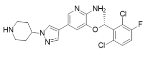
| SMILES |
O(C1C(N)=NC=C(C2C=NN(C3CCNCC3)C=2)C=1)[C@@H](C1C(Cl)=CC=C(F)C=1Cl)C
|
| InChi Key |
KTEIFNKAUNYNJU-GFCCVEGCSA-N
|
| InChi Code |
InChI=1S/C21H22Cl2FN5O/c1-12(19-16(22)2-3-17(24)20(19)23)30-18-8-13(9-27-21(18)25)14-10-28-29(11-14)15-4-6-26-7-5-15/h2-3,8-12,15,26H,4-7H2,1H3,(H2,25,27)/t12-/m1/s1
|
| 化学名 |
3-[(1R)-1-(2,6-dichloro-3-fluorophenyl)ethoxy]-5-(1-piperidin-4-ylpyrazol-4-yl)pyridin-2-amine
|
| 别名 |
PF-2341066; PF2341066; PF02341066; PF-02341066; PF 2341066; Crizotinib; PF 02341066; US trade name: Xalkori
|
| HS Tariff Code |
2934.99.9001
|
| 存储方式 |
Powder -20°C 3 years 4°C 2 years In solvent -80°C 6 months -20°C 1 month |
| 运输条件 |
Room temperature (This product is stable at ambient temperature for a few days during ordinary shipping and time spent in Customs)
|
| 溶解度 (体外实验) |
|
|||
|---|---|---|---|---|
| 溶解度 (体内实验) |
配方 1 中的溶解度: ≥ 1.25 mg/mL (2.78 mM) (饱和度未知) in 10% DMSO + 90% Corn Oil (这些助溶剂从左到右依次添加,逐一添加), 澄清溶液。
例如,若需制备1 mL的工作液,可将100 μL 12.5 mg/mL 澄清 DMSO 储备液加入900 μL 玉米油中,混合均匀。 配方 2 中的溶解度: ≥ 1 mg/mL (2.22 mM) (饱和度未知) in 10% DMSO + 40% PEG300 + 5% Tween80 + 45% Saline (这些助溶剂从左到右依次添加,逐一添加), 澄清溶液。 例如,若需制备1 mL的工作液,可将 100 μL 10.0 mg/mL澄清DMSO储备液加入400 μL PEG300中,混匀;然后向上述溶液中加入50 μL Tween-80,混匀;加入450 μL生理盐水定容至1 mL。 *生理盐水的制备:将 0.9 g 氯化钠溶解在 100 mL ddH₂O中,得到澄清溶液。 View More
配方 3 中的溶解度: ≥ 1 mg/mL (2.22 mM) (饱和度未知) in 10% DMSO + 90% (20% SBE-β-CD in Saline) (这些助溶剂从左到右依次添加,逐一添加), 澄清溶液。 配方 4 中的溶解度: 5% DMSO+30% PEG 300+dd H2O: 5 mg/mL 配方 5 中的溶解度: 20 mg/mL (44.41 mM) in 50% PEG300 50% Saline (这些助溶剂从左到右依次添加,逐一添加), 悬浮液; 需要超声助溶并加热至 40°C。 *生理盐水的制备:将 0.9 g 氯化钠溶解在 100 mL ddH₂O中,得到澄清溶液。 1、请先配制澄清的储备液(如:用DMSO配置50 或 100 mg/mL母液(储备液)); 2、取适量母液,按从左到右的顺序依次添加助溶剂,澄清后再加入下一助溶剂。以 下列配方为例说明 (注意此配方只用于说明,并不一定代表此产品 的实际溶解配方): 10% DMSO → 40% PEG300 → 5% Tween-80 → 45% ddH2O (或 saline); 假设最终工作液的体积为 1 mL, 浓度为5 mg/mL: 取 100 μL 50 mg/mL 的澄清 DMSO 储备液加到 400 μL PEG300 中,混合均匀/澄清;向上述体系中加入50 μL Tween-80,混合均匀/澄清;然后继续加入450 μL ddH2O (或 saline)定容至 1 mL; 3、溶剂前显示的百分比是指该溶剂在最终溶液/工作液中的体积所占比例; 4、 如产品在配制过程中出现沉淀/析出,可通过加热(≤50℃)或超声的方式助溶; 5、为保证最佳实验结果,工作液请现配现用! 6、如不确定怎么将母液配置成体内动物实验的工作液,请查看说明书或联系我们; 7、 以上所有助溶剂都可在 Invivochem.cn网站购买。 |
| 制备储备液 | 1 mg | 5 mg | 10 mg | |
| 1 mM | 2.2205 mL | 11.1027 mL | 22.2054 mL | |
| 5 mM | 0.4441 mL | 2.2205 mL | 4.4411 mL | |
| 10 mM | 0.2221 mL | 1.1103 mL | 2.2205 mL |
1、根据实验需要选择合适的溶剂配制储备液 (母液):对于大多数产品,InvivoChem推荐用DMSO配置母液 (比如:5、10、20mM或者10、20、50 mg/mL浓度),个别水溶性高的产品可直接溶于水。产品在DMSO 、水或其他溶剂中的具体溶解度详见上”溶解度 (体外)”部分;
2、如果您找不到您想要的溶解度信息,或者很难将产品溶解在溶液中,请联系我们;
3、建议使用下列计算器进行相关计算(摩尔浓度计算器、稀释计算器、分子量计算器、重组计算器等);
4、母液配好之后,将其分装到常规用量,并储存在-20°C或-80°C,尽量减少反复冻融循环。
计算结果:
工作液浓度: mg/mL;
DMSO母液配制方法: mg 药物溶于 μL DMSO溶液(母液浓度 mg/mL)。如该浓度超过该批次药物DMSO溶解度,请首先与我们联系。
体内配方配制方法:取 μL DMSO母液,加入 μL PEG300,混匀澄清后加入μL Tween 80,混匀澄清后加入 μL ddH2O,混匀澄清。
(1) 请确保溶液澄清之后,再加入下一种溶剂 (助溶剂) 。可利用涡旋、超声或水浴加热等方法助溶;
(2) 一定要按顺序加入溶剂 (助溶剂) 。
Targeted Treatment for ALK Positive Patients Who Have Previously Been Treated for Non-squamous Non-small Cell Lung Cancer
CTID: NCT03737994
Phase: Phase 2 Status: Active, not recruiting
Date: 2024-11-13
Mol Cancer Ther. 2012 Jul;11(7):1557-64. |
Mol Cancer Ther. 2012 Jul;11(7):1557-64. |
") |
 ARGX-111
 3-Fluoro-desmethyl-cabozantinib-C3-O-C3-PEG-acid
 MET Y1230D Recombinant Human Active Protein Kinase
 MET D1228N Recombinant Human Active Protein Kinase
InvivoChem的所有产品仅用于作科学研究,不面向患者销售
Copyright 2020 InvivoChem LLC | All Rights Reserved 粤ICP备20063088号-1


 COA
COA
")
")
 463611831
