| 规格 | 价格 | 库存 | 数量 |
|---|---|---|---|
| 5mg |
|
||
| 10mg |
|
||
| 50mg |
|
||
| 100mg |
|
||
| Other Sizes |
|

| 靶点 |
Fluorescent dye
|
|---|---|
| 体外研究 (In Vitro) |
原液制备
1. 蛋白准备 为了获得最佳标记效率,请将蛋白(抗体)浓度配制为 2 mg/mL。 (1) 蛋白溶液的 pH 值应为 8.5±0.5。如果 pH 低于 8.0,请使用 1 M 碳酸氢钠进行调整。 (2) 如果蛋白浓度低于 2 mg/mL,标记效率会显著降低。为了获得最佳标记效率,最终蛋白浓度范围应为 2-10 mg/mL。 (3) 蛋白必须置于不含伯胺(如 Tris 或甘氨酸)和铵离子的缓冲液中,否则会影响标记效率。 2. 染料准备 用无水 DMSO 稀释 CY 染料,制备 10 mM 储备溶液。使用玻璃管或旋涡混合器充分混合。 注意:建议将 CY 储备液分装后于 -20°C 或 -80°C 避光保存。 使用前需用缩合液(500 μg/mL)活化染料,然后进行后续标记实验。 3. 染料工作液用量计算 标记反应所需的 CY 染料用量取决于待标记蛋白的量,CY 染料与蛋白的最佳摩尔比约为 10。 示例:如果需要标记的蛋白为 500 μL 2 mg/mL 的 IgG(MW=150,000),并用 100 μL DMSO 溶解 1 mg CY 染料,则以 CY3-NHS ester 为例,所需 CY 体积的详细计算如下: (1) mmol (IgG) = mg/mL (IgG) × mL (IgG) / MW (IgG) = 2 mg/mL × 0.5 mL / 150,000 mg/mmol = 6.7×10^-6 mmol (2) mmol (CY3-NHS ester) = mmol (IgG) × 10 = 6.7×10^-6 mmol × 10 = 6.7×10^-5 mmol (3) μL (CY3-NHS ester) = mmol (CY3-NHS ester) × MW (CY3-NHS ester) / mg/μL (CY3-NHS ester) = 6.7×10^-5 mmol × 917.05 mg/mmol / 0.01 mg/μL 使用方法 1. 标记反应 (1) 取计算好的新鲜配制的 10 mM CY 染料并活化(约 10 μL 母液与 50 μL 500 μg/mL 缩合液混合)。将活化后的染料缓慢加入 0.5 mL 蛋白样品溶液中,轻轻摇匀混合,然后短暂离心将样品收集在反应管底部。避免剧烈混匀,以防止蛋白变性或失活。 (2) 将反应管置于避光处,在室温条件下轻轻摇晃孵育 60 分钟。每隔 10-15 分钟,轻轻颠倒反应管几次,以确保反应物充分混合并提高标记效率。 2. 蛋白纯化与脱盐 以下方案以使用 Sephadex G-25 柱纯化染料-蛋白偶联物为例。 (1) 按照生产说明书制备 Sephadex G-25 柱。 (2) 将反应混合物加至 Sephadex G-25 柱顶部。 (3) 当样品运行到树脂表面下方时,立即加入 PBS(pH 7.2-7.4)。 (4) 继续向柱中加入 PBS(pH 7.2-7.4)以完成纯化。收集含有目标染料-蛋白偶联物的组分。 |
| 体内研究 (In Vivo) |
Cy5.5 标记的因子 VIIa 专为肿瘤成像而设计。标记有这些抑制蛋白的 Cy5.5 定位于肿瘤异种移植物至少 14 天,而未结合的 Cy5.5 则不定位于任何异种移植物。这种可视化 VEC 中抗组织因子的方法可用于检测初始肿瘤和转移、监测和体内治疗反应 [1]。与 Cy5.5 标记的乳铁蛋白相关的 pH/温度敏感磁性纳米导体(Cy5.5-Lf-MPNA 纳米导体)被开发为一种有前景的成像剂,用于星状肿瘤的术前 MRI 和术中指纹成像 [2]。
胶质瘤是最常见的原发性脑肿瘤,在广泛的人群中导致不成比例的发病率和死亡率。从以往的临床实践来看,胶质瘤边缘的定义是手术切除的关键。为了勾勒出胶质瘤的确切边界,并在手术前计划阶段和手术切除阶段为医生提供指导作用,开发了与Cy5.5标记的乳铁蛋白结合的pH/温度敏感磁性纳米凝胶(Cy5.5-Lf-MPNA纳米凝胶)作为一种有前景的造影剂。由于其pH/温度敏感性,Cy5.5-Lf-MPNA纳米凝胶在不同pH和温度下的亲水性/疏水性和尺寸会发生变化。在生理条件下(pH 7.4,37°C),Cy5.5-Lf-MPNA纳米凝胶具有亲水性和溶胀性,可以延长血液循环时间。在肿瘤组织的酸性环境(pH 6.8,37°C)中,Cy5.5-Lf-MPNA纳米凝胶变得疏水并收缩,更容易在肿瘤组织中积累并被肿瘤细胞内化。此外,乳铁蛋白是胶质瘤的有效靶向配体,具有主动的肿瘤靶向能力。对患有原位胶质瘤的大鼠的体内研究表明,由于Lf的主动靶向功能和通过调整纳米凝胶的亲水性/疏水性来增强细胞摄取,使用Cy5.5-Lf-MPNA纳米凝胶可以获得高灵敏度和特异性的MR/荧光成像。细胞毒性试验和组织病理学分析显示,Cy5.5-Lf-MPNA纳米凝胶具有良好的生物相容性,有望开发成为胶质瘤术前MRI和术中荧光成像的特异性和高灵敏度造影剂[4]。 |
| 细胞实验 |
癌症细胞皮下接种[2]
将U87EGFRviii神经胶质瘤细胞、MiaPaCa和ASPC-1胰腺癌症细胞以106个细胞/0.1mL接种,并将KB-V1 SCC细胞以3×10⁶个细胞+0.1mL接种于皮下悬浮于PBS中。当所有肿瘤直径达到0.5-1.0cm时,将含有约0.03mg Cy5.5/0.1mL/小鼠的Cy5.5-FFRck-fVIIa或未偶联的Cy5.5的等分试样静脉注射到无胸腺裸鼠的侧尾静脉中。 Cy5.5与因子VIIa、抗TF抗体、FFRck fVIIa和紫杉醇FFRck fHIIa[2]的结合 将因子VIIa(5 mg/mL)、FFRck fVIIa(ASIS,批号NLDP013:7 mg/mL)和抗TF抗体(1 mg/mL)溶解在蒸馏水中,并在2升0.1 M碳酸钠缓冲液(pH8.8)中透析48小时。将Cy5.5(10mg)溶解在3mL 100%DMSO中。根据制造商的说明进行计算,将Cy5.5的等分试样以大约指示的Cy5.5:蛋白质比率添加到以下蛋白质中:fVIIa(1.5:1)、FFRck fVIIa(2:1)、紫杉醇FFRck fHIIa(2:1)和抗TF抗体(2:1)。在室温下将混合物轻轻搅拌1-1.5小时。通过预先用0.1 M碳酸钠缓冲液(pH 8.8)平衡的Sephadex G25-150柱将所得的Cy5.5蛋白偶联物与未偶联的Cy5.5分离。在一个典型的实验中,将1.8 mg fVIIa在0.6 ml 0.1M碳酸氢钠缓冲液(pH8.8)中的溶液与1 mg Cy5.5单NHS酯在0.3 ml DMSO中的溶液在室温下孵育1小时。使用Sephadex G25-150柱(8 ml)分离Cy5.5-fVIIa和游离Cy5.5染料。对于含有Cy5.5-fVIIa的组分2-6,收集0.3 ml(0.324 ml=6滴)/组分(1滴=54μL)。然后以1ml/级分洗脱无色级分7-14。从组分15-21中洗脱游离的Cy5.5染料,然后洗脱。A280和A678的吸光度读数确定了含有Cy5.5-fVIIa(蛋白质)和游离Cy5.5染料(无蛋白质)的组分。使用Micro-BCA蛋白检测试剂盒测定蛋白质含量较高的组分并合并。合并组分(1 mL总体积)的蛋白质浓度通常为0.7 mg/mL。使用fVIIa和Cy5.5染料的消光系数分别为1.39×10 5 M-1cm-1和2.5×10⁵ M-1cm-1,计算出Cy5.5与fVIIa的比值为1.24:1。Cy5.5与抗TF抗体的比例计算为1.86:1,使用抗体的消光系数1.7×10⁵ M-1cm-1,按照制造商手册确定。 体外细胞毒性[4] 根据先前的报告[14],C6细胞和NIH/3T3小鼠胚胎成纤维细胞系(NIH/3T)用于细胞存活率研究。以稀释系列(含0、25、50、75和100µg/mL Fe的细胞培养基)加入含有Cy5.5-Lf MPNA纳米凝胶或MPNA纳米胶囊的培养基。对照是不含纳米凝胶的培养基。孵育24、48和72小时后,向孔中加入20μL 3-(4,5-二甲基噻唑-2-基)-2,5-二苯基溴化四唑(MTT)(5mg/mL)。孵育4小时后,在培养箱中用100μL异丁醇溶解甲酰胺晶体过夜。在560nm下,在微孔板读数器(1420多标记计数器)上读取每个孔的吸光度。通过[A]试验/[A]对照×100%计算与含有不含纳米颗粒的细胞培养基的对照孔相关的相对细胞存活率(%),其中[A]试验是受试细胞的吸光度值,[A]对照是对照组的吸光度值。平均结果由6个样本计算得出。 |
| 动物实验 |
In vivo biocompatibility [4]
The normal rats were randomly divided into four groups (n = 9). The rats in one group were left without any treatment as the control. The rats in other three groups were injected with saline, Cy5.5-Lf-MPNA nanogels and MPNA nanogels (12 mg Fe/kg body weight) via the tail vein, respectively. At the time of the 21st day post-injection, rats were euthanized. 4 mL of blood was collected from femoral artery and sent to the clinical laboratory of Huazhong University of Science and Technology Hospital for the important biological function analysis immediately. Meanwhile, rats were perfused with sodium chloride (250 mL), and various tissues (heart, liver, spleen, lung, kidney and brain) were collected for histological examination. All the tissues were stained with H&E according to the standard clinical laboratory protocol and reviewed by a pathologist with expertise in veterinary pathology. Imaging Cy5.5 near infrared in vivo [2] Imaging of Cy5.5-labeled fVIIa, FFRck-fVIIa, paclitaxel-FFRck-fVIIa and anti-TF antibody was monitored over time by detecting Cy5.5 in the whole animal according to the instructions of the IVIS Lumina Imaging System 100 Series (Xenogen). Standard filter set pairs for Cy5.5 were selected in the Filter Lock box and ensured that the excitation (615-665 nm) and emission (695-770 nm) filters were properly paired for Cy5.5. Imaging was carried out daily for up to 26 days after the injection (Figures 2-5). Mice were anesthetized by an intraperitoneal injection of the mixture of ketamine (50 mg/mL), xylazine (20 mg/mL) and sterile distilled water mixed at a ratio of 8, 1 and 9 volumes according to the IACUC approved protocol at Emory University. In Figure 5, tumors and normal organs were individually dissected and imaged. Cy5.5 was imaged at 2 days after the i.v. injection using the IVIS Imaging System 100 Series located in the Department of Animal Facility according to the manufacturer’s instructions. |
| 参考文献 |
|
| 其他信息 |
Several classes of small organic molecules exhibit properties that make them suitable for fluorescence in vivo imaging. The most promising candidates are cyanines, squaraines, boron dipyrromethenes, porphyrin derivatives, hydroporphyrins, and phthalocyanines. The recent designing and synthetic efforts have been dedicated to improving their optical properties (shift the absorption and emission maxima toward longer wavelengths and increase the brightness) as well as increasing their stability and water solubility. The most notable advances include development of encapsulated cyanine dyes with increased stability and water solubility, squaraine rotaxanes with increased stability, long-wavelength-absorbing boron dipyrromethenes, long-wavelength-absorbing porphyrin and hydroporphyrin derivatives, and water-soluble phthalocyanines. Recent advances in luminescence and bioluminescence have made self-illuminating fluorophores available for in vivo applications. Development of new types of hydroporphyrin energy-transfer dyads gives the promise for further advances in in vivo multicolor imaging. [1]
We have developed a specific technique for imaging cancer in vivo using Cy5.5-labeled factor VIIa (fVIIa), clotting-deficient FFRck-fVIIa, paclitaxel-FFRck-fVIIa, and anti-tissue factor (TF) antibody. FVIIa is the natural ligand for TF. We took advantage of the fact that vascular endothelial cells (VECs) in cancer, but not normal tissue, aberrantly express TF due to its induction by vascular endothelial growth factor (VEGF). Under physiological conditions, TF is expressed by stromal cells and outer blood vessel layers (smooth muscle and adventitia), but not by VECs. We hypothesized that labeled fVIIa or anti-TF antibodies could be used to image the tumor vasculature in vivo. To test this, Cy5.5-labeled fVIIa, FFRck-fVIIa, paclitaxel-FFRck-fVIIa, and anti-TF antibody were developed and administered to athymic nude mice carrying xenografts including glioma U87EGFRviii, pancreatic cancer ASPC-1 and Mia PaCa-2, and squamous cell carcinoma KB-V1. Cy5.5 labeled with these targeting proteins specifically localized to the tumor xenografts for at least 14 days but unconjugated Cy5.5 did not localize to any xenografts or organs. This method of imaging TF in the tumor VECs may be useful in detecting primary tumors and metastases as well as monitoring in vivo therapeutic responses. [2] In this review paper, some of the important fundamentals in the chemistry of cyanine dyes were explained. This include topics like structure and resonance forms of cyanine dyes, naturally occurring cyanine dyes, different classes of cyanine dyes and formation mechanisms of cyanine dyes. This covers methine cyanine dyes, apocyanine dyes, styryl cyanine dyes (hemicyanine dyes), aza-styryl cyanine dyes)aza-hemicyanine dyes(, merocyanine dyes (acyclic merocyanine dyes and cyclic merocyanine dyes) squarylium cyanine dyes (aromatic squarylium cyanine dyes and heterocyclic squarylium cyanine dyes), spectral sensitization evaluation of cyanine dyes, solvatochromic evaluation of cyanine dyes, halochromic evaluation of cyanine dyes, cyanine dyes for CD-R and DVD-R, cyanine dyes as fluorescent labels for nucleic acid research, mechanisms of dimethine cyanine dyes and mechanisms of apocyanine dyes. In addition, in the introduction section of this review paper some light is focussed on some important uses and applications of cyanine dyes. This special and/or specific type of collective review in the fundamentals, principles, knowledge and/or the understanding of cyanine dyes chemistry has been paid little attention and is lacking in the chemistry literature. [3] Progress in pharmaceutical development is highly-dependent on preclinical in vivo animal studies. Small animal imaging is invaluable for the identification of new disease markers and the evaluation of drug efficacy. Here, we report for the first time the use of a three-dimensional fluorescence bioimager called FLuorescence Emission Computed Tomography (FLECT) for the detection of a novel recombinant fluoroprobe that is safe, easily prepared on a large scale and stably stored prior to scan. This novel fluoroprobe (Targ-Cy7) comprises a single-chain antibody-fragment (scFvTarg), which binds exclusively to activated-platelets, conjugated to a near-infrared (NIR) dye, Cy7, for detection. Upon mouse carotid artery injury, the injected fluoroprobe circulates and binds within the platelet-rich thrombus. This specific in vivo binding of the fluoroprobe to the thrombus, compared to its non-targeting control-fluoroprobe, is detected by the FLECT imager. The analyzed FLECT image quantifies the NIR signal and localizes it to the site of vascular injury. The detected fluorescence is further verified using a two-dimensional IVIS® Lumina scanner, where significant NIR fluorescence is detected in vivo at the thrombotic site, and ex vivo, at the injured carotid artery. Furthermore, fluorescence levels in various organs have also been quantified for biodistribution, with the highest fluoroprobe uptake shown to be in the injured artery. Subsequently, this live animal imaging technique is successfully employed to monitor the response of the induced thrombus to treatment over time. This demonstrates the potential of using longitudinal FLECT scanning to examine the efficacy of candidate drugs in preclinical settings. Besides intravascular thrombosis, we have shown that this non-invasive FLECT-imaging can also detect in vivo pulmonary embolism. Overall, this report describes a novel fluorescence-based preclinical imaging modality that uses an easy-to-prepare and non-radioactive recombinant fluoroprobe. This represents a unique tool to study mechanisms of thromboembolic diseases and it will strongly facilitate the in vivo testing of antithrombotic drugs. Furthermore, the non-radiation nature, low-cost, high sensitivity, and the rapid advancement of optical scanning technologies make this fluorescence imaging an attractive development for future clinical applications. [5] |
| 分子式 |
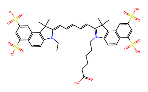
C41H44N2O14S4
|
|---|---|
| 分子量 |
917.053067207336
|
| 精确质量 |
916.168
|
| CAS号 |
210892-23-2
|
| 相关CAS号 |
Cy5.5 acetate;Cy5.5 TEA;Cy5.5-SE;442912-55-2
|
| PubChem CID |
131704516
|
| 外观&性状 |
Purple to purplish red solid powder
|
| LogP |
10.819
|
| tPSA |
297.38
|
| 氢键供体(HBD)数目 |
4
|
| 氢键受体(HBA)数目 |
15
|
| 可旋转键数目(RBC) |
13
|
| 重原子数目 |
61
|
| 分子复杂度/Complexity |
2250
|
| 定义原子立体中心数目 |
0
|
| SMILES |
CCN\1C2=C(C3=C(C=C2)C(=CC(=C3)S(=O)(=O)O)S(=O)(=O)[O-])C(/C1=C/C=C/C=C/C4=[N+](C5=C(C4(C)C)C6=C(C=C5)C(=CC(=C6)S(=O)(=O)O)S(=O)(=O)O)CCCCCC(=O)O)(C)C
|
| InChi Key |
LIZDKDDCWIEQIN-UHFFFAOYSA-N
|
| InChi Code |
InChI=1S/C41H44N2O14S4/c1-6-42-31-18-16-27-29(21-25(58(46,47)48)23-33(27)60(52,53)54)38(31)40(2,3)35(42)13-9-7-10-14-36-41(4,5)39-30-22-26(59(49,50)51)24-34(61(55,56)57)28(30)17-19-32(39)43(36)20-12-8-11-15-37(44)45/h7,9-10,13-14,16-19,21-24H,6,8,11-12,15,20H2,1-5H3,(H4-,44,45,46,47,48,49,50,51,52,53,54,55,56,57)
|
| 化学名 |
(2Z)-2-[(2E,4E)-5-[3-(5-carboxypentyl)-1,1-dimethyl-6,8-disulfobenzo[e]indol-3-ium-2-yl]penta-2,4-dienylidene]-3-ethyl-1,1-dimethyl-8-sulfobenzo[e]indole-6-sulfonate
|
| 别名 |
210892-23-2; Cy5.5; 1H-Benz[e]indolium, 2-[5-[3-(5-carboxypentyl)-1,3-dihydro-1,1-dimethyl-6,8-disulfo-2H-benz[e]indol-2-ylidene]-1,3-pentadien-1-yl]-3-ethyl-1,1-dimethyl-6,8-disulfo-, inner salt;
|
| HS Tariff Code |
2934.99.9001
|
| 存储方式 |
Powder -20°C 3 years 4°C 2 years In solvent -80°C 6 months -20°C 1 month 注意: 本产品在运输和储存过程中需避光。 |
| 运输条件 |
Room temperature (This product is stable at ambient temperature for a few days during ordinary shipping and time spent in Customs)
|
| 溶解度 (体外实验) |
DMSO : ~100 mg/mL (~109.05 mM)
H2O : ~5 mg/mL (~5.45 mM) |
|---|---|
| 溶解度 (体内实验) |
配方 1 中的溶解度: ≥ 4.17 mg/mL (4.55 mM) (饱和度未知) in 10% DMSO + 40% PEG300 + 5% Tween80 + 45% Saline (这些助溶剂从左到右依次添加,逐一添加), 澄清溶液。
例如,若需制备1 mL的工作液,可将100 μL 41.7 mg/mL澄清的DMSO储备液加入到400 μL PEG300中,混匀;再向上述溶液中加入50 μL Tween-80,混匀;然后加入450 μL生理盐水定容至1 mL。 *生理盐水的制备:将 0.9 g 氯化钠溶解在 100 mL ddH₂O中,得到澄清溶液。 配方 2 中的溶解度: ≥ 4.17 mg/mL (4.55 mM) (饱和度未知) in 10% DMSO + 90% (20% SBE-β-CD in Saline) (这些助溶剂从左到右依次添加,逐一添加), 澄清溶液。 例如,若需制备1 mL的工作液,可将 100 μL 41.7 mg/mL 澄清 DMSO 储备液加入 900 μL 20% SBE-β-CD 生理盐水溶液中,混匀。 *20% SBE-β-CD 生理盐水溶液的制备(4°C,1 周):将 2 g SBE-β-CD 溶解于 10 mL 生理盐水中,得到澄清溶液。 View More
配方 3 中的溶解度: 5 mg/mL (5.45 mM) in PBS (这些助溶剂从左到右依次添加,逐一添加), 澄清溶液; 超声助溶 (<60°C). 1、请先配制澄清的储备液(如:用DMSO配置50 或 100 mg/mL母液(储备液)); 2、取适量母液,按从左到右的顺序依次添加助溶剂,澄清后再加入下一助溶剂。以 下列配方为例说明 (注意此配方只用于说明,并不一定代表此产品 的实际溶解配方): 10% DMSO → 40% PEG300 → 5% Tween-80 → 45% ddH2O (或 saline); 假设最终工作液的体积为 1 mL, 浓度为5 mg/mL: 取 100 μL 50 mg/mL 的澄清 DMSO 储备液加到 400 μL PEG300 中,混合均匀/澄清;向上述体系中加入50 μL Tween-80,混合均匀/澄清;然后继续加入450 μL ddH2O (或 saline)定容至 1 mL; 3、溶剂前显示的百分比是指该溶剂在最终溶液/工作液中的体积所占比例; 4、 如产品在配制过程中出现沉淀/析出,可通过加热(≤50℃)或超声的方式助溶; 5、为保证最佳实验结果,工作液请现配现用! 6、如不确定怎么将母液配置成体内动物实验的工作液,请查看说明书或联系我们; 7、 以上所有助溶剂都可在 Invivochem.cn网站购买。 |
| 制备储备液 | 1 mg | 5 mg | 10 mg | |
| 1 mM | 1.0905 mL | 5.4523 mL | 10.9045 mL | |
| 5 mM | 0.2181 mL | 1.0905 mL | 2.1809 mL | |
| 10 mM | 0.1090 mL | 0.5452 mL | 1.0905 mL |
1、根据实验需要选择合适的溶剂配制储备液 (母液):对于大多数产品,InvivoChem推荐用DMSO配置母液 (比如:5、10、20mM或者10、20、50 mg/mL浓度),个别水溶性高的产品可直接溶于水。产品在DMSO 、水或其他溶剂中的具体溶解度详见上”溶解度 (体外)”部分;
2、如果您找不到您想要的溶解度信息,或者很难将产品溶解在溶液中,请联系我们;
3、建议使用下列计算器进行相关计算(摩尔浓度计算器、稀释计算器、分子量计算器、重组计算器等);
4、母液配好之后,将其分装到常规用量,并储存在-20°C或-80°C,尽量减少反复冻融循环。
计算结果:
工作液浓度: mg/mL;
DMSO母液配制方法: mg 药物溶于 μL DMSO溶液(母液浓度 mg/mL)。如该浓度超过该批次药物DMSO溶解度,请首先与我们联系。
体内配方配制方法:取 μL DMSO母液,加入 μL PEG300,混匀澄清后加入μL Tween 80,混匀澄清后加入 μL ddH2O,混匀澄清。
(1) 请确保溶液澄清之后,再加入下一种溶剂 (助溶剂) 。可利用涡旋、超声或水浴加热等方法助溶;
(2) 一定要按顺序加入溶剂 (助溶剂) 。