| 规格 | 价格 | 库存 | 数量 |
|---|---|---|---|
| 10 mM * 1 mL in DMSO |
|
||
| 1mg |
|
||
| 5mg |
|
||
| 10mg |
|
||
| 25mg |
|
||
| 50mg |
|
||
| 100mg |
|
||
| 250mg |
|
||
| 500mg |
|
||
| Other Sizes |
|
| 靶点 |
Natural substrate of luciferase (Luc) enzyme
|
|---|---|
| 体外研究 (In Vitro) |
1. 注意事项:
a) D-荧光素盐(钠盐或钾盐)在水性缓冲液(pH 6.1-6.5)中表现出高达100mM的高溶解度。储备溶液可以用不含ATP的水制备,并在-20°C的避光/黑暗中储存。游离酸必须用适当的碱中和才能溶解。在较高的pH值下,荧光素在碱性催化下会形成脱氢荧光素,并外消旋为L-异构体(L-荧光素)。 b) D-荧光素可用于任何现有的报告分析或ATP分析系统。 c) 如果测试ATP,请戴手套并使用无ATP容器,以尽量减少所有可能的ATP污染源。仅使用无菌无ATP水和试剂。使用高压灭菌水制备所有试剂。 2.实验方案:该方案仅供参考,可根据您的具体要求进行调整 以下方案是D-Luciferin钠/钾盐的制备示例。它适用于大多数细胞类型和体内动物使用。 2.1体外生物发光图像分析示例 a) 在无菌水中制备100 mM(100-200X)荧光素储备溶液。混合均匀。立即使用或一次性分装,在-20°C下避光储存,避免冻融循环。 b) 在预热的组织培养基中制备0.5-1mM D-荧光素工作溶液。 c) 从培养的细胞中吸出培养基。 d) 向细胞中加入荧光素工作溶液,在成像前在37°C下孵育5-10分钟。 2.2体内生物发光图像分析示例 a) 在DPBS中制备15mg/mL荧光素储备溶液,不含Mg2+和Ca2+。混合均匀。 b) 过滤器通过0.2μM过滤器从溶液中去除细菌。立即使用或一次性分装,并在-20°C下避光储存,以避免冻融循环。 c) 成像前10-15分钟,腹腔注射150mg/kg(或10μL/g荧光素储备溶液)动物体重的荧光素。 注意:应在每种动物模型上进行荧光素动力学研究,以确定峰值信号时间。 2.3荧光素报告基因检测示例 a) 在无菌水中制备100 mM荧光素储备溶液。立即使用或一次性分装,在-20°C下避光储存,避免冻融循环。 b) 在pH 7.8的25 mM Tricine缓冲液中制备1 mM D-荧光素工作溶液和3 mM ATP、1 mM DTT和15 mM MgSO4。 c) 将5-10μL细胞裂解液转移到微孔板上。使用裂解试剂或不含裂解物的缓冲溶液作为空白。 d) 根据制造商的说明,向发光计中注入荧光素工作溶液。 e) 立即注入200μL荧光素工作溶液,积分时间为10秒。 |
| 体内研究 (In Vivo) |
目前最流行的方法是生物发光(BLI),它使用D-荧光素底物和萤火虫荧光素酶(Fluc)作为报告基因。通过绘制总体信号强度与 D-荧光素注射后的时间量的关系图来创建时间-强度曲线。除了峰值信号之外,峰值信号的替代信号被识别为注射 D-荧光素后预定时间间隔(5、10、15 和 20 分钟)的信号。为了描述 D-荧光素注射后时间变化的模式,给定时间-强度曲线中的信号根据曲线中的峰值信号进行标准化 [3]。每克体重使用 10 μL D-荧光素原液(腹膜内或静脉注射)。由于常规剂量为 150 mg/kg,20 g 注射液通常应含有 200 μL。将 D-荧光素(钾盐或钠盐)溶液溶解至终浓度 15 mg/mL,将其解冻并在 dPBS(透明钙盐或镁盐)中稀释。用 5–10 mL 无菌 H2O 润湿 0.22 µM 过滤器,然后沥干。 ..将 D-荧光素溶液通过已生产的 0.22 µM 注射器过滤器。
|
| 酶活实验 |
D-荧光素是所有萤光素酶的天然底物,这些萤光素酶催化生物发光昆虫产生光。本综述涵盖了D-荧光素和衍生物或类似物的合成,它们是美国萤火虫Photinus pyralis荧光素酶的底物或抑制剂,Photinus piralis是体外和光学成像技术中更常用的酶[1]。
|
| 动物实验 |
注射D-荧光素后,峰值信号或预定固定时间点的信号可用于体内生物发光成像的定量分析。我们对皮下肿瘤小鼠皮下注射D-荧光素后进行了多次连续生物发光成像。每次测量的峰值时间在细胞接种后早期逐渐缩短,这可能是由于肿瘤内血管的逐渐建立所致,并在第10天达到约10分钟的平台期。尽管固定时间点的信号与峰值信号之间存在高度相关性,但早期5分钟或10分钟时的信号强度(以峰值信号进行归一化)较低,这导致对肿瘤生长的估计过高。注射D-荧光素后信号的时间进程可能随细胞接种后时间的变化而变化,在确定用于定量生物发光肿瘤监测的成像方案时应考虑这种变化。[2]
|
| 参考文献 |
|
| 其他信息 |
生物发光这一迷人现象及其在光学成像领域广泛的生物技术应用,使得D-荧光素及其类似物的化学性质极具研究价值。这超越了White等人于1971年提出的早期预期:“化学合成的激发态在生物发光中起着至关重要的作用;它们在生物学的其他领域也必将发挥重要作用。”¹ 相关苯并噻唑体系的合成方法与近四十年前的原始研究相比并没有太大变化。然而,其化学原理仍然可靠,并用于制备D-荧光素及其相关化合物。这些化合物可作为荧光素酶的底物,应用于光学成像技术等应用中,而光学成像技术目前已广泛应用于细胞和小动物的临床前分子成像。用于此目的的经典酶PpyLuc发出的黄绿色光具有宽发射光谱,最大发射波长为560 nm,且背景生物发光较低。这使得利用PpyLuc进行体内生物发光成像成为一种简便且高灵敏度的小动物分子成像方法。调控发射光的波长是一个重要的研究目标,目前主要通过对荧光素酶氨基酸残基进行定点诱变来实现。虽然通过化学方法修饰D-荧光素的结构或许也能达到类似的效果,但对于D-荧光素这种相对简单的结构而言,可进行的修饰并不多。事实上,目前仅合成了少数几种化合物,并且只有少数化合物被证实能够使PpyLuc催化的生物发光发生红移。然而,本文所列举的实例表明,除了对酶进行生物化学改造之外,合成有机化学的巧妙构思和创新应用也能为这一引人入胜的研究领域带来新的突破。最后,D-荧光素的其他类似物可能满足体内光学成像新应用的预期,正如最近的报道所揭示的那样,这些报道阐明了多种化合物(包括肽或功能化聚乙二醇)与D-6′-氨基荧光素的氨基结合的可能性。[1]
染色实例1: D-荧光素钠可用作荧光素酶的底物进行体内成像。 方法:用于生物发光成像。 1)麻醉小鼠,然后向小鼠注射D-荧光素钠(75 mg/kg)进行成像。 2)使用生物发光成像系统进行成像。 染色实例2: D-荧光素钠可用作荧光素酶的底物进行体内成像,以监测肿瘤生长。 方法:用于生物发光成像。 1)将D-荧光素钠(150 mg/kg;腹腔注射)注射到小鼠体内。 2). 使用生物发光成像系统进行成像。 染色示例 3: D-荧光素钠可用作荧光素酶的底物,用于分裂荧光素酶 (LUC) 检测。 方法:用于分裂荧光素酶 (LUC) 检测。 1). 将植物样品(叶片)与 D-荧光素钠(1 mM;10 分钟)和荧光素孵育。 2). 使用 Photek 相机采集信号和图像。 染色示例 4: D-荧光素钠可用作荧光素酶的底物,用于体内成像以监测肿瘤生长。 方法:用于生物发光成像。 1).向小鼠腹腔注射D-荧光素钠(150 mg/kg)。 2). 使用IVIS Lumina XRMS系列进行生物发光成像。 |
| 分子式 |
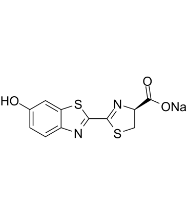
C11H7N2NAO3S2
|
|---|---|
| 分子量 |
302.29
|
| 精确质量 |
301.979
|
| 元素分析 |
C, 43.71; H, 2.33; N, 9.27; Na, 7.61; O, 15.88; S, 21.21
|
| CAS号 |
103404-75-7
|
| 相关CAS号 |
D-Luciferin;2591-17-5;D-Luciferin potassium;115144-35-9
|
| PubChem CID |
2733762
|
| 外观&性状 |
Typically exists as Light yellow to yellow solids at room temperature
|
| 沸点 |
473.7ºC at 760mmHg
|
| 闪点 |
240.3ºC
|
| 蒸汽压 |
2.78E-10mmHg at 25°C
|
| LogP |
0.049
|
| tPSA |
139.15
|
| 氢键供体(HBD)数目 |
1
|
| 氢键受体(HBA)数目 |
7
|
| 可旋转键数目(RBC) |
2
|
| 重原子数目 |
19
|
| 分子复杂度/Complexity |
396
|
| 定义原子立体中心数目 |
1
|
| SMILES |
S1C(C2=NC3C([H])=C([H])C(=C([H])C=3S2)O[H])=N[C@@]([H])(C(=O)[O-])C1([H])[H].[Na+]
|
| InChi Key |
BZNVUYVALNTPBG-WJCSTRGMSA-M
|
| InChi Code |
InChI=1S/C11H8N2O3S2.Na/c14-5-1-2-6-8(3-5)18-10(12-6)9-13-7(4-17-9)11(15)16/h1-3,7,13H,4H2,(H,15,16)/q+1/p-1/b10-9+/t7-/m1./s1
|
| 化学名 |
sodium (S,E)-2-(6-oxobenzo[d]thiazol-2(6H)-ylidene)thiazolidine-4-carboxylate
|
| 别名 |
D-Luciferin Sodium; D-Luciferin sodium salt; Sodium (S)-2-(6-hydroxybenzo[d]thiazol-2-yl)-4,5-dihydrothiazole-4-carboxylate; D-Luciferin Sodium; D-Luciferin, Sodium Salt; D-Luciferin (sodium); C11H7N2NaO3S2; D-Luciferin sodium salt monohydrate;
|
| HS Tariff Code |
2934.99.9001
|
| 存储方式 |
Powder -20°C 3 years 4°C 2 years In solvent -80°C 6 months -20°C 1 month 注意: 请将本产品存放在密封且受保护的环境中(例如氮气保护),避免吸湿/受潮和光照。 |
| 运输条件 |
Room temperature (This product is stable at ambient temperature for a few days during ordinary shipping and time spent in Customs)
|
| 溶解度 (体外实验) |
H2O : ~250 mg/mL (~826.99 mM)
DMSO : ~100 mg/mL (~330.80 mM) |
|---|---|
| 溶解度 (体内实验) |
配方 1 中的溶解度: ≥ 2.5 mg/mL (8.27 mM) (饱和度未知) in 10% DMSO + 40% PEG300 + 5% Tween80 + 45% Saline (这些助溶剂从左到右依次添加,逐一添加), 澄清溶液。
例如,若需制备1 mL的工作液,可将100 μL 25.0 mg/mL澄清DMSO储备液加入到400 μL PEG300中,混匀;然后向上述溶液中加入50 μL Tween-80,混匀;加入450 μL生理盐水定容至1 mL。 *生理盐水的制备:将 0.9 g 氯化钠溶解在 100 mL ddH₂O中,得到澄清溶液。 配方 2 中的溶解度: ≥ 2.5 mg/mL (8.27 mM) (饱和度未知) in 10% DMSO + 90% (20% SBE-β-CD in Saline) (这些助溶剂从左到右依次添加,逐一添加), 澄清溶液。 例如,若需制备1 mL的工作液,可将 100 μL 25.0 mg/mL澄清DMSO储备液加入900 μL 20% SBE-β-CD生理盐水溶液中,混匀。 *20% SBE-β-CD 生理盐水溶液的制备(4°C,1 周):将 2 g SBE-β-CD 溶解于 10 mL 生理盐水中,得到澄清溶液。 View More
配方 3 中的溶解度: 100 mg/mL (330.80 mM) in PBS (这些助溶剂从左到右依次添加,逐一添加), 澄清溶液; 超声助溶. 1、请先配制澄清的储备液(如:用DMSO配置50 或 100 mg/mL母液(储备液)); 2、取适量母液,按从左到右的顺序依次添加助溶剂,澄清后再加入下一助溶剂。以 下列配方为例说明 (注意此配方只用于说明,并不一定代表此产品 的实际溶解配方): 10% DMSO → 40% PEG300 → 5% Tween-80 → 45% ddH2O (或 saline); 假设最终工作液的体积为 1 mL, 浓度为5 mg/mL: 取 100 μL 50 mg/mL 的澄清 DMSO 储备液加到 400 μL PEG300 中,混合均匀/澄清;向上述体系中加入50 μL Tween-80,混合均匀/澄清;然后继续加入450 μL ddH2O (或 saline)定容至 1 mL; 3、溶剂前显示的百分比是指该溶剂在最终溶液/工作液中的体积所占比例; 4、 如产品在配制过程中出现沉淀/析出,可通过加热(≤50℃)或超声的方式助溶; 5、为保证最佳实验结果,工作液请现配现用! 6、如不确定怎么将母液配置成体内动物实验的工作液,请查看说明书或联系我们; 7、 以上所有助溶剂都可在 Invivochem.cn网站购买。 |
| 制备储备液 | 1 mg | 5 mg | 10 mg | |
| 1 mM | 3.3081 mL | 16.5404 mL | 33.0808 mL | |
| 5 mM | 0.6616 mL | 3.3081 mL | 6.6162 mL | |
| 10 mM | 0.3308 mL | 1.6540 mL | 3.3081 mL |
1、根据实验需要选择合适的溶剂配制储备液 (母液):对于大多数产品,InvivoChem推荐用DMSO配置母液 (比如:5、10、20mM或者10、20、50 mg/mL浓度),个别水溶性高的产品可直接溶于水。产品在DMSO 、水或其他溶剂中的具体溶解度详见上”溶解度 (体外)”部分;
2、如果您找不到您想要的溶解度信息,或者很难将产品溶解在溶液中,请联系我们;
3、建议使用下列计算器进行相关计算(摩尔浓度计算器、稀释计算器、分子量计算器、重组计算器等);
4、母液配好之后,将其分装到常规用量,并储存在-20°C或-80°C,尽量减少反复冻融循环。
计算结果:
工作液浓度: mg/mL;
DMSO母液配制方法: mg 药物溶于 μL DMSO溶液(母液浓度 mg/mL)。如该浓度超过该批次药物DMSO溶解度,请首先与我们联系。
体内配方配制方法:取 μL DMSO母液,加入 μL PEG300,混匀澄清后加入μL Tween 80,混匀澄清后加入 μL ddH2O,混匀澄清。
(1) 请确保溶液澄清之后,再加入下一种溶剂 (助溶剂) 。可利用涡旋、超声或水浴加热等方法助溶;
(2) 一定要按顺序加入溶剂 (助溶剂) 。
InvivoChem的所有产品仅用于作科学研究,不面向患者销售
Copyright 2020 InvivoChem LLC | All Rights Reserved 粤ICP备20063088号-1


 COA
COA
 463611831
