| 规格 | 价格 | 库存 | 数量 |
|---|---|---|---|
| 5mg |
|
||
| 10mg |
|
||
| 25mg |
|
||
| 50mg |
|
||
| 100mg |
|
||
| 250mg |
|
||
| 500mg |
|
||
| Other Sizes |
|
| 靶点 |
FAK
|
|---|---|
| 体外研究 (In Vitro) |
体外活性:在紫杉烷敏感 (SKOV3ip1) 和紫杉烷耐药 (SKOV3-TR) 细胞系中,Defactinib 显着抑制 pFAK (Tyr397) 表达。 Defactinib 和紫杉醇的组合可协同减少 SKOV3ip1、SKOV3-TR、HeyA8 和 HeyA8-MDR 细胞的增殖并增加细胞凋亡。 Defactinib 和 Y15 的组合可协同降低甲状腺癌细胞系的活力、克隆性和细胞附着。激酶测定:Defactinib 以时间和剂量依赖性方式抑制 Tyr397 位点的 FAK 磷酸化。 Defactinib 和紫杉醇的组合显着降低了增殖并增加了细胞凋亡,从而使肿瘤重量减少了 92.7% 至 97.9%。 RPPA 数据显示,Defactinib 降低了紫杉烷耐药细胞系中 AKT 和 YB-1 的水平。 FAK 抑制通过降低 YB-1 磷酸化以及随后以 AKT 依赖性方式降低 CD44 来增强紫杉烷抗性细胞的化疗敏感性。在人类卵巢癌样本中,核 FAK 表达与核 YB-1 表达增加相关 (χ2) = 37.7; P < .001)。核 FAK 和 YB-1 共表达与统计学上显着较差的中位总生存期相关(24.9 个月 vs 67.3 个月;风险比 = 2.64;95% 置信区间 = 1.38 至 5.05;P = .006)。细胞测定:用增加浓度的 Defactinib 处理卵巢癌细胞 96 小时,然后进行 MTT 测定。结果通过三次重复实验得到证实。
抑制FAK对PTX敏感性的影响[1] 我们首先测试了Defactinib/VS-6063对FAK磷酸化的体外作用。在所有细胞系中,VS-6063以剂量依赖的方式显著抑制了pFAK(Tyr397)的表达(图1A;补充图1A,可在线获得)。VS-6063在3小时内抑制了pFAK(Tyr397)的表达,48小时后表达逐渐恢复(图1B;补充图1B和2,可在线获得)。补充图3(可在线获得)代表了一个典型的实验,其中对三次实验的统计分析表明,在其他测试残基(Tyr 576/577、Tyr 925或Tyr 861)上,FAK磷酸化没有统计学上的显著变化。由于Pyk2和FAK在中心催化结构域中的相似性约为60%,我们还测试了Pyk2中的Tyr402、Tyr 579/580和Tyr 881。仅在HeyA8细胞中观察到Tyr402的磷酸化抑制,VS-6063(1µM)处理1小时后,HeyA8 MDR细胞系中的磷酸化残基没有变化。 VS-6063/Defactinib作为单一药物(0-1µM)不影响任何细胞或其紫杉烷抗性对应物的生长(补充图4,在线提供)。然而,PTX和VS-6063(1µM)的组合比单独使用任何一种药物都更有效。与单独使用PTX相比,与VS-6063联合使用PTX的细胞毒性高2.1至4.9倍(图1C;补充图1C,可在线获得)。采用Chou-Talalay法(20)用组合指数评估PTX和VS-6063的组合效果。PTX和VS-6063之间的相互作用在HeyA8和HeyA8 MDR细胞中具有协同作用(图1D)。HeyA8和HeyA8-MDR以1:1000和1:10的比例同时暴露于PTX和VS-6063剂量对细胞生长具有协同抑制作用,组合指数分别为0.953和0.705。在SKOV3ip1或SKOV3-TR细胞中,PTX和VS-6063之间没有观察到协同作用,但观察到对增殖的加性抑制作用(数据未显示)。 接下来,我们测试了基于VS-6063/Defactinib的疗法对增殖和凋亡的影响。与单独使用PTX相比,VS-6063(1µM)与PTX的组合(PTX对SKOV3和HeyA8细胞的中位抑制浓度[IC50]水平分别为8.5和6.3 nmol/L)降低了紫杉烷敏感和紫杉烷抗性细胞的增殖率(图1E;补充图1D,在线提供)。对于细胞凋亡,PTX和VS-6063联合使用的效果最大(图1F;补充图1E,可在线获得)。这些发现表明,VS-6063与PTX联合使用至少对细胞增殖和凋亡具有相加作用。与对照组(34.3%±0.3%;P<0.001)相比,PTX将HeyA8细胞中G2期细胞群的比例增加到51.0%±0.4%,与VS-6063治疗联合使用时,G2期细胞的比例在统计学上显著增加到60.8%±0.9%(与PTX相比,P<0.001)。在PTX组或联合治疗组中,HeyA8 MDR细胞均未被阻滞在G2期(P=0.05)(图1G;补充图1F,可在线获得)。 FAK抑制对下游信号的影响[1] 由于已知FAK通过β1整合素发出信号,我们首先测试了β1整合素水平是否受到Defactinib/VS-6063的影响,但没有发现统计学上的显著变化(补充图8,在线提供)。为了鉴定紫杉烷抗性细胞系中FAK下游的潜在信号通路,使用并分析了RPPA(DAVID生物信息学资源;http://david.abcc.ncifcrf.gov/).在VS-6063治疗组中,与未治疗组相比,所分析的161种蛋白质中有53种在两种耐药细胞系中显示出统计学上的显著变化(P<0.05)(图3A;补充表1,可在线获得)。我们发现AKT通路是参与最多的通路,包括pFAK(Tyr 397)、pAKT(Thr308)、GSK-3α/β(Ser21/9)、p27(T198)、p70S6K(Thr389)、PRAS40(T246)和pYB-1(Ser102)。其中,pYB-1(Ser102)是统计学上降低最显著的蛋白质(Defactinib/VS-6063治疗降低了36.6%;P<0.001)。鉴于YB-1在致癌和耐药途径中的潜在作用,我们在后续研究中重点研究了FAK和YB-1之间的潜在关系。图3B显示,PTX增加了核YB-1的表达,而PTX与VS-6063联合使用降低了其在HeyA8 MDR细胞中的表达。免疫荧光研究也揭示了类似的发现(图3C)。 FAK抑制抑制ESCC细胞的恶性进展[2] 由于FAK在几种ESCC细胞系中被过度激活,包括KYSE150、KYSE180、KYSE30、KYSE410、KYSE450、KYSE510、Colo680和KYSE70,9,我们使用MTS测定法测量了Defactinib对这些ESCC细胞株的生长抑制作用。如图1A所示,Defactinib治疗72小时 h剂量依赖性地降低了这些指示的ESCC细胞系的存活率(图1A)。我们使用Transwell试验进一步观察了defactinib在KYSE410和KYSE510中的抗侵袭或迁移能力。如图1B、C所示,defactinib剂量依赖性地抑制了指定ESCC细胞的迁移和侵袭。综上所述,这些结果表明,defactinib在ESCC细胞中具有优异的抗肿瘤作用。 接下来,FAK通过与PI3K催化亚基-p85相互作用增强PI3K/AKT通路的活性。如图2E所示,在4或24小时后,F、Defactinib剂量依赖性地破坏了FAK和PI3K p85亚基之间的相互作用 h治疗。总之,PI3K/AKT通路抑制的动力学和幅度表明,这种作用在defactinib反应中起着重要作用。 Defactinib对下游基因网络的有效抑制 为了进一步了解Defactinib治疗引起的分子效应,我们分析了用10 μM defactinib 4至24 h(图3A),并重点研究了Defactinib治疗中显著下调的转录本。如图3B-J所示,令人惊讶的是,在4或24小时后,几个靶基因在相同的功能集中大量富集,如化疗耐药性(图3B)、细胞周期(图3C)、生长因子、细胞因子和趋化因子(图3D)、恶性进展(图3E)、细胞可塑性(图3F)、热休克蛋白家族(图3G)、代谢(图3H)、癌基因(图3I)或转录因子(图3J) h defactinib治疗。对参与这些功能集的代表性基因的详细分析表明,其中许多基因在4或24小时后被defactinib持续抑制 h处理(表S1)。 |
| 体内研究 (In Vivo) |
在 PTX 敏感和 PTX 耐药模型中,Defactinib(50 mg/kg po)可增强紫杉醇对肿瘤生长的抑制作用。
VS-6063/的体内效应德法替尼[1] Defactinib/VS-6063剂量为25mg/kg,每天两次或更高,在3小时时统计学上显著抑制了pFAK(Tyr397),24小时后表达恢复(补充图5,在线提供)。因此,选择每天两次25mg/kg的Defactinib/VS-6063作为后续治疗实验的给药方案。在治疗实验中,将腹腔内携带HeyA8肿瘤的雌性裸鼠随机分为4组(每组n=10):1)每天口服两次载体,每周腹腔注射磷酸盐缓冲盐水(对照组);2)VS-6063/Defactinib25mg/kg口服,每日两次;3)每周腹腔注射PTX;4)VS-6063 25mg/kg每日口服两次,PTX每周腹腔注射一次。在HeyA8模型中,PTX单药治疗使肿瘤重量减轻了87.4%,联合治疗导致肿瘤重量减轻最大,减少了97.9%(与PTX相比P=0.05)(图2,a和B)。在SKOV3ip1模型中,与PTX相比,联合组的肿瘤重量减轻了92.7%(P<0.001)。 Defactinib对体内肿瘤恶性肿瘤的抑制有效[2] 我们进一步使用了三种异种移植模型,包括皮下肿瘤细胞接种模型(评估肿瘤生长)、腘淋巴结转移模型(评估瘤细胞的淋巴结转移能力)或肺定植模型(评估癌细胞的转移能力),以全面观察Defactinib介导的体内抗肿瘤作用。我们将KYSE410和KYSE510细胞皮下接种到BALB/c小鼠的右侧腹。当异种移植物达到约100 mm3,我们用defactinib(25mg/kg/天,口服)治疗动物约连续3周,并观察肿瘤生长。如图5A所示,治疗2周后,与对照组相比,Defactinib组的肿瘤生长明显延迟。由defactinib介导的肿瘤生长消退持续到第3周。此外,治疗3周后,Defactinib有效地阻断了指定肿瘤组织中FAK的激活(图S4)。腘淋巴结转移模型的结果显示,与对照组相比,FAK抑制显著降低了defactinib治疗组淋巴结中ESCC细胞的体积(图5B)。我们在肺定植模型中进一步评估了Defactinib对ESCC进展的影响。动物静脉注射指定的ESCC细胞。如图5C所示,defactinib治疗组肺部肿瘤巢的数量明显低于对照组。图5D的结果显示,与对照治疗相比,defactinib显著延长了携带ESCC肿瘤的动物的存活期。IHC检测显示,达法替尼显著降低了指定肿瘤中增殖生物标志物Ki-67(图5E)和血管生成生物标志物CD31(图5F)的表达。重要的是,对心脏、肝脏、脾脏和肾脏组织的组织学分析显示,对照组和Defactinib治疗组之间没有明显变化,表明Defactinib对正常组织没有产生显著的毒性作用(图6A)。Defactinib和对照治疗组的体重没有明显差异(图6B)。 |
| 酶活实验 |
以依赖时间和剂量的方式,defactinib 阻止了 Tyr397 位点的 FAK 磷酸化。德法替尼和紫杉醇一起显着减少增殖并增强细胞凋亡,导致肿瘤重量减少 92.7% 至 97.9%。 RPPA 的数据表明,在紫杉烷耐药细胞系中,德法替尼降低了 AKT 和 YB-1 水平。在紫杉烷抗性细胞中,FAK 抑制通过依赖于 AKT 的机制提高了化疗敏感性,并减少了 YB-1 磷酸化,从而减少了 CD44。核 YB-1 表达增加 (χ2) = 37.7; P <.001) 与人类卵巢癌样本中的核 FAK 表达相关。中位总生存期显着较差(24.9 个月与 67.3 个月;风险比 = 2.64;95% 置信区间 = 1.38 至 5.05;P = 0.006)与核 FAK 和 YB-1 的共表达有关。
核因子-κB转录活性的酶联免疫吸附分析[2] 根据制造商的说明,使用人核细胞因子-κB(NF-κB)p65转录因子活性检测试剂盒来评估NF-κB的活性。简而言之,将核提取物、DNA结合缓冲液、DTT和转录因子活性测定试剂加入96孔(100μl/孔)中2 h在室温下。用洗涤缓冲液洗涤四次后,向96孔板中加入NF-κB p65一抗溶液(100μl/孔)1 h在室温下。然后,依次加入辣根过氧化物酶偶联的二抗、TMB一步法底物试剂和终止溶液。NF-κB p65活性的光学密度值在450 使用微孔板阅读器读取nm。这个实验重复了五次。 微阵列分析[2] 安捷伦SurePrint G3人类基因表达v3 8 × 60 K微阵列用于评估下游基因的变化。简而言之,提取KYSE410细胞的总核糖核酸(RNA)并将其转录为互补的脱氧核糖核酸(cDNA)。然后,用花青-3-CTP标记cDNA,与微阵列杂交,用安捷伦扫描仪G2505C洗涤和扫描。使用特征提取软件提取荧光强度数据,然后使用Genespring获取原始数据。上调或下调基因的阈值是倍数变化 ≥2,p值为 ≤.05. |
| 细胞实验 |
MTT 测定是在用浓度不断增加的 defactinib 对卵巢癌细胞进行处理 96 小时后进行的。一式三份进行的实验验证了结果。
体外基因沉默[1] YB-1小干扰RNA(siRNA)1(靶序列5′-CCUGUGGCGUCGACCACA-3′)和siRNA2(靶序列5′-GUCCAGUUCAAGGCAGUA-3′),并用于抑制卵巢癌症细胞系中YB-1的表达。如前所述,使用与来自基本局部比对搜索工具(BLAST)搜索的任何已知人类mRNA不具有序列同源性的非突变siRNA作为对照。 蛋白质印迹分析[1] 如前所述制备培养细胞的裂解物。通常,30μg蛋白质通过10%十二烷基硫酸钠聚丙烯酰胺凝胶电泳分离,并转移到硝化纤维膜上。免疫印迹的更多细节见补充方法(在线提供)。 细胞活力、增殖和凋亡测定[1] 如前所述,进行了存活率[3-(4,5-二甲基噻唑-2-基)-2,5-二苯基溴化四唑(MTT)]、增殖(5-乙基il-2’-脱氧尿苷)和凋亡(膜联蛋白-V藻红蛋白[PE]/7AAD染色)测定。 细胞增殖/存活率测定[2] 使用3-(4,5-二甲基噻唑-2-基)-5-(3-羧甲氧基苯基)-2-(4-磺基苯基)-2H-四唑内盐(MTS)法评估了Defactinib对ESCC细胞的抗增殖作用。简言之,3 × 103 100个指示细胞 μl RPMI 1640培养基接种在96孔板中。24小时后,细胞被附着,并用不同剂量的Defactinib(0-25μM)处理72小时 h.然后,丢弃培养基,用MTS溶液孵育细胞1小时 h.在490℃下用分光光度法扫描板 nm,活细胞数量与甲赞产物呈正相关。 井外迁移/侵入分析[2] 使用具有含8-μm孔径的聚碳酸酯膜插入物的Transwell室系统测定细胞的迁移。所示ESCC细胞(~1× 10⁵ 细胞)在上室中接种200 μl不含FBS的RPMI 1640培养基,在下腔室中加入1 ml含有20%FBS的RPMI 1640培养基。24之后 h、 将上部腔室在甲醇中固定10分钟 用2%结晶紫溶液染色30分钟 min,用棉签去除上腔中的非迁移细胞。然后在显微镜下拍摄迁移细胞。这个实验重复了五次。 对于经孔侵入试验,上室的膜上预先涂有50 μl 2.5 mg/ml基质胶溶液。其他实验条件与迁移测定相似。 |
| 动物实验 |
小鼠:通过腹腔注射SKOV3ip1、SKOV3-TR、HeyA8和HeyA8-MDR细胞来评估defactinib的抗肿瘤作用。肿瘤细胞注射一周后,将小鼠随机分为四组,每组十只(对照组、PTX单药组、Defactinib单药组和PTX联合Defactinib组)。三至四周后开始治疗。每周腹腔注射PTX(SKOV3ip1和SKOV3-TR组为2 mg/kg,HeyA8和HeyA8-MDR组为2.5 mg/kg);建议每日两次口服defactinib(25 mg/kg)。对照组小鼠每周腹腔注射一次HBSS,而对照组小鼠每日两次腹腔注射溶剂。在第35天(SKOV3ip1或SKOV3-TR)、第28天(HeyA8或HeyA8-MDR)或任何小鼠出现濒死状态时,处死小鼠。每日观察治疗副作用。记录小鼠的总重量、肿瘤质量和发生率以及肿瘤结节的数量。肿瘤处理采用三种方法:福尔马林固定、石蜡包埋和液氮速冻(OCT)。
4周龄雌性BALB/c裸鼠购自北京维通利华实验动物技术有限公司,饲养于标准无特定病原体(SPF)条件下。所有动物实验均按照北京大学肿瘤医院伦理委员会批准的方案进行。为了评估Defactinib的抗肿瘤生长能力,将指定的食管鳞状细胞癌(ESCC)细胞(1 × 10⁶/100 μl PBS/只小鼠)皮下接种到每只小鼠(n = 5/组)的右侧腹部。当肿瘤体积达到约100 mm³时,小鼠接受defactinib(25 mg/kg/天,口服)治疗,持续3周。defactinib的剂量和给药方案参考了之前的报道17。为了评估肿瘤组织中FAK的活性,使用人磷酸化FAK(Tyr397)试剂盒。具体操作步骤按照制造商的说明进行。简而言之,将肿瘤裂解液加入酶联免疫吸附测定板的孔中,于37°C孵育2小时。然后,弃去裂解液,在相应的孔中加入检测抗体溶液,以检测磷酸化FAK(Tyr397)的表达。[2] 采用腘窝淋巴结转移模型检测了Defactinib对食管鳞状细胞癌(ESCC)细胞的抗淋巴转移作用。该模型通过将指定ESCC细胞(1 × 10⁶/100 μl PBS/只小鼠,n = 5/组)注射到小鼠足垫中建立。测量并使用公式:长 × 宽² × 0.5计算肿瘤和淋巴结的体积。[2] 采用肺转移模型评估了Defactinib对ESCC转移的影响。将指定ESCC细胞(1 × 10⁵/100 μl PBS/只小鼠,n = 5/组)静脉注射到小鼠体内,并用defactinib治疗。注射后4周,处死小鼠并取出肺组织进行苏木精-伊红(H&E)染色。[2] 生存分析的治疗方法与上述皮下异种移植模型一致。当肿瘤负荷直径超过1 cm³或达到绝对生存事件数时,记录为一次生存事件。为进行肿瘤组织中Ki-67和CD31表达的免疫组织化学(IHC)染色评估,将肿瘤组织固定于10%中性缓冲福尔马林溶液中24小时。然后,将肿瘤组织进行石蜡包埋,并切取5 μm厚的切片。 Ki-67 和 CD31 抗体(稀释度为 1:500)用于 IHC 染色。[2] 为了评估Defactinib对小鼠的毒性,收集皮下异种移植模型小鼠的肝脏、心脏、脾脏和肾脏等器官组织进行 H&E 染色。[2] |
| 药代性质 (ADME/PK) |
药代动力学[https://pubmed.ncbi.nlm.nih.gov/27025608/]
VS-6063/Defactinib 吸收迅速,口服 200–600 mg BID 后,中位达峰时间 (Tmax) 为给药后 2.0 小时(范围 0.5–4.0 小时)。血浆 VS-6063 暴露量(Cmax 和 AUC)的增加并非剂量比例增加,且在所评估的整个剂量范围内,平均 AUC0-12 和 AUC0-24 值保持相对稳定(图 1)。剂量超过 400 mg BID 并未导致 VS-6063 暴露量显著增加。在第1天和第15天,400 mg和600 mg每日两次给药组的Cmax值相似,且在200-600 mg每日两次给药剂量范围内,平均AUC值相对一致。在第1天和第15天,清除率似乎随剂量增加而增加,这与暴露量增加幅度小于剂量比例相符。所有给药方案和两个药代动力学研究日的半衰期中位数均为2.3至4.3小时。第1天,200 mg、400 mg和600 mg剂量组的平均CL/F值分别为45.6、105和204 L/h。第15天,200 mg、400 mg和600 mg剂量组的平均CL/F值分别为32.1、70和123 L/h(表3)。所有患者的尿液中均检测到了VS-6063,且在所有剂量组中均表现一致。平均肾清除率(CLr)值范围为0.0855至0.179 L/h,占总给药剂量的百分比范围为0.0439%至0.356%。所有9名受试者均检测到了所评估的4种VS-6063/Defactinib代谢物(M2、M3、M4和M5)的全身浓度。在两个药代动力学采样日,所有剂量组所有代谢物的血浆达峰时间(Tmax)中位数均在给药后2.0至4.0小时之间。根据代谢物相对于VS-6063的血浆峰浓度(Cmax)和AUC0-12值,M2是最丰富的代谢物,其次是M4、M3,最后是M5。 M2 和 M4 的暴露量均高于母体化合物暴露量的 10%,而 M3 和 M5 的暴露量则低于母体化合物暴露量的 10%。在尿液中,M2 代谢物含量最高,且其排泄量高于母体化合物。 |
| 毒性/毒理 (Toxicokinetics/TK) |
安全性和耐受性 [https://pubmed.ncbi.nlm.nih.gov/27025608/]
表2总结了至少两名受试者发生的治疗相关不良事件(AE)。最常见的AE为非结合型高胆红素血症(7例,78%)、疲乏(6例,67%)、食欲下降(4例,44%)和腹泻(3例,33%)。200 mg剂量组中仅有1例患者出现3级非结合型高胆红素血症。所有其他毒性均可控,且主要为轻度(1级或2级)。未发生导致死亡或严重不良事件(SAE)的AE,也未发生导致提前退出研究的AE。所有剂量组均未报告剂量限制性毒性(DLT)。高胆红素血症通常无症状,且多在开始治疗后的前两周内出现。1级或2级非结合型高胆红素血症患者可继续用药,但治疗期间胆红素水平波动较大。所有剂量组均报告了高胆红素血症,其中200 mg剂量组3例(100%),400 mg剂量组2例(67%),600 mg剂量组2例(67%)。200 mg剂量组有一例高胆红素血症为3级。该患者于第7天出现1-2级高胆红素血症;3级高胆红素血症于第42天出现,停用研究药物后6天缓解。所有高胆红素血症报告均被认为与Defactinib相关。所有受试者均未出现ALT或AST高于正常值上限的情况。最常见的胃肠道不良事件是腹泻(3例,33%)和恶心(2例,22%)。400 mg剂量组有1例(33%)受试者报告腹泻,600 mg剂量组有2例(67%)受试者报告腹泻。200 mg剂量组有1例(33%)受试者报告恶心,600 mg剂量组也有1例(33%)受试者报告恶心。两例恶心均为轻度,3例腹泻中有2例也为轻度;600 mg剂量组有1例受试者出现中度腹泻。所有剂量组均未观察到任何具有临床意义的心电图参数变化,且所有受试者的QTc间期均未≥500 ms,QTc间期较基线增加也未超过30 ms。 |
| 参考文献 |
|
| 其他信息 |
盐酸达法替尼是达法替尼的盐酸盐形式,达法替尼是一种口服生物利用度高的小分子黏着斑激酶 (FAK) 抑制剂,具有潜在的抗血管生成和抗肿瘤活性。达法替尼抑制 FAK,从而可能阻止整合素介导的多个下游信号转导通路的激活,包括 RAS/MEK/ERK 和 PI3K/Akt 通路,进而抑制肿瘤细胞的迁移、增殖、存活和肿瘤血管生成。酪氨酸激酶 FAK 是整合素的信号转导分子,通常通过与细胞外基质 (ECM) 中的整合素结合而被激活,但在多种肿瘤细胞类型中可能上调并持续激活。
达法替尼已被研究用于治疗恶性胸膜间皮瘤。 达法替尼是一种口服生物利用度高的小分子黏着斑激酶 (FAK) 抑制剂,具有潜在的抗血管生成和抗肿瘤活性。 Defactinib 可抑制 FAK,从而阻止整合素介导的多个下游信号转导通路(包括 RAS/MEK/ERK 和 PI3K/Akt 通路)的激活,进而抑制肿瘤细胞的迁移、增殖、存活和血管生成。酪氨酸激酶 FAK 是整合素的信号转导分子,通常通过与细胞外基质 (ECM) 中的整合素结合而被激活,但在多种肿瘤细胞类型中可能上调并持续激活。 另见:盐酸 Defactinib(注释已移至)。 背景 我们之前发现,抑制黏着斑激酶 (FAK) 可增强卵巢癌对紫杉烷类药物的敏感性;然而,其机制尚不完全清楚。 方法 我们对一种新型 FAK 抑制剂 (VS-6063) 进行了表征,研究了紫杉烷耐药和紫杉烷敏感的卵巢癌模型的生物学反应。我们利用反相蛋白芯片(RPPA)鉴定了紫杉烷类耐药细胞系中的新型下游靶点。此外,我们还分析了105例卵巢癌样本中FAK和YB-1的核内和胞质表达与临床病理数据之间的相关性。所有统计检验均为双侧检验,P值采用Student t检验或Fisher精确检验计算。 结果 我们发现VS-6063以时间和剂量依赖的方式抑制FAK在Tyr397位点的磷酸化。VS-6063与紫杉醇联合用药显著降低了细胞增殖并促进了细胞凋亡,导致肿瘤重量减少了92.7%至97.9%。RPPA数据显示,VS-6063降低了紫杉烷类耐药细胞系中AKT和YB-1的表达水平。 FAK抑制剂通过AKT依赖性途径降低YB-1磷酸化水平,进而降低CD44表达,从而增强紫杉烷类耐药细胞的化疗敏感性。在人卵巢癌样本中,核FAK表达与核YB-1表达增加相关(χ² = 37.7;P < .001)。核FAK和YB-1共表达与中位总生存期显著缩短相关(24.9个月 vs 67.3个月;风险比 = 2.64;95%置信区间 = 1.38至5.05;P = .006)。 结论 我们发现了一条新的通路,即使用VS-6063抑制FAK可通过AKT依赖性途径克服YB-1介导的紫杉醇耐药性。这些发现对旨在靶向FAK的临床试验具有重要意义。[1] 由于缺乏靶向药物,食管鳞状细胞癌(ESCC)的临床治疗效果并不理想。黏着斑激酶(FAK)是一种非受体酪氨酸激酶,参与多种肿瘤发生过程,近期研究表明其是ESCC治疗中一个有前景的靶点。本研究发现,特异性FAK抑制剂defactinib能够有效抑制ESCC细胞的恶性增殖。机制上,defactinib以剂量和时间依赖的方式诱导磷脂酰肌醇-3-激酶(PI3K)与FAK的解离,从而阻断蛋白激酶B(AKT)信号通路,并抑制多种癌基因的表达,例如SOX2、MYC、EGFR、MET、MDM2和TGFBR2,这些癌基因的表达已通过微阵列和实时聚合酶链式反应(RT-PCR)检测得到证实。具体而言,体外实验表明,FAK抑制介导的PI3K/AKT信号通路及其下游食管鳞状细胞癌(ESCC)特异性生物标志物的抑制作用可持续24小时,从而保证了治疗的持久性和疗效。重要的是,defactinib抑制了肿瘤生长和转移能力,并提高了异种移植动物的总体生存期,且未产生明显的全身毒性。我们的数据表明,FAK抑制可通过有效抑制PI3K/AKT通路及其下游分子网络,为ESCC提供一种优秀的靶向治疗方法。[2] |
| 分子式 |
C20H22CLF3N8O3S
|
|---|---|
| 分子量 |
546.95
|
| 精确质量 |
546.118
|
| 元素分析 |
C, 43.92; H, 4.05; Cl, 6.48; F, 10.42; N, 20.49; O, 8.78; S, 5.86
|
| CAS号 |
1073160-26-5
|
| 相关CAS号 |
Defactinib;1073154-85-4; 1073160-26-5; 1345713-71-4
|
| PubChem CID |
25117347
|
| 外观&性状 |
White to off-white solid powder
|
| LogP |
4.165
|
| tPSA |
153.71
|
| 氢键供体(HBD)数目 |
4
|
| 氢键受体(HBA)数目 |
13
|
| 可旋转键数目(RBC) |
8
|
| 重原子数目 |
36
|
| 分子复杂度/Complexity |
804
|
| 定义原子立体中心数目 |
0
|
| SMILES |
Cl[H].S(C([H])([H])[H])(N(C([H])([H])[H])C1C(C([H])([H])N([H])C2C(C(F)(F)F)=C([H])N=C(N([H])C3C([H])=C([H])C(C(N([H])C([H])([H])[H])=O)=C([H])C=3[H])N=2)=NC([H])=C([H])N=1)(=O)=O
|
| InChi Key |
RCHQNUQAHJNRBY-UHFFFAOYSA-N
|
| InChi Code |
InChI=1S/C20H21F3N8O3S.ClH/c1-24-18(32)12-4-6-13(7-5-12)29-19-28-10-14(20(21,22)23)16(30-19)27-11-15-17(26-9-8-25-15)31(2)35(3,33)34;/h4-10H,11H2,1-3H3,(H,24,32)(H2,27,28,29,30);1H
|
| 化学名 |
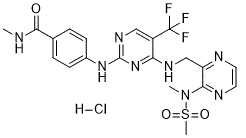
N-methyl-4-[[4-[[3-[methyl(methylsulfonyl)amino]pyrazin-2-yl]methylamino]-5-(trifluoromethyl)pyrimidin-2-yl]amino]benzamide;hydrochloride
|
| 别名 |
VS-6063 HCl; PF-04554878 HCl; Defactinib hydrochloride; Defactinib HCl; VS6063 HCl; VS 6063 HCl; VS-6063 HCl; PF04554878 HCl; PF 04554878 HCl; PF4554878 HCl; PF-4554878 HCl; PF4554878 HCl; 1073160-26-5; Defactinib hydrochloride [USAN]; UNII-L2S469LM49; VS-6063 HYDROCHLORIDE; L2S469LM49; Defactinib hydrochloride (USAN); DEFACTINIB HYDROCHLORIDE [WHO-DD];
|
| HS Tariff Code |
2934.99.9001
|
| 存储方式 |
Powder -20°C 3 years 4°C 2 years In solvent -80°C 6 months -20°C 1 month 注意: 请将本产品存放在密封且受保护的环境中,避免吸湿/受潮。 |
| 运输条件 |
Room temperature (This product is stable at ambient temperature for a few days during ordinary shipping and time spent in Customs)
|
| 溶解度 (体外实验) |
|
|||
|---|---|---|---|---|
| 溶解度 (体内实验) |
配方 1 中的溶解度: ≥ 0.67 mg/mL (1.22 mM) (饱和度未知) in 10% DMSO + 40% PEG300 + 5% Tween80 + 45% Saline (这些助溶剂从左到右依次添加,逐一添加), 澄清溶液。
例如,若需制备1 mL的工作液,可将100 μL 6.7 mg/mL澄清DMSO储备液加入400 μL PEG300中,混匀;然后向上述溶液中加入50 μL Tween-80,混匀;加入450 μL生理盐水定容至1 mL。 *生理盐水的制备:将 0.9 g 氯化钠溶解在 100 mL ddH₂O中,得到澄清溶液。 配方 2 中的溶解度: ≥ 0.67 mg/mL (1.22 mM) (饱和度未知) in 10% DMSO + 90% (20% SBE-β-CD in Saline) (这些助溶剂从左到右依次添加,逐一添加), 澄清溶液。 例如,若需制备1 mL的工作液,可将 100 μL 6.7mg/mL澄清的DMSO储备液加入到900μL 20%SBE-β-CD生理盐水中,混匀。 *20% SBE-β-CD 生理盐水溶液的制备(4°C,1 周):将 2 g SBE-β-CD 溶解于 10 mL 生理盐水中,得到澄清溶液。 View More
配方 3 中的溶解度: ≥ 0.67 mg/mL (1.22 mM) (饱和度未知) in 10% DMSO + 90% Corn Oil (这些助溶剂从左到右依次添加,逐一添加), 澄清溶液。 配方 4 中的溶解度: 5% DMSO+50% PEG 300+5% Tween 80+ddH2O: 5mg/mL 1、请先配制澄清的储备液(如:用DMSO配置50 或 100 mg/mL母液(储备液)); 2、取适量母液,按从左到右的顺序依次添加助溶剂,澄清后再加入下一助溶剂。以 下列配方为例说明 (注意此配方只用于说明,并不一定代表此产品 的实际溶解配方): 10% DMSO → 40% PEG300 → 5% Tween-80 → 45% ddH2O (或 saline); 假设最终工作液的体积为 1 mL, 浓度为5 mg/mL: 取 100 μL 50 mg/mL 的澄清 DMSO 储备液加到 400 μL PEG300 中,混合均匀/澄清;向上述体系中加入50 μL Tween-80,混合均匀/澄清;然后继续加入450 μL ddH2O (或 saline)定容至 1 mL; 3、溶剂前显示的百分比是指该溶剂在最终溶液/工作液中的体积所占比例; 4、 如产品在配制过程中出现沉淀/析出,可通过加热(≤50℃)或超声的方式助溶; 5、为保证最佳实验结果,工作液请现配现用! 6、如不确定怎么将母液配置成体内动物实验的工作液,请查看说明书或联系我们; 7、 以上所有助溶剂都可在 Invivochem.cn网站购买。 |
| 制备储备液 | 1 mg | 5 mg | 10 mg | |
| 1 mM | 1.8283 mL | 9.1416 mL | 18.2832 mL | |
| 5 mM | 0.3657 mL | 1.8283 mL | 3.6566 mL | |
| 10 mM | 0.1828 mL | 0.9142 mL | 1.8283 mL |
1、根据实验需要选择合适的溶剂配制储备液 (母液):对于大多数产品,InvivoChem推荐用DMSO配置母液 (比如:5、10、20mM或者10、20、50 mg/mL浓度),个别水溶性高的产品可直接溶于水。产品在DMSO 、水或其他溶剂中的具体溶解度详见上”溶解度 (体外)”部分;
2、如果您找不到您想要的溶解度信息,或者很难将产品溶解在溶液中,请联系我们;
3、建议使用下列计算器进行相关计算(摩尔浓度计算器、稀释计算器、分子量计算器、重组计算器等);
4、母液配好之后,将其分装到常规用量,并储存在-20°C或-80°C,尽量减少反复冻融循环。
计算结果:
工作液浓度: mg/mL;
DMSO母液配制方法: mg 药物溶于 μL DMSO溶液(母液浓度 mg/mL)。如该浓度超过该批次药物DMSO溶解度,请首先与我们联系。
体内配方配制方法:取 μL DMSO母液,加入 μL PEG300,混匀澄清后加入μL Tween 80,混匀澄清后加入 μL ddH2O,混匀澄清。
(1) 请确保溶液澄清之后,再加入下一种溶剂 (助溶剂) 。可利用涡旋、超声或水浴加热等方法助溶;
(2) 一定要按顺序加入溶剂 (助溶剂) 。
| NCT Number | Recruitment | interventions | Conditions | Sponsor/Collaborators | Start Date | Phases |
| NCT04439331 | Active Recruiting |
Drug: Defactinib Hydrochloride | Refractory Lymphoma Advanced Lymphoma |
National Cancer Institute (NCI) |
August 12, 2015 | Phase 2 |
| NCT04720417 | Active Recruiting |
Drug: Defactinib Hydrochloride Procedure: Biopsy |
Metastatic Uveal Melanoma | Thomas Jefferson University | January 26, 2021 | Phase 2 |
| NCT02465060 | Active Recruiting |
Drug: Defactinib Drug: Defactinib Hydrochloride |
Lymphoma Melanoma |
National Cancer Institute (NCI) |
August 17, 2015 | Phase 2 |
|
|---|
|
|
 In vitro biological effects of VS-6063 on taxane-sensitive and taxane-resistant cell lines.J Natl Cancer Inst.2013 Oct 2;105(19):1485-95. |
|---|
 In vivo effects of VS-6063 combined with paclitaxel (PTX).J Natl Cancer Inst.2013 Oct 2;105(19):1485-95. |
 VS-6063 restores YB-1–mediated paclitaxel (PTX) resistance.J Natl Cancer Inst.2013 Oct 2;105(19):1485-95. |
 VS-6063 downregulated YB-1 phosphorylation and nuclear translocation in taxane-resistant cells by an AKT-dependent pathway.J Natl Cancer Inst.2013 Oct 2;105(19):1485-95. |
|---|
 FAK inhibition or silencing of YB-1 downregulates the expression of CD44.J Natl Cancer Inst.2013 Oct 2;105(19):1485-95. |
Impact of pFAK (Tyr 397) and pYB-1 (Ser 102) expressions on patient survival.J Natl Cancer Inst.2013 Oct 2;105(19):1485-95. |
InvivoChem的所有产品仅用于作科学研究,不面向患者销售
Copyright 2020 InvivoChem LLC | All Rights Reserved 粤ICP备20063088号-1


 COA
COA


 463611831
