| 规格 | 价格 | 库存 | 数量 |
|---|---|---|---|
| 10 mM * 1 mL in DMSO |
|
||
| 1mg |
|
||
| 5mg |
|
||
| 10mg |
|
||
| 25mg |
|
||
| 50mg |
|
||
| 100mg |
|
||
| 250mg |
|
||
| 500mg |
|
||
| 1g |
|
||
| Other Sizes |
|
| 靶点 |
Topoisomerase I (IC50 = 0.8 μM); Topoisomerase II (IC50 = 2.67 μM); Daunorubicins/Doxorubicins; HIV-1; DNA topoisomerase II (induces DNA double-strand breaks) [5][9]
Histone eviction from chromatin [5] |
|---|---|
| 体外研究 (In Vitro) |
体外活性:阿霉素是一种蒽环类抗生素,通常被认为在两个基本水平上发挥其抗肿瘤活性:改变 DNA 并产生自由基,通过 DNA 损伤引发癌细胞凋亡。阿霉素可以通过插入 DNA 链来阻断 DNA 的合成,并抑制 DNA 拓扑异构酶 II (TOP2)。当细胞快速增殖并表达高水平 TOP2 时,阿霉素最有效。此外,多柔比星还可以通过产生神经酰胺(通过激活 p53 或其他下游途径(例如 JNK)促进细胞凋亡)、丝氨酸苏氨酸蛋白酶降解 Akt、线粒体释放细胞色素 c、增加 FasL(死亡受体 Fas/CD95 配体)来触发细胞凋亡。 ) mRNA 的产生,以及自由基的产生。用 GSNO(亚硝基谷胱甘肽)预处理可抑制多柔比星耐药乳腺癌细胞系 MCF7/Dx 的耐药性,同时增强蛋白质谷胱甘肽化和多柔比星在细胞核中的积累。阿霉素诱导的 G2/M 检查点阻滞归因于细胞周期蛋白 G2 (CycG2) 表达升高以及共济失调毛细血管扩张突变 (ATM) 以及 ATM 和 Rad3 相关 (ATR) 信号通路中蛋白质的磷酸化修饰。阿霉素抑制 AMP 激活蛋白激酶 (AMPK),导致 SIRT1 功能障碍、p53 积累以及小鼠胚胎成纤维细胞 (MEF) 和心肌细胞的细胞死亡增加,而 AMPK 的预抑制可进一步使其敏化。阿霉素引起显着的热休克反应,并且抑制或沉默热休克蛋白可增强阿霉素在神经母细胞瘤细胞中的凋亡作用。在没有可测量的蛋白酶体抑制的情况下,纳摩尔多柔比星治疗神经母细胞瘤细胞会导致一组特定蛋白质发生剂量依赖性过度泛素化,并导致泛素化酶(如乳酸脱氢酶和 α-烯醇酶)活性丧失,其蛋白质泛素化模式与蛋白酶体抑制剂硼替佐米相似,表明阿霉素也可能通过破坏蛋白质来发挥作用。细胞测定:用增加浓度的阿霉素(0.1、0.3、0.5和1.0 μg/ml,分别等于0.17、0.52、0.85和1.71 μM)处理H9c2细胞2小时,或用0.3 μg/ml(等于0.52μM)的阿霉素在不同的时间点。 Doxorubicin 以时间和剂量依赖性方式诱导 AMPKα (Thr 172) 及其下游乙酰辅酶 A 羧化酶 (ACC、Ser 79) 强烈磷酸化。 AMPKα 磷酸化在多柔比星处理 1 小时后变得明显,并进一步持续至少 6 小时。 LKB1(AMPK 可能的上游激酶)在 H9c2 细胞中也被阿霉素激活。
- 阿霉素对MDA-MB-231乳腺癌细胞(IC50=0.1μM)和MCF-7细胞(IC50=0.05μM)显示强细胞毒性(72小时暴露)[4] - 通过激活p53/p21通路诱导人心肌祖细胞衰老(0.5μM时SA-β-gal+细胞增至80%)[1] - 在耐药细胞NCI/ADR-RES中与Bcl-2 siRNA协同作用(50nM时联合指数=0.3)[4] - 在H9c2心肌细胞中产生活性氧(1μM时增加2倍)[3] |
| 体内研究 (In Vivo) |
在体内,阿霉素与腺病毒 MnSOD (AdMnSOD) 加 1,3-双(2-氯乙基)-1-亚硝基脲 (BCNU) 联合使用,在减少 MB231 肿瘤体积和延长小鼠存活方面具有最大效果。尽管其使用受到其产生的慢性和急性毒副作用的限制,但阿霉素对于治疗乳腺癌和食道癌、儿童实体瘤、骨肉瘤、卡波西肉瘤、软组织肉瘤以及霍奇金和非霍奇金淋巴瘤至关重要。
- 阿霉素(5mg/kg/周×4,静脉注射)使MDA-MB-231移植瘤体积减少70%[7] - 磁性纳米粒制剂(3mg/kg,静脉注射)使肿瘤药物浓度比游离药物提高5倍[7] - 累积剂量(15mg/kg)诱导大鼠慢性心脏毒性:LVEF下降40%,纤维化面积25%[2] |
| 酶活实验 |
纯化的人DNA拓扑异构酶I通过在0-2.0μM阿霉素存在下用超螺旋pHC624 DNA进行酶滴定来定量测定。在溴化乙锭存在下,通过琼脂糖凝胶电泳解析超螺旋和松弛的DNA,并通过扫描微密度测定法定量超螺旋DNA转化为松弛DNA的百分比。在不同浓度的阿霉素下测量DNA拓扑异构酶I活性的抑制作用。阿霉素在0.8微M的IC50值(抑制总活性的50%所需的浓度)下抑制酶活性。对结构相关的蒽环类抗肿瘤药物柔红霉素也观察到类似的抑制作用。这些结果表明,蒽环类药物在体内引起DNA损伤和细胞毒性的浓度下抑制人类DNA拓扑异构酶I活性[11]。
- 拓扑异构酶II解连环实验:酶与阿霉素(0.1-10μM)和kinetoplast DNA在37°C孵育30分钟。DNA断裂通过琼脂糖凝胶电泳检测[5] - Caspase-3活性测定:用阿霉素(1μM)处理的细胞裂解液与DEVD-pNA底物孵育。405nm检测裂解产物[1] |
| 细胞实验 |
细胞培养[7]
LS141原代人细胞系来源于患有高级腹膜后去分化脂肪肉瘤的患者,MPNST细胞来源于患有大腿高级周围神经鞘肿瘤的患者。这些生长在补充有15%热灭活胎牛血清加青霉素和链霉素的RPMI1640中。[7] 菌落测定[7] MPNST细胞依次用阿霉素、黄必利或这两种药物的组合处理。选择MPNST细胞是因为LS141(和其他CDK4依赖性)细胞在体外对CDK4抑制非常敏感,因此组合研究是不间断的。MPNST细胞以每板1000个细胞/100 mm2的密度进行三次接种。接种24小时后,用阿霉素(D,15nM)、黄必利(F,150nM)的IC50、无药物培养基(对照)或两种药物的组合同时或依次处理细胞24小时。处理后,去除含药物的培养基,并使细胞生长10天以形成集落。将所得菌落用0.01%结晶紫染色30分钟,并使用自动菌落计数器计数菌落。结果以未治疗对照的百分比表示,实验结果的统计学意义通过双侧t检验确定。 - 凋亡实验:阿霉素处理(0.1-5μM)48小时后Annexin V/PI染色,流式细胞仪定量[4] - 活性氧检测:DCFH-DA负载的H9c2细胞经阿霉素(0.5-5μM)处理。488/525nm检测荧光[3] - 衰老实验:0.5μM阿霉素处理72小时后SA-β-gal染色,显微镜计数蓝染细胞[1] |
| 动物实验 |
雌性无胸腺裸鼠皮下注射MB231细胞;3 mg/kg/天;瘤内给药
雌性无胸腺裸鼠皮下注射MB231细胞 体内研究:将LS141异种移植瘤直接植入重症联合免疫缺陷(SCID)小鼠体内。当肿瘤体积达到100 mm³时,每组五只小鼠分别接受最大耐受剂量(MTD)的黄酮吡啶醇(9 mg/kg)、阿霉素(0.9 mg/kg)或阿霉素(0.7 mg/kg)后给予黄酮吡啶醇(7 mg/kg)治疗,并在选定的时间点(1、4和7小时)再次给药。此外,一组动物接受相反顺序的治疗,即先给予黄酮吡啶醇,再给予阿霉素,两次给药间隔7小时。所有治疗均采用腹腔注射,每周两次,共5次。每隔2至3天用游标卡尺测量肿瘤大小,并根据公式π/6 × (大直径) × (小直径)²计算肿瘤体积。根据实验,在不同时间点比较各组小鼠的肿瘤体积,并采用双侧t检验确定实验结果的统计学意义。由于肿瘤的侵袭性强,无法估算动物的生存数据。通过体重减轻监测毒性。这些研究均按照《实验动物护理原则》进行,并经IACUC批准。 - 异种移植模型:将MDA-MB-231细胞皮下植入裸鼠体内。 阿霉素(5 mg/kg,溶于生理盐水)每周静脉注射一次,共4周。每两周测量一次肿瘤大小[7] - 心脏毒性模型:大鼠腹腔注射阿霉素(每周累积剂量2.5 mg/kg,共6周)。治疗前后进行超声心动图检查[2] - 生物分布:荷瘤小鼠注射99mTc标记的脂质体阿霉素(3 mg/kg)。24小时后采集器官进行γ计数[10] |
| 药代性质 (ADME/PK) |
吸收、分布和排泄
在艾滋病相关卡波西肉瘤患者中,给予10 mg/m2脂质体阿霉素后,计算得出Cmax和AUC值分别为4.12 ± 0.215 μg/mL和277 ± 32.9 μg/mL•h。 大约40%的剂量在5天内出现在胆汁中,而同期仅有5%至12%的药物及其代谢物出现在尿液中。在尿液中,7天内回收的阿霉素剂量不足3%。 阿霉素的稳态分布容积范围为809 L/m²至1214 L/m²。 阿霉素的血浆清除率范围为324 mL/min/m²至809 mL/min/m²,主要通过代谢和胆汁排泄。阿霉素的清除率也存在性别差异,男性高于女性(1088 mL/min/m² vs. 433 mL/min/m²)。在给予剂量范围为 10 mg/m² 至 75 mg/m² 的盐酸多柔比星后,2 岁以上儿童的血浆清除率估计为 1540 mL/min/m²,2 岁以下婴儿的血浆清除率估计为 813 mL/min/m²。未包封的盐酸多柔比星在胃酸中不稳定,动物研究表明该药物几乎不被胃肠道吸收。该药物对组织具有极强的刺激性,因此必须静脉给药。在患有艾滋病相关卡波西肉瘤的患者中,静脉输注单次10或20 mg/m²剂量的脂质体盐酸阿霉素后,15分钟输注后血浆中阿霉素(主要与脂质体结合)的平均峰浓度分别为4.33或10.1 μg/mL,30分钟输注后分别为4.12或8.34 μg/mL。在患有艾滋病相关卡波西肉瘤的成人患者中,静脉输注40 mg/m²剂量的脂质体盐酸阿霉素15分钟后,血浆峰浓度平均为20.1 μg/mL。 非包封(常规)盐酸阿霉素表现出线性药代动力学特征;聚乙二醇稳定的脂质体盐酸阿霉素在10-20 mg/m²的剂量范围内也表现出剂量比例线性药代动力学特征。据报道,脂质体包裹的阿霉素在50 mg/m²剂量下的药代动力学呈非线性。预计在50 mg/m²剂量下,其消除半衰期将比20 mg/m²剂量下更长,清除率将更低,血浆浓度-时间曲线下面积的增加幅度将大于剂量比例。将盐酸阿霉素包封于聚乙二醇(PEG)稳定的(隐形)脂质体中,可显著改变其与传统静脉制剂(即未包封药物)相比的药代动力学,导致药物在周围组织中的分布减少,在卡波西肉瘤病灶中的分布增加,以及血浆清除率降低。 常规注射给药的阿霉素广泛分布于血浆和组织中。静脉注射后仅30秒,即可在肝脏、肺、心脏和肾脏中检测到阿霉素。阿霉素可被细胞吸收并与细胞成分结合,尤其是核酸。常规静脉注射给药的盐酸阿霉素的分布容积约为700-1100升/平方米。未包封的多柔比星与血浆蛋白的结合率约为50-85%…… 静脉注射脂质体包裹的多柔比星盐酸盐后,其在卡波西肉瘤病灶中的分布程度高于在健康皮肤中的分布程度。单次静脉注射20 mg/m²的脂质体包裹的多柔比星盐酸盐后,卡波西肉瘤病灶中的多柔比星浓度比健康皮肤中的浓度高19倍(范围:3-53倍);然而,该研究并未考虑病灶或健康皮肤中的血药浓度。此外,静脉注射脂质体包裹的阿霉素后,其在卡波西肉瘤病灶中的分布量比静脉注射同等剂量的常规(非包裹)阿霉素高5.2-11.4倍。脂质体包裹增强阿霉素在卡波西肉瘤病灶中分布的机制尚未完全阐明,但已有研究表明,含有胶体金作为标记物的类似聚乙二醇(PEG)稳定脂质体能够进入动物体内的卡波西肉瘤样病灶。脂质体也可能通过卡波西肉瘤内皮细胞间隙渗出。药物进入病灶后,脂质体降解并使其在原位变得可渗透,药物很可能在局部释放。 有关多柔比星(共16种)的更多吸收、分布和排泄(完整)数据,请访问HSDB记录页面。 代谢/代谢物 多柔比星可通过三种代谢途径进行代谢:单电子还原、双电子还原和脱糖基化。然而,大约一半的剂量以原形从体内排出。双电子还原是多柔比星的主要代谢途径。在该代谢途径中,阿霉素经多种酶还原为仲醇阿霉素醇,这些酶包括醇脱氢酶[NADP(+)]、羰基还原酶[NADPH]1、羰基还原酶[NADPH]3和醛酮还原酶1家族成员C3。胞质和线粒体中的多种氧化还原酶促进单电子还原,生成阿霉素半醌自由基。这些酶包括线粒体和胞质NADPH脱氢酶、黄嘌呤氧化酶和一氧化氮合酶。该半醌代谢物可被重新氧化为阿霉素,但同时会产生活性氧(ROS)和过氧化氢。导致阿霉素相关不良反应(尤其是心脏毒性)的主要原因是该途径产生的活性氧(ROS),而非阿霉素半醌的形成。脱糖基化是一条次要的代谢途径,仅占阿霉素代谢的1%至2%。在胞质NADPH醌脱氢酶、黄嘌呤氧化酶和NADPH-细胞色素P450还原酶的催化下,阿霉素可被还原为阿霉素脱氧苷元或水解为阿霉素羟基苷元。 非包膜阿霉素经NADPH依赖性醛酮还原酶代谢为亲水性的13-羟基代谢物阿霉素醇,后者具有抗肿瘤活性,是阿霉素的主要代谢产物;这些还原酶存在于大多数细胞中,甚至可能存在于所有细胞中,尤其是在红细胞、肝脏和肾脏中。虽然尚未完全确定,但阿霉素醇似乎也是该药物心脏毒性作用的罪魁祸首。据报道,静脉注射单次10~50 mg/m²剂量的聚乙二醇稳定脂质体包裹的盐酸阿霉素后,血浆中阿霉素醇的浓度极低或无法检测(即0.8~26.2 ng/mL);目前尚不清楚这种脂质体包裹的蒽环类药物是否比传统(非包裹)药物的心脏毒性更低,并且目前针对非包裹药物的常规预防措施也应适用于脂质体制剂。使用聚乙二醇(PEG)稳定脂质体注射剂后,观察到阿霉素主要代谢物的血浆浓度显著降低或消失,这表明药物在循环过程中可能并未从脂质体中大量释放,或者部分阿霉素可能被释放,但阿霉素醇的消除速率远高于释放速率;未用PEG稳定脂质体包裹的盐酸阿霉素则代谢为阿霉素醇。 其他无治疗活性的代谢物包括难溶于水的苷元,如阿霉素酮(阿霉素)和7-脱氧阿霉素酮(17-脱氧阿霉素),以及它们的结合物。这些苷元在微粒体中通过NADPH依赖的细胞色素还原酶介导的氨基糖部分裂解而形成。多柔比星经酶促还原为7-脱氧苷元是其发挥细胞毒性作用的关键,因为该过程会产生羟自由基,从而导致广泛的细胞损伤和死亡。对于非包封的多柔比星,给药后5分钟内,血浆中超过20%的药物以代谢物的形式存在;30分钟后达到70%;4小时后达到75%;24小时后达到90%。……目前已鉴定出至少6种代谢物,其中最主要的是阿霉素醇。该产物是由白细胞和红细胞中存在的一种酶(推测也存在于恶性组织中)还原C13位酮基而产生的。 阿霉素可转化为阿霉素醇、苷元和其他衍生物。 有关阿霉素(共6种代谢物)的更多代谢/代谢物(完整)数据,请访问HSDB记录页面。 阿霉素可通过三种代谢途径进行代谢:单电子还原、双电子还原和脱糖基化。然而,大约一半的剂量以原形从体内排出。双电子还原生成阿霉素醇,一种仲醇。该途径被认为是主要的代谢途径。单电子还原由多种氧化还原酶催化,生成阿霉素半醌自由基。这些酶包括线粒体和胞质NADPH脱氢酶、黄嘌呤氧化酶和一氧化氮合酶。脱糖基化是次要的代谢途径(1-2%的剂量通过此途径代谢)。由此产生的代谢产物是脱氧苷元或羟基苷元,分别通过还原或水解形成。可能参与此途径的酶包括黄嘌呤氧化酶、NADPH-细胞色素P450还原酶和胞质NADPH脱氢酶。 排泄途径:40%的剂量在5天内经胆汁排出。5-12%的药物及其代谢物在同一时间段内经尿液排出。尿液中回收的剂量中,阿霉素醇的含量不足3%。 半衰期:末端半衰期 = 20 - 48 小时。 生物半衰期 阿霉素的末端半衰期为 20 至 48 小时。阿霉素的分布半衰期约为 5 分钟。对于脂质体制剂,在 AIDS 相关卡波西肉瘤患者中,10 mg/m² 阿霉素的第一相半衰期和第二相半衰期分别计算为 4.7 ± 1.1 小时和 52.3 ± 5.6 小时。 未包封的阿霉素及其代谢物的血浆浓度呈双相或三相下降。在三相模型的第一阶段,未包封的阿霉素迅速代谢,推测是通过肝脏首过效应。大部分代谢似乎在给药前就已完成。在三相模型中,未包封的阿霉素及其代谢物迅速分布到血管外间隙,阿霉素的血浆半衰期约为0.2-0.6小时,其代谢物的血浆半衰期约为3.3小时。随后,阿霉素及其代谢物的血浆浓度维持在相对较长的水平,这可能是由于组织结合所致。在第二阶段,未包封的阿霉素的血浆半衰期为16.7小时,其代谢物的血浆半衰期为31.7小时。在双相模型中,据报道初始分布半衰期平均约为 5-10 分钟,末端消除半衰期平均约为 30 小时。 脂质体包裹的盐酸阿霉素的血浆浓度似乎以双相方式下降。在艾滋病相关卡波西肉瘤患者中,静脉注射单次10~40 mg/m²剂量的盐酸多柔比星脂质体后,多柔比星的初始血浆半衰期(t1/2α)平均为3.76~5.2小时,而末端消除半衰期(t1/2β)平均为39.1~55小时。 约5分钟的初始分布半衰期表明多柔比星被组织快速吸收,而其从组织中缓慢消除则体现在20~48小时的末端半衰期上。 患者体内阿霉素的血浆半衰期约为17小时,而其代谢物的血浆半衰期约为32小时。 - 人体血浆半衰期为20~48小时,分布容积为25 L/kg [11] - 血浆蛋白结合率90% [11] - 主要代谢产物:阿霉素醇(活性成分)、苷元 [9] - 胆汁排泄 >60% [9] |
| 毒性/毒理 (Toxicokinetics/TK) |
化疗药物阿霉素(DOX)具有剂量依赖性心脏毒性,可导致心力衰竭。本研究旨在评估大鼠接受三种不同给药方案(DOX1:单次腹腔注射10 mg/kg DOX;DOX2:每日腹腔注射1 mg/kg DOX,持续10天;DOX3:每周腹腔注射2 mg/kg DOX,持续5周)后心脏毒性的发生时间和程度。每周进行经胸超声心动图检查,以评估三组大鼠心脏毒性的发生时间和程度。DOX1组大鼠在第28天死亡率达到80%,而DOX2组和DOX3组大鼠分别在第107天和第98天达到80%的死亡率。在DOX1组中,第2周时射血分数下降了30%;在DOX2组中,第13周时下降了55%;在DOX3组中,第13周时下降了42%。此外,DOX1组和DOX3组的心脏功能存在明显差异,而DOX2组和DOX3组的心脏功能相似。这些结果表明,与单次10 mg/kg多柔比星推注相比,在数天(DOX2)或数周(DOX3)内给药可提高生存率,并表现出更典型的多柔比星诱导扩张型心肌病症状,尽管发病较晚。[J Am Assoc Lab Anim Sci.] 2007年7月;46(4):20-32.]
妊娠期和哺乳期影响 ◉ 哺乳期用药概述 大多数资料认为,在母亲接受抗肿瘤药物治疗期间,尤其是蒽环类药物(如多柔比星)治疗期间,哺乳是禁忌的。间歇性治疗期间,如果哺乳期适当,或许可以安全地进行母乳喂养;然而,由于活性代谢物多柔比星醇在乳汁中的浓度高且持续时间长,因此很难确定合适的哺乳期。一些研究建议在给药后5至10天停止哺乳。而最近采用最坏情况的药代动力学模型表明,初乳期后需要13天才能最大限度地减少全身和肠道毒性。 化疗可能会对母乳的正常微生物群和化学成分产生不利影响。孕期接受化疗的女性更容易出现哺乳困难。 ◉ 对母乳喂养婴儿的影响 一位女性在妊娠27周时被诊断出患有B细胞淋巴瘤。妊娠34周零4天时引产,并开始接受利妥昔单抗、环磷酰胺、多柔比星、长春新碱和泼尼松的标准方案治疗,剂量未明确,疗程为21天,从产后第2天开始。她将乳汁挤出并丢弃,在每个疗程的前10天用捐赠母乳喂养婴儿,然后在下一个疗程开始前的剩余10天进行母乳喂养。这10天的母乳喂养暂停期是根据长春新碱的半衰期(约3个半衰期)确定的。据报道,在完成4个疗程的化疗后,她的婴儿健康状况良好,发育正常,未出现任何并发症。 ◉ 对哺乳和母乳的影响 一项针对接受过儿童恶性肿瘤化疗的青少年男性的研究发现,接受多柔比星治疗与血清催乳素浓度升高相关。 一名在妊娠中期被诊断出患有霍奇金淋巴瘤的女性,在妊娠晚期接受了3个疗程的化疗,并在产后4周恢复化疗。在重新开始化疗后的16周内,分别在化疗前后15至30分钟采集乳汁样本。化疗方案包括多柔比星40毫克、博来霉素16单位、长春碱9.6毫克和达卡巴嗪600毫克,每2周一次,每次2小时给药。研究人员将该女性乳汁中的微生物群落和代谢谱与8名未接受化疗的健康女性的乳汁进行了比较。该患者母乳中的微生物群落与健康女性的显著不同,鲍曼不动杆菌属(Acinetobacter sp.)、黄单胞菌科(Xanthomonadacea)和嗜麦芽窄食单胞菌属(Stenotrophomonas sp.)的丰度增加,而双歧杆菌属(Bifidobacterium sp.)和真杆菌属(Eubacterium sp.)的丰度降低。接受治疗的女性母乳中的多种化学成分也存在显著差异,其中最显著的是DHA和肌醇含量降低。 一项电话随访研究对74名在妊娠中晚期于同一中心接受癌症化疗的女性进行了调查,以确定她们产后母乳喂养的成功率。结果显示,仅有34%的女性能够纯母乳喂养婴儿,66%的女性报告存在母乳喂养困难。相比之下,22名在妊娠期间确诊癌症但未接受化疗的母亲的母乳喂养成功率高达91%。其他具有统计学意义的相关性包括:1. 有哺乳困难的母亲平均接受 5.5 个化疗周期,而无哺乳困难的母亲平均接受 3.8 个化疗周期;2. 有哺乳困难的母亲在妊娠期间平均提前 3.4 周接受第一个化疗周期。在接受含多柔比星方案的 62 名女性中,39 名有哺乳困难。 - 心脏毒性:累积剂量 >400 mg/m² 时,左心室射血分数 (LVEF) 下降 >10% [9] - 骨髓抑制:最低值出现在 10-14 天(白细胞计数 <2000 个/mm³)[11] - 小鼠 LD50 = 10 mg/kg(单次静脉注射)[6] - 肝毒性:累积剂量 15 mg/kg 时,ALT 升高 3 倍 [2] |
| 参考文献 |
[1]. Cancer Res. 2009 May 15;69(10):4294-300. [2]. Food Chem Toxicol. 2010 Jun;48(6):1425-38. [3]. Biochem J. 2011 Dec 1;440(2):175-83. [4]. Br J Cancer. 2011 Mar 15;104(6):957-67. [5]. J Biol Chem. 2012 Jun 29;287(27):22838-53. [6]. J Biol Chem. 2012 Mar 9;287(11):8001-12. [7]. Clin Cancer Res. 2012 May 1;18(9):2638-47. [8]. FEBS J. 2012 Jun;279(12):2182-91. [9]. Nat Rev Cancer. 2009 May;9(5):338-50. |
| 其他信息 |
根据一个由科学和健康专家组成的独立委员会的说法,盐酸多柔比星(阿霉素)可能致癌。根据州或联邦政府的标签要求,它可能具有发育毒性和男性生殖毒性。
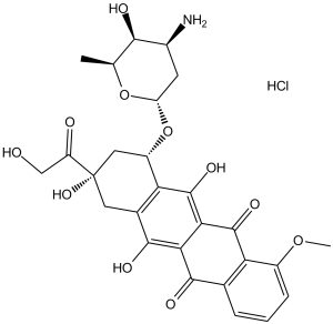
盐酸阿霉素呈橙红色细针状。其水溶液在酸性pH值下呈黄橙色,在中性pH值下呈橙红色,pH值高于9时呈紫蓝色。(NTP, 1992) 盐酸多柔比星是一种蒽环类抗生素。 盐酸多柔比星(脂质体)是一种抗肿瘤处方药,已获得美国食品药品监督管理局 (FDA) 的批准,用于治疗某些类型的癌症,包括卵巢癌、多发性骨髓瘤和艾滋病相关的卡波西肉瘤。 卡波西肉瘤是由人疱疹病毒8型 (HHV-8) 感染引起的。 HHV-8感染可能是HIV的机会性感染(OI)。 盐酸阿霉素是阿霉素的盐酸盐,阿霉素是一种蒽环类抗生素,具有抗肿瘤活性。阿霉素是从链霉菌(Streptomyces peucetius var. caesius)中分离得到的,是柔红霉素的羟基化同系物。阿霉素可插入DNA双螺旋的碱基对之间,从而阻止DNA复制,最终抑制蛋白质合成。此外,阿霉素抑制拓扑异构酶II,导致DNA复制过程中可裂解的酶-DNA连接复合物增加并稳定,进而阻止双链断裂后核苷酸链的连接。阿霉素还会形成氧自由基,导致细胞膜脂质过氧化,从而产生细胞毒性;氧自由基的形成也是蒽环类抗生素毒性的促成因素,特别是其对心脏和皮肤血管的影响。 抗肿瘤抗生素,提取自链霉菌(Streptomyces peucetius)。它是柔红霉素的羟基衍生物。 另见:阿霉素(具有活性部分)。 药物适应症 聚乙二醇化脂质体塞来昔布适用于成人:作为单药治疗,用于治疗转移性乳腺癌患者,尤其适用于心脏风险增加的患者;或用于治疗一线铂类化疗失败的晚期卵巢癌女性患者;与硼替佐米联合用于治疗至少接受过一种既往治疗且已接受或不适合进行骨髓移植的进展性多发性骨髓瘤患者。用于治疗CD4计数低(<200个CD4淋巴细胞/mm3)且伴有广泛黏膜皮肤或内脏病变的艾滋病相关卡波西肉瘤(KS)患者。Celdoxome聚乙二醇化脂质体可用作一线全身化疗,或用于既往接受过至少包含以下两种药物的联合全身化疗后病情进展或不耐受的艾滋病相关KS患者的二线化疗:长春花生物碱、博来霉素和标准阿霉素(或其他蒽环类药物)。 Caelyx聚乙二醇化脂质体适用于:作为单药治疗,用于存在心脏风险增加的转移性乳腺癌患者;用于治疗一线铂类化疗方案失败的晚期卵巢癌女性患者;与硼替佐米联合用于治疗至少接受过一种先前治疗且已接受或不适合接受骨髓移植的进展性多发性骨髓瘤患者;用于治疗CD4计数低的艾滋病相关卡波西肉瘤(KS)患者( Myocet脂质体与环磷酰胺联合使用,适用于成年女性转移性乳腺癌的一线治疗。 治疗乳腺癌和卵巢癌。 治疗肝细胞癌。 阿霉素是一种脱氧己糖苷、蒽环类抗生素、氨基糖苷类抗生素、四并苯醌类抗生素、对醌类抗生素、伯α-羟基酮和叔α-羟基酮。它是大肠杆菌的代谢产物。它是阿霉素(1+)的共轭碱。它来源于四并苯的氢化物。 盐酸阿霉素(脂质体)是一种抗肿瘤处方药。经美国食品药品监督管理局 (FDA) 批准,用于治疗某些类型的癌症,包括卵巢癌、多发性骨髓瘤和艾滋病相关的卡波西肉瘤。 卡波西肉瘤是由人疱疹病毒8型 (HHV-8) 感染引起的。HHV-8 感染可能是 HIV 的机会性感染 (OI)。 阿霉素是一种细胞毒性蒽环类抗生素,于 1970 年与另一种细胞毒性药物柔红霉素一起从链霉菌 (Streptomyces peucetius var. caesius) 的培养物中分离出来。虽然它们都含有糖苷配基和糖部分,但阿霉素的侧链末端是伯醇基,而柔红霉素的侧链末端是甲基。尽管其详细的分子机制尚未完全阐明,但普遍认为阿霉素通过 DNA 嵌入发挥作用,最终导致 DNA 损伤。活性氧的产生。由于其疗效显著且作用广泛,阿霉素于1974年获得FDA批准,用于治疗多种癌症,包括但不限于乳腺癌、肺癌、胃癌、卵巢癌、甲状腺癌、非霍奇金淋巴瘤和霍奇金淋巴瘤、多发性骨髓瘤、肉瘤以及儿童癌症。然而,阿霉素的主要副作用之一是心脏毒性,因此心脏功能不全的患者不宜使用,并且一旦达到最大耐受累积剂量,就需要停止治疗。 阿霉素是一种蒽环类拓扑异构酶抑制剂。阿霉素的作用机制是作为拓扑异构酶抑制剂。 据报道,阿霉素存在于棘孢霉(Talaromyces aculeatus)、褐孢霉(Hamigera fusca)和其他有相关数据的生物体中。 阿霉素是一种蒽环类抗生素。抗肿瘤活性。阿霉素是从链霉菌(Streptomyces peucetius var. caesius)中分离得到的,是柔红霉素的羟基化类似物。阿霉素可插入DNA双螺旋的碱基对之间,从而阻止DNA复制,最终抑制蛋白质合成。此外,阿霉素抑制拓扑异构酶II,导致DNA复制过程中可裂解的酶-DNA连接复合物增加并稳定,进而阻止双链断裂后核苷酸链的连接。阿霉素还会形成氧自由基,导致细胞膜脂质过氧化,从而产生细胞毒性;氧自由基的形成也是蒽环类抗生素毒性(特别是心脏和皮肤血管毒性)的成因之一。 阿霉素仅存在于使用或服用过该药物的个体体内。它是一种从链霉菌中提取的抗肿瘤抗生素。链霉菌(Streptomyces peucetius)是柔红霉素的羟基衍生物。[PubChem]阿霉素通过多种作用机制发挥抗有丝分裂和细胞毒活性:阿霉素通过插入碱基对之间与DNA形成复合物,并通过稳定DNA-拓扑异构酶II复合物来抑制拓扑异构酶II的活性,从而阻止拓扑异构酶II催化的连接-再连接反应中的再连接部分。 从链霉菌(Streptomyces peucetius)中提取的抗肿瘤抗生素。它是柔红霉素的羟基衍生物。 另见:盐酸阿霉素(有盐形式);佐普瑞林阿霉素(其活性部分);佐普瑞林醋酸阿霉素(其活性部分)。 药物适应症 阿霉素适用于治疗急性淋巴细胞白血病、急性髓系白血病、霍奇金淋巴瘤和非霍奇金淋巴瘤、转移性乳腺癌、转移性肾母细胞瘤、转移性神经母细胞瘤、转移性软组织和骨肉瘤、转移性卵巢癌、转移性膀胱移行细胞癌、转移性甲状腺癌、转移性胃癌和转移性支气管癌等肿瘤性疾病。多柔比星还适用于原发性乳腺癌切除术后出现腋窝淋巴结转移的女性患者的辅助治疗。对于脂质体剂型,多柔比星适用于治疗铂类化疗后进展或复发的卵巢癌、既往全身化疗失败或不耐受此类治疗的艾滋病相关卡波西肉瘤,以及既往未接受过硼替佐米治疗的多发性骨髓瘤患者,需与硼替佐米联合使用。并且至少接受过一种既往治疗。 FDA 标签 佐斯凯蒂尔聚乙二醇化脂质体是一种用于治疗成人以下类型癌症的药物:â欧元¢ 乳腺癌已扩散至身体其他部位,且患者有心脏问题风险。唑斯凯蒂尔聚乙二醇化脂质体可单独用于治疗该疾病;â欧元¢ 晚期卵巢癌,指既往接受过包括铂类抗癌药物在内的治疗但疗效不佳的女性;â欧元¢ 多发性骨髓瘤(骨髓白细胞癌),适用于既往接受过至少一种其他治疗且病情进展,并已接受或不适合接受骨髓移植的患者。聚乙二醇化脂质体唑斯替尔与硼替佐米(另一种抗癌药物)联合使用;â欧元¢ 卡波西肉瘤常见于免疫系统严重受损的艾滋病患者。卡波西肉瘤是一种癌症,会导致异常组织在皮肤下、潮湿的体表或内脏器官上生长。Zolsketil 聚乙二醇化脂质体含有活性成分阿霉素,是一种‘“混合医学”。这意味着它类似于一种……€˜参考药物“含有相同的活性成分阿霉素”。然而,在佐斯凯替聚乙二醇化脂质体中,活性成分被包裹在称为脂质体的微小脂肪球中,而阿霉素则没有这种情况。 Caelyx®聚乙二醇化脂质体适用于:作为单药疗法,用于治疗存在心脏风险增加的转移性乳腺癌患者;用于治疗一线铂类化疗方案失败的晚期卵巢癌女性患者;与硼替佐米联合用于治疗至少接受过一种既往治疗且已接受或不适合接受骨髓移植的进展性多发性骨髓瘤患者;用于治疗CD4计数低的艾滋病相关卡波西肉瘤(KS)患者( Myocet脂质体与环磷酰胺联合使用,适用于成年女性转移性乳腺癌的一线治疗。 治疗乳腺癌和卵巢癌。 作用机制 通常认为,阿霉素通过两种主要机制发挥其抗肿瘤活性:嵌入DNA和破坏拓扑异构酶介导的修复以及自由基介导的细胞损伤。阿霉素可通过蒽醌环嵌入DNA,该环通过与DNA碱基形成氢键来稳定复合物。阿霉素的嵌入可向多核苷酸结构引入扭转应力,从而破坏核小体结构,导致核小体脱落和替换。此外,阿霉素-DNA复合物可干扰阿霉素通过抑制拓扑异构酶II的活性,阻止拓扑异构酶介导的DNA断裂的修复,从而抑制DNA复制和转录,并诱导细胞凋亡。此外,阿霉素可被微粒体NADPH-细胞色素P-450还原酶代谢为半醌自由基,该自由基在氧气存在下可被重新氧化生成氧自由基。已知活性氧可通过多种机制造成细胞损伤,包括脂质过氧化和膜损伤、DNA损伤、氧化应激和细胞凋亡。虽然该途径产生的自由基可被过氧化氢酶和超氧化物歧化酶灭活,但肿瘤细胞和心肌细胞往往缺乏这些酶,这解释了阿霉素对癌细胞的有效性以及其导致心脏毒性的倾向。 盐酸阿霉素是一种抗肿瘤抗生素,其药理作用与柔红霉素相似。尽管该药物具有抗感染特性,但其细胞毒性限制了其作为抗感染药物的应用。阿霉素抗肿瘤作用的确切机制和/或主要机制尚未完全阐明。该药物的细胞毒性作用似乎源于一个复杂的多种作用机制,包括:阿霉素经电子还原代谢活化后产生的自由基、药物嵌入DNA、诱导DNA断裂和染色体畸变,以及药物引起的细胞膜改变。体外研究表明,阿霉素处理细胞后,细胞凋亡(程序性细胞死亡)也可能参与了该药物的作用机制。这些机制以及其他机制(例如螯合金属离子形成药物-金属络合物)也可能导致该药物的心脏毒性。 阿霉素经酶促单电子和双电子还原生成相应的半醌和二氢醌。 7-脱氧糖苷配基是由酶促单电子还原反应生成的,生成的半醌自由基与氧气反应,引发一系列级联反应生成羟基自由基;该自由基可与DNA、RNA、细胞膜和蛋白质反应,导致细胞死亡。阿霉素经双电子还原生成的二氢醌也可由两个半醌反应生成。在氧气存在下,二氢醌反应生成过氧化氢;在氧气不存在下,二氢醌失去糖基,生成醌甲化物,这是一种对DNA亲和力较低的单功能烷基化剂。二氢醌和醌甲化物对阿霉素细胞毒性的贡献尚不明确。实验证据表明,阿霉素通过插入碱基对之间与DNA形成复合物,导致模板紊乱和空间位阻,从而抑制DNA合成和DNA依赖的RNA合成。阿霉素还能抑制蛋白质合成。阿霉素在整个细胞周期(包括间期)均具有活性。 蒽环类药物的多种诱导作用可能导致心脏毒性的发生。在动物实验中,蒽环类药物选择性抑制心肌中β-肌动蛋白、肌钙蛋白、肌球蛋白轻链2和肌酸激酶M亚型的基因表达,这可能导致与蒽环类药物诱导的心脏毒性相关的肌原纤维丢失。蒽环类药物诱导心脏毒性的其他潜在原因包括钙超载引起的心肌细胞损伤、心肌肾上腺素能功能改变、血管活性胺的释放以及促炎细胞因子的释放。有限的数据表明,钙通道阻滞剂(例如,异戊二烯胺)或β-肾上腺素能阻滞剂可能预防钙超载……有研究表明,蒽环类药物诱导心脏毒性的主要原因是自由基损伤。 DNA。 蒽环类药物可嵌入DNA,螯合金属离子形成药物-金属复合物,并通过氧化还原反应产生氧自由基。蒽环类药物含有醌结构,可通过NADPH依赖性反应发生还原反应,生成半醌自由基,进而引发氧自由基级联反应。代谢产物阿霉素醇似乎是导致心脏毒性的罪魁祸首,而心脏可能由于抗氧化剂浓度相对较低而特别容易受到自由基损伤。……药物螯合金属离子(尤其是铁)会形成阿霉素-金属复合物,该复合物可催化活性氧自由基的生成,并且是一种强氧化剂,即使在没有氧自由基的情况下也能引发脂质过氧化。该反应不受自由基清除剂的抑制,可能是蒽环类药物引起心脏毒性的主要机制。 本研究探讨了阿霉素对大鼠心脏活性氧代谢的影响。结果显示,阿霉素在心脏匀浆、肌浆网、线粒体和胞质溶胶(心脏损伤的主要部位)中均产生了氧自由基。心脏肌球体和线粒体组分中的超氧化物生成增加。显然,阿霉素在与药物诱导组织损伤相同的心肌细胞区室中产生的自由基,可能通过超过心脏线粒体和肌浆网的氧自由基解毒能力而损伤心脏。 - 蒽环类抗生素,可嵌入DNA并抑制拓扑异构酶II [9] - FDA黑框警告:具有心脏毒性 [9] - 临床应用:乳腺癌、淋巴瘤、肉瘤 [9] - 耐药机制:P-糖蛋白外排、谷胱甘肽结合[8] |
| 分子式 |
C27H29NO11.HCL
|
|
|---|---|---|
| 分子量 |
579.98
|
|
| 精确质量 |
579.15
|
|
| 元素分析 |
C, 55.91; H, 5.21; Cl, 6.11; N, 2.42; O, 30.34
|
|
| CAS号 |
25316-40-9
|
|
| 相关CAS号 |
25316-40-9 (Doxorubicin HCl); 23214-92-8
|
|
| PubChem CID |
443939
|
|
| 外观&性状 |
Red to orange solid powder
|
|
| 沸点 |
810.3ºC at 760 mmHg
|
|
| 熔点 |
216ºC
|
|
| 闪点 |
443.8ºC
|
|
| 蒸汽压 |
9.64E-28mmHg at 25°C
|
|
| 来源 |
Streptomyces peucetius var. Caesius
|
|
| LogP |
1.503
|
|
| tPSA |
206.07
|
|
| 氢键供体(HBD)数目 |
7
|
|
| 氢键受体(HBA)数目 |
12
|
|
| 可旋转键数目(RBC) |
5
|
|
| 重原子数目 |
40
|
|
| 分子复杂度/Complexity |
977
|
|
| 定义原子立体中心数目 |
6
|
|
| SMILES |
Cl[H].O([C@@]1([H])C([H])([H])[C@@]([H])([C@@]([H])([C@]([H])(C([H])([H])[H])O1)O[H])N([H])[H])[C@]1([H])C2C(=C3C(C4C(=C([H])C([H])=C([H])C=4C(C3=C(C=2C([H])([H])[C@@](C(C([H])([H])O[H])=O)(C1([H])[H])O[H])O[H])=O)OC([H])([H])[H])=O)O[H]
|
|
| InChi Key |
MWWSFMDVAYGXBV-RUELKSSGSA-N
|
|
| InChi Code |
InChI=1S/C27H29NO11.ClH/c1-10-22(31)13(28)6-17(38-10)39-15-8-27(36,16(30)9-29)7-12-19(15)26(35)21-20(24(12)33)23(32)11-4-3-5-14(37-2)18(11)25(21)34;/h3-5,10,13,15,17,22,29,31,33,35-36H,6-9,28H2,1-2H3;1H/t10-,13-,15-,17-,22+,27-;/m0./s1
|
|
| 化学名 |
(7S,9S)-7-[(2R,4S,5S,6S)-4-amino-5-hydroxy-6-methyloxan-2-yl]oxy-6,9,11-trihydroxy-9-(2-hydroxyacetyl)-4-methoxy-8,10-dihydro-7H-tetracene-5,12-dione;hydrochloride
|
|
| 别名 |
|
|
| HS Tariff Code |
2934.99.9001
|
|
| 存储方式 |
Powder -20°C 3 years 4°C 2 years In solvent -80°C 6 months -20°C 1 month 注意: 请将本产品存放在密封且受保护的环境中(例如氮气保护),避免吸湿/受潮和光照。 |
|
| 运输条件 |
Room temperature (This product is stable at ambient temperature for a few days during ordinary shipping and time spent in Customs)
|
| 溶解度 (体外实验) |
|
|||
|---|---|---|---|---|
| 溶解度 (体内实验) |
配方 1 中的溶解度: ≥ 2.75 mg/mL (4.74 mM) (饱和度未知) in 5% DMSO + 40% PEG300 + 5% Tween80 + 50% Saline (这些助溶剂从左到右依次添加,逐一添加), 澄清溶液。
*生理盐水的制备:将 0.9 g 氯化钠溶解在 100 mL ddH₂O中,得到澄清溶液。 配方 2 中的溶解度: ≥ 2.08 mg/mL (3.59 mM) (饱和度未知) in 10% DMSO + 40% PEG300 + 5% Tween80 + 45% Saline (这些助溶剂从左到右依次添加,逐一添加), 澄清溶液。 例如,若需制备1 mL的工作液,可将 100 μL 20.8 mg/mL澄清的DMSO储备液加入到400 μL PEG300中,混匀;再向上述溶液中加入50 μL Tween-80,混匀;然后加入450 μL生理盐水定容至1 mL。 *生理盐水的制备:将 0.9 g 氯化钠溶解在 100 mL ddH₂O中,得到澄清溶液。 View More
配方 3 中的溶解度: ≥ 2.08 mg/mL (3.59 mM) (饱和度未知) in 10% DMSO + 90% (20% SBE-β-CD in Saline) (这些助溶剂从左到右依次添加,逐一添加), 澄清溶液。 1、请先配制澄清的储备液(如:用DMSO配置50 或 100 mg/mL母液(储备液)); 2、取适量母液,按从左到右的顺序依次添加助溶剂,澄清后再加入下一助溶剂。以 下列配方为例说明 (注意此配方只用于说明,并不一定代表此产品 的实际溶解配方): 10% DMSO → 40% PEG300 → 5% Tween-80 → 45% ddH2O (或 saline); 假设最终工作液的体积为 1 mL, 浓度为5 mg/mL: 取 100 μL 50 mg/mL 的澄清 DMSO 储备液加到 400 μL PEG300 中,混合均匀/澄清;向上述体系中加入50 μL Tween-80,混合均匀/澄清;然后继续加入450 μL ddH2O (或 saline)定容至 1 mL; 3、溶剂前显示的百分比是指该溶剂在最终溶液/工作液中的体积所占比例; 4、 如产品在配制过程中出现沉淀/析出,可通过加热(≤50℃)或超声的方式助溶; 5、为保证最佳实验结果,工作液请现配现用! 6、如不确定怎么将母液配置成体内动物实验的工作液,请查看说明书或联系我们; 7、 以上所有助溶剂都可在 Invivochem.cn网站购买。 |
| 制备储备液 | 1 mg | 5 mg | 10 mg | |
| 1 mM | 1.7242 mL | 8.6210 mL | 17.2420 mL | |
| 5 mM | 0.3448 mL | 1.7242 mL | 3.4484 mL | |
| 10 mM | 0.1724 mL | 0.8621 mL | 1.7242 mL |
1、根据实验需要选择合适的溶剂配制储备液 (母液):对于大多数产品,InvivoChem推荐用DMSO配置母液 (比如:5、10、20mM或者10、20、50 mg/mL浓度),个别水溶性高的产品可直接溶于水。产品在DMSO 、水或其他溶剂中的具体溶解度详见上”溶解度 (体外)”部分;
2、如果您找不到您想要的溶解度信息,或者很难将产品溶解在溶液中,请联系我们;
3、建议使用下列计算器进行相关计算(摩尔浓度计算器、稀释计算器、分子量计算器、重组计算器等);
4、母液配好之后,将其分装到常规用量,并储存在-20°C或-80°C,尽量减少反复冻融循环。
计算结果:
工作液浓度: mg/mL;
DMSO母液配制方法: mg 药物溶于 μL DMSO溶液(母液浓度 mg/mL)。如该浓度超过该批次药物DMSO溶解度,请首先与我们联系。
体内配方配制方法:取 μL DMSO母液,加入 μL PEG300,混匀澄清后加入μL Tween 80,混匀澄清后加入 μL ddH2O,混匀澄清。
(1) 请确保溶液澄清之后,再加入下一种溶剂 (助溶剂) 。可利用涡旋、超声或水浴加热等方法助溶;
(2) 一定要按顺序加入溶剂 (助溶剂) 。
Treatment of Acute Lymphoblastic Leukemia in Children
CTID: NCT00400946
Phase: Phase 3 Status: Completed
Date: 2024-11-27
") |
|---|
") |
 SDOX
SDOX
 Topoisomerase I/II inhibitor 2
 Camptothecin-20(S)-O-propionate hydrate (Camptothecin-20-O-propionate hydrate)
 Topoisomerase I inhibitor 9
InvivoChem的所有产品仅用于作科学研究,不面向患者销售
Copyright 2020 InvivoChem LLC | All Rights Reserved 粤ICP备20063088号-1


 COA
COA
 463611831
