| 规格 | 价格 | 库存 | 数量 |
|---|---|---|---|
| 1mg |
|
||
| 5mg |
|
||
| 10mg |
|
||
| 25mg |
|
||
| 50mg |
|
||
| 100mg |
|
||
| Other Sizes |
|

| 靶点 |
LDHA/lactate dehydrogenase A (Ki =8 μM)
|
|---|---|
| 体外研究 (In Vitro) |
丙酮辅酶 A 丙酮酰酶是 FX-11 (9 μM) [2] 的底物,它被磷酸化以证明 AMP 的激活。在 P493 细胞中,FX-11 抑制糖酵解并改变细胞能量补充。在 BxPc-3 和 MIA PaCa-2 细胞中,FX-11(0-100 μM,72 小时)限制细胞生长 [3]。
|
| 体内研究 (In Vivo) |
FX-11(42 μg/小鼠;IP,每天一次,持续 10-14 天)抑制 P493 肿瘤的形成 [2]。 FX-11(0–2 mg/kg,IP,每日一次,持续三周)
在这项研究中,研究人员研究了PKM2激活剂TEPP-46和LDHA抑制剂FX-11是否可以联合抑制胰腺癌临床前模型的体外和体内肿瘤生长。他们评估了用TEPP-46、FX-11或两者联合处理后的PKM2和LDHA表达、酶活性和细胞增殖率。在体内通过评估肿瘤生长情况、血浆和肿瘤中PK和LDHA活性以及治疗后肿瘤组织中PKM2、LDHA和Ki-67的表达来验证疗效。双重治疗在体内协同抑制胰腺癌细胞增殖,显著延缓肿瘤生长,无明显毒性。TEPP-46和FX-11处理后,血浆和肿瘤组织中PK和LDHA酶活性增加,LDHA酶活性降低,肿瘤中PKM2和LDHA表达降低,表现为肿瘤体积和增殖减少。靶向糖酵解酶如PKM2和LDHA是治疗胰腺癌的一种很有前景的治疗方法。[2] |
| 细胞实验 |
蛋白质印迹分析 [2]
细胞类型: P493 细胞 测试浓度: 9 μM 孵育时间: 24 hrs(小时)、48 hrs(小时) 实验结果: ATP 水平降低,伴随着 AMP 激酶的激活及其底物乙酰辅酶 A 羧化酶的磷酸化。 细胞增殖测定 [3] 细胞类型: BxPc-3 和 MIA PaCa-2 细胞 测试浓度: 0-100 µM 孵育持续时间:72 hrs(小时) 实验结果:以浓度依赖性方式降低细胞代谢活性,显示显着降低细胞增殖,BxPc-3 和 MIA PaCa-2 细胞的 IC50 值分别为 49.27 µM 和 60.54 µM。 |
| 动物实验 |
动物/疾病模型:雄性SCID(重症联合免疫缺陷)小鼠和RH-Foxn1nu(裸鼠)(人P493 B细胞异种移植瘤)[2]
剂量:42 μg/只小鼠(2.1 mg/kg) 给药途径:腹腔注射;可延缓肿瘤生长[3]。每日一次,持续10-14天。 实验结果:显著抑制肿瘤生长和肿瘤异种移植瘤的进展。 动物/疾病模型:免疫缺陷CD-1小鼠(6-8周龄;20-25克,每组n=5)[3] 剂量:2 mg/kg,1 mg/kg+15 mg/kg TEPP-46,2 mg/kg+30 mg/kg TEPP-46 给药途径:腹腔注射(ip)(100 µL),每日一次,持续3周 实验结果:血浆和肿瘤裂解液中的LDHA活性显著降低;增殖标志物Ki-67表达显著降低;肿瘤切片中增殖指数显著下降;肿瘤生长显著延迟。 |
| 参考文献 |
|
| 其他信息 |
由于基因改变和肿瘤缺氧,许多癌细胞会大量摄取葡萄糖,并通过乳酸脱氢酶A (LDHA) 生成乳酸。LDHA由c-Myc和缺氧诱导因子(HIF-1)的靶基因编码。既往研究表明,LDHA表达降低与肿瘤起始有关,但其在肿瘤维持和进展中的作用尚未明确。此外,通过干扰或反义RNA降低LDHA表达如何抑制肿瘤发生机制尚不清楚。本研究发现,通过siRNA降低LDHA表达或使用小分子抑制剂FX11 [3-二羟基-6-甲基-7-(苯甲基)-4-丙基萘-1-羧酸]抑制LDHA活性,均可降低ATP水平,并诱导显著的氧化应激和细胞死亡,而抗氧化剂N-乙酰半胱氨酸可部分逆转这些作用。此外,我们证实FX11能够抑制大型人类淋巴瘤和胰腺癌异种移植瘤的进展。当与NAD(+)合成抑制剂FK866联合使用时,FX11可诱导淋巴瘤消退。因此,使用FX11抑制LDHA是治疗LDHA依赖性肿瘤的一种可行且耐受性良好的方法。我们的研究记录了一种针对瓦博格效应的治疗方法,并表明氧化应激和癌症代谢表型是癌症生物学中需要考虑的关键方面,可用于靶向癌症能量代谢的治疗。[2]
近年来,利用癌细胞代谢作为抗癌治疗策略已引起广泛关注。早在20世纪20年代,德国科学家奥托·瓦博格就观察到癌组织对葡萄糖的旺盛消耗和高水平的有氧糖酵解,这种现象现在被称为瓦博格效应。如今,我们认识到瓦博格效应是由多种复杂因素介导的,包括胰岛素非依赖性葡萄糖转运蛋白GLUT-1的过度表达以及多种糖酵解酶(如乳酸脱氢酶A (LDH-A))的过度表达。作为糖酵解的末端酶,LDH-A催化丙酮酸可逆转化为乳酸,并在该过程中将NADH氧化为NAD+。该反应产生的乳酸大部分被分泌到肿瘤微环境中,酸化周围组织,帮助肿瘤逃避免疫细胞的攻击。NADH氧化为NAD+可在氧化代谢缺失或功能减弱的情况下补充NAD+,从而维持糖酵解持续产生ATP。细胞培养和体内研究表明,LDH-A敲低(使用RNA干扰)可显著降低细胞和肿瘤的增殖,这表明LDH-A可能是一个可行的抗癌靶点。尽管目前存在多种体外LDH-A抑制剂,但仍需要一种高效、选择性强的小分子抑制剂,能够在细胞内和体内均发挥作用。本文报道了N-羟基吲哚类LDH-A抑制剂的开发和生物学评估,其中包括一系列新型的双重Warburg靶向葡萄糖偶联LDH-A抑制剂,这些抑制剂由Hergenrother实验室和Minutolo实验室合作开发。本文还讨论了用于评估NHI系列化合物的相对细胞摄取、细胞乳酸生成以及与13C葡萄糖竞争细胞进入的新型检测方法的开发。此外,本文还报道了最有前景的NHI系列化合物与文献报道的体外LDH-A抑制剂的直接细胞活性比较。最后,本文还讨论了利用CETSA和DARTS技术直接研究化合物与细胞裂解液和完整细胞中LDH-A相互作用的研究[1]。 |
| 分子式 |
C22H22O4MO
|
|---|---|
| 分子量 |
350.4077
|
| 精确质量 |
350.152
|
| 元素分析 |
C, 75.41; H, 6.33; O, 18.26
|
| CAS号 |
213971-34-7
|
| PubChem CID |
10498042
|
| 外观&性状 |
White to off-white solid powder
|
| LogP |
4.8
|
| tPSA |
77.76
|
| 氢键供体(HBD)数目 |
3
|
| 氢键受体(HBA)数目 |
4
|
| 可旋转键数目(RBC) |
5
|
| 重原子数目 |
26
|
| 分子复杂度/Complexity |
473
|
| 定义原子立体中心数目 |
0
|
| InChi Key |
LVPYVYFMCKYFCZ-UHFFFAOYSA-N
|
| InChi Code |
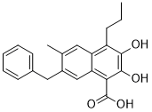
InChI=1S/C22H22O4/c1-3-7-16-17-10-13(2)15(11-14-8-5-4-6-9-14)12-18(17)19(22(25)26)21(24)20(16)23/h4-6,8-10,12,23-24H,3,7,11H2,1-2H3,(H,25,26)
|
| 化学名 |
7-Benzyl-2,3-dihydroxy-6-methyl-4-propylnaphthalene-1-carboxylic acid
|
| 别名 |
FX 11; FX11; LDHA Inhibitor FX11; FX11; 7-benzyl-2,3-dihydroxy-6-methyl-4-propyl-naphthalene-1-carboxylic Acid; 2,3-Dihydroxy-6-methyl-7-(phenylmethyl)-4-propyl-1-naphthalenecarboxylic Acid; CHEMBL126519; 7-benzyl-2,3-dihydroxy-6-methyl-4-propylnaphthalene-1-carboxylic acid; FX-11
|
| HS Tariff Code |
2934.99.9001
|
| 存储方式 |
Powder -20°C 3 years 4°C 2 years In solvent -80°C 6 months -20°C 1 month |
| 运输条件 |
Room temperature (This product is stable at ambient temperature for a few days during ordinary shipping and time spent in Customs)
|
| 溶解度 (体外实验) |
DMSO : ~250 mg/mL (~713.45 mM)
|
|---|---|
| 溶解度 (体内实验) |
配方 1 中的溶解度: ≥ 2.08 mg/mL (5.94 mM) (饱和度未知) in 10% DMSO + 40% PEG300 + 5% Tween80 + 45% Saline (这些助溶剂从左到右依次添加,逐一添加), 澄清溶液。
例如,若需制备1 mL的工作液,可将100 μL 20.8 mg/mL澄清DMSO储备液加入400 μL PEG300中,混匀;然后向上述溶液中加入50 μL Tween-80,混匀;加入450 μL生理盐水定容至1 mL。 *生理盐水的制备:将 0.9 g 氯化钠溶解在 100 mL ddH₂O中,得到澄清溶液。 配方 2 中的溶解度: ≥ 2.08 mg/mL (5.94 mM) (饱和度未知) in 10% DMSO + 90% Corn Oil (这些助溶剂从左到右依次添加,逐一添加), 澄清溶液。 例如,若需制备1 mL的工作液,可将 100 μL 20.8 mg/mL 澄清 DMSO 储备液加入到 900 μL 玉米油中并混合均匀。 请根据您的实验动物和给药方式选择适当的溶解配方/方案: 1、请先配制澄清的储备液(如:用DMSO配置50 或 100 mg/mL母液(储备液)); 2、取适量母液,按从左到右的顺序依次添加助溶剂,澄清后再加入下一助溶剂。以 下列配方为例说明 (注意此配方只用于说明,并不一定代表此产品 的实际溶解配方): 10% DMSO → 40% PEG300 → 5% Tween-80 → 45% ddH2O (或 saline); 假设最终工作液的体积为 1 mL, 浓度为5 mg/mL: 取 100 μL 50 mg/mL 的澄清 DMSO 储备液加到 400 μL PEG300 中,混合均匀/澄清;向上述体系中加入50 μL Tween-80,混合均匀/澄清;然后继续加入450 μL ddH2O (或 saline)定容至 1 mL; 3、溶剂前显示的百分比是指该溶剂在最终溶液/工作液中的体积所占比例; 4、 如产品在配制过程中出现沉淀/析出,可通过加热(≤50℃)或超声的方式助溶; 5、为保证最佳实验结果,工作液请现配现用! 6、如不确定怎么将母液配置成体内动物实验的工作液,请查看说明书或联系我们; 7、 以上所有助溶剂都可在 Invivochem.cn网站购买。 |
| 制备储备液 | 1 mg | 5 mg | 10 mg | |
| 1 mM | 2.8538 mL | 14.2690 mL | 28.5380 mL | |
| 5 mM | 0.5708 mL | 2.8538 mL | 5.7076 mL | |
| 10 mM | 0.2854 mL | 1.4269 mL | 2.8538 mL |
1、根据实验需要选择合适的溶剂配制储备液 (母液):对于大多数产品,InvivoChem推荐用DMSO配置母液 (比如:5、10、20mM或者10、20、50 mg/mL浓度),个别水溶性高的产品可直接溶于水。产品在DMSO 、水或其他溶剂中的具体溶解度详见上”溶解度 (体外)”部分;
2、如果您找不到您想要的溶解度信息,或者很难将产品溶解在溶液中,请联系我们;
3、建议使用下列计算器进行相关计算(摩尔浓度计算器、稀释计算器、分子量计算器、重组计算器等);
4、母液配好之后,将其分装到常规用量,并储存在-20°C或-80°C,尽量减少反复冻融循环。
计算结果:
工作液浓度: mg/mL;
DMSO母液配制方法: mg 药物溶于 μL DMSO溶液(母液浓度 mg/mL)。如该浓度超过该批次药物DMSO溶解度,请首先与我们联系。
体内配方配制方法:取 μL DMSO母液,加入 μL PEG300,混匀澄清后加入μL Tween 80,混匀澄清后加入 μL ddH2O,混匀澄清。
(1) 请确保溶液澄清之后,再加入下一种溶剂 (助溶剂) 。可利用涡旋、超声或水浴加热等方法助溶;
(2) 一定要按顺序加入溶剂 (助溶剂) 。
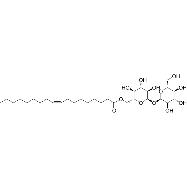 6-O-Oleoyltrehalose
 NCI-006
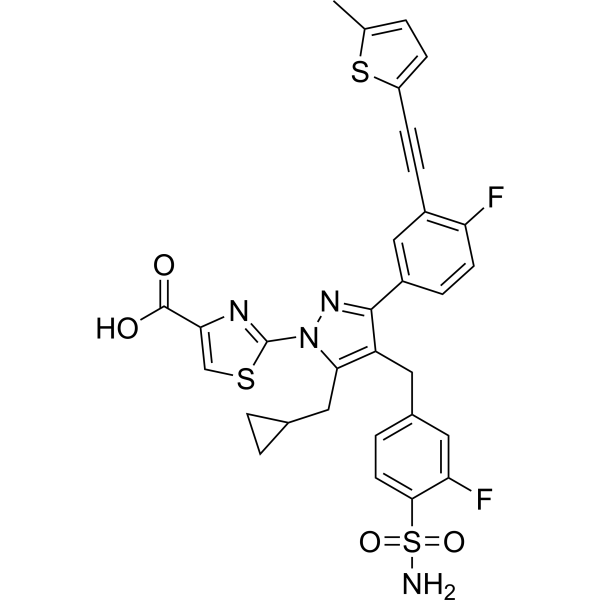
 LDHA-IN-10
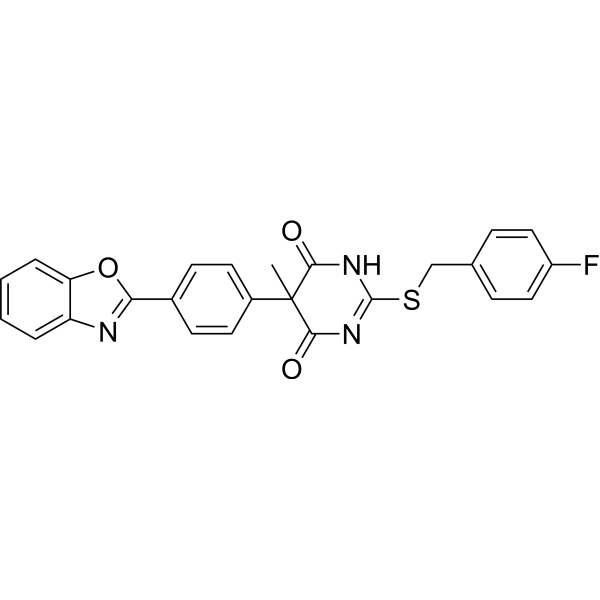
 Bisadinrone A
InvivoChem的所有产品仅用于作科学研究,不面向患者销售
Copyright 2020 InvivoChem LLC | All Rights Reserved 粤ICP备20063088号-1


 COA
COA
 463611831
