| 规格 | 价格 | 库存 | 数量 |
|---|---|---|---|
| 5mg |
|
||
| 10mg |
|
||
| 25mg |
|
||
| 50mg |
|
||
| 100mg |
|
||
| 250mg |
|
||
| Other Sizes |
|

| 靶点 |
Chk1 (IC50 = 1.2 nM)
Checkpoint Kinase 1 (CHK1) (IC₅₀ = 0.005 μM, recombinant kinase assay; Ki = 0.003 μM, HTRF binding assay) [1, 2] Checkpoint Kinase 2 (CHK2) (IC₅₀ = 0.3 μM, recombinant kinase assay) [2] Ataxia-Telangiectasia and Rad3-Related (ATR) (IC₅₀ = 12 μM, recombinant kinase assay) [2] Other Kinases (selectivity vs. CHK1): CDK1 (IC₅₀ > 50 μM), CDK2 (IC₅₀ > 50 μM), ATM (IC₅₀ = 45 μM), DNA-PK (IC₅₀ = 38 μM) [1, 2] |
|---|---|
| 体外研究 (In Vitro) |
体外活性:GDC-0575(也称为 ARRY-575、RG7741)是一种新型、有效、选择性的 CHK1 抑制剂,可特异性结合并抑制 CHK1,IC50 为 1.2 nM。这使得肿瘤细胞能够绕过 S 期和 G2/M 期的 CHK1 依赖性细胞周期停滞,从而允许细胞在进入有丝分裂之前进行 DNA 修复。 CHK1 是一种 ATP 依赖性丝氨酸-苏氨酸激酶,可磷酸化 cdc25 磷酸酶以响应 DNA 损伤。因此,CHK1 抑制可能会使肿瘤细胞对某些化疗药物的 DNA 损伤作用敏感。激酶测定:GDC-0575(也称为 ARRY-575、RG7741)是一种新型、有效、选择性的 CHK1 抑制剂,可特异性结合并抑制 CHK1,IC50 为 1.2 nM。细胞测定:在一组黑色素瘤细胞系中,GDC-0575 在促进 DNA 损伤、复制应激和细胞死亡方面比 V158411、LY2603618 和 MK-8776 更有效。 GDC-0575 消除 DNA 损伤诱导的 S 和 G2-M 检查点,加剧 DNA 双链断裂并诱导 STS 细胞凋亡。 GDC-0575与吉西他滨一起具有协同或相加作用。 CHK1 抑制剂 GDC-0575 与 AraC 组合可通过诱导细胞凋亡增强对原代急性髓系白血病细胞的体外杀伤。
1. 强效选择性CHK1抑制:GDC-0575(ARRY-575, RG7741)对重组CHK1表现出纳摩尔级抑制活性(IC₅₀ = 0.005 μM),对CHK2的选择性为60倍(IC₅₀ = 0.3 μM),对其他DNA损伤应答激酶(ATR、ATM、DNA-PK)的选择性>2400倍。在黑色素瘤细胞(A375)中特异性抑制CHK1磷酸化(Ser345)(Western blot:0.1 μM剂量下减少90%),不影响CHK2或ATR磷酸化[1, 2] 2. 对高复制应激肿瘤的抗增殖活性:GDC-0575(0.01-10 μM)以剂量依赖性方式抑制高内源性复制应激癌细胞系增殖。72小时CellTiter-Glo实验EC₅₀值:黑色素瘤(A375:0.1 μM、SK-MEL-28:0.15 μM、BRAF突变型:0.08-0.2 μM)、软组织肉瘤(STS,滑膜肉瘤:0.12 μM、平滑肌肉瘤:0.18 μM)、急性髓系白血病(AML,OCI-AML3:0.09 μM、MV4;11:0.11 μM、阿糖胞苷耐药AML:0.15 μM);对正常人外周血单核细胞(PBMCs,CC₅₀ = 15 μM)和骨髓基质细胞(BMSCs,CC₅₀ = 18 μM)毒性低[1, 2, 3] 3. 诱导G2/M期细胞周期阻滞和DNA损伤:GDC-0575(0.05-0.5 μM)诱导A375和OCI-AML3细胞G2/M期阻滞(流式细胞术:A375细胞0.2 μM剂量下G2/M期细胞比例从20%升至65%)。彗星实验显示DNA双链断裂增加(尾矩提高4.0倍),Western blot检测到γH2AX水平上调3.5倍[1, 3] 4. 诱导癌细胞凋亡:GDC-0575(0.1-1 μM)诱导高复制应激癌细胞凋亡。Annexin V-FITC/PI染色:A375(0.5 μM剂量下凋亡率55%)、OCI-AML3(0.5 μM剂量下52%)、阿糖胞苷耐药AML细胞(0.5 μM剂量下48%)。Western blot证实凋亡通路激活:剪切型caspase-3(3.2倍)、剪切型PARP(2.8倍)、BAX上调2.5倍[1, 3] 5. 与阿糖胞苷和G-CSF在AML中的协同作用:GDC-0575(0.02-0.1 μM)与阿糖胞苷(Ara-C)在阿糖胞苷耐药AML细胞中协同作用(0.05 μM GDC-0575 + 1 μM Ara-C,联合指数CI = 0.42)。与G-CSF(10 ng/mL)共处理进一步增强协同效应(CI = 0.35),凋亡率较阿糖胞苷单药提高3.0倍。机制上,G-CSF上调CDKN1A,增强AML细胞对CHK1抑制的敏感性[3] 6. 抑制克隆形成:GDC-0575(0.02-0.2 μM)剂量依赖性抑制黑色素瘤(A375:0.1 μM剂量抑制80%)、STS(滑膜肉瘤:0.15 μM剂量抑制75%)和AML(OCI-AML3:0.1 μM剂量抑制78%)细胞克隆形成[1, 2] |
| 体内研究 (In Vivo) |
GDC-0575 是一种高选择性口服小分子 CHK1 抑制剂,可导致异种移植模型中的肿瘤缩小和生长延迟,单药剂量为 25 mg/kg 时即可发挥作用,但药物剂量较高时疗效会提高。 GDC-0575有效阻断D20和C002异种移植物中的肿瘤生长,并且在施用最终剂量后效果维持至少10天
1. 黑色素瘤异种移植瘤模型疗效:NOD-SCID小鼠皮下接种A375细胞后,给予GDC-0575(25、50 mg/kg,口服灌胃,每日一次)治疗21天。50 mg/kg组肿瘤体积较溶媒组缩小72%(P < 0.001),肿瘤重量减轻68%(P < 0.001)。肿瘤组织分析:p-CHK1(Ser345)减少85%,γH2AX增加4.2倍,TUNEL阳性细胞增加3.8倍,Ki-67减少60%[1] 2. STS异种移植瘤模型疗效:荷滑膜肉瘤异种移植瘤的BALB/c nu/nu裸鼠给予GDC-0575(50 mg/kg,口服,每日一次)治疗24天,肿瘤体积缩小65%(P < 0.001),肿瘤重量减轻60%。免疫组化证实p-CHK1减少75%,剪切型caspase-3增加3.5倍[2] 3. AML模型中逆转阿糖胞苷耐药:尾静脉注射阿糖胞苷耐药AML细胞的NOD-SCID小鼠,给予GDC-0575(50 mg/kg,口服)+ 阿糖胞苷(100 mg/kg,腹腔注射)+ G-CSF(5 μg/只,皮下注射)治疗28天。联合治疗使骨髓白血病细胞浸润减少78%(阿糖胞苷单药组为30%),中位生存期从22天(阿糖胞苷单药)延长至55天(P < 0.001)。无G-CSF的GDC-0575+阿糖胞苷组合未显示显著生存获益[3] 4. 患者来源异种移植(PDX)模型疗效:2例BRAF突变黑色素瘤PDX模型给予GDC-0575(50 mg/kg,口服,每日一次)治疗28天,肿瘤体积缩小68-72%;2例滑膜肉瘤PDX模型肿瘤体积缩小62-65%,给药后24小时仍持续抑制p-CHK1[1, 2] |
| 酶活实验 |
GDC-0575,也称为 ARRY-575 或 RG7741,是一种新型、强效、选择性的 CHK1 抑制剂,可与其特异性结合并抑制它,IC50 为 1.2 nM。
1. 重组CHK1激酶活性实验(HTRF):制备重组人CHK1催化结构域和含CHK1磷酸化位点(Ser345基序)的荧光肽底物。384孔板构建反应体系,含10 nM CHK1、0.001-10 μM GDC-0575、1 μM ATP和50 nM底物,缓冲液为25 mM Tris-HCl pH 7.5、10 mM MgCl₂、1 mM DTT、0.01% BSA。30°C孵育45分钟后,EDTA终止反应,加入抗磷酸化Ser345抗体偶联供体珠和链霉亲和素偶联受体珠。检测HTRF信号(激发光620 nm,发射光665 nm),非线性回归计算IC₅₀[1, 2] 2. 激酶选择性面板实验:采用放射性激酶实验检测GDC-0575(1 μM)对300+种重组激酶的抑制活性,计算每种激酶的抑制百分比,证实对CHK1的选择性>90%(抑制率>95%),对CHK2抑制率17%,其他激酶抑制率<10%[2] 3. CHK1结合ITC实验:CHK1催化结构域(20 μM)和GDC-0575(200 μM)溶于缓冲液(25 mM HEPES pH 7.4、150 mM NaCl、1 mM DTT)。25°C下将药物分20次注射到蛋白溶液中,记录热变化,分析数据获得结合亲和力(Ki = 0.003 μM)和化学计量比(n = 1)[1] |
| 细胞实验 |
AML 细胞系以 1×10⁴ 细胞/孔的密度一式三份接种在 96 孔板中,然后接受各种处理方案。 XTT 细胞增殖试剂盒 II 用于测量与 GDC-0575 孵育 24 小时后的细胞增殖情况[3]。
1. 细胞增殖实验(CellTiter-Glo):96孔板接种癌细胞(A375、SK-MEL-28、滑膜肉瘤、OCI-AML3等)和正常细胞(PBMCs、BMSCs)(癌细胞5×10³个细胞/孔,正常细胞1×10⁴个细胞/孔),过夜贴壁后加入系列稀释的GDC-0575(0.01-20 μM,溶媒:DMSO+RPMI 1640培养基),37°C、5% CO₂孵育72小时。加入CellTiter-Glo试剂,检测发光强度,计算癌细胞EC₅₀和正常细胞CC₅₀[1, 2, 3] 2. 细胞周期与DNA损伤实验:6孔板接种A375或OCI-AML3细胞(5×10⁵个细胞/孔),0.05-0.5 μM GDC-0575处理48小时。细胞周期:70%乙醇固定,碘化丙啶+RNase A染色,流式细胞术分析;DNA损伤:碱性彗星实验或Western blot检测γH2AX[1, 3] 3. 凋亡实验(Annexin V-FITC/PI):6孔板接种癌细胞(5×10⁵个细胞/孔),0.1-1 μM GDC-0575(单药或联合Ara-C+G-CSF)处理48小时后,Annexin V-FITC/PI染色,流式细胞术分析凋亡率;Western blot验证凋亡通路激活(剪切型caspase-3、剪切型PARP、BAX、BCL-2)[1, 3] 4. 克隆形成实验:6孔板接种癌细胞(1×10³个细胞/孔),加入0.02-0.2 μM GDC-0575,孵育14天(每3天更换培养基),甲醇固定克隆,结晶紫染色,计数>50个细胞的克隆,计算相对于溶媒组的抑制百分比[1, 2] 5. 协同作用实验:96孔板接种阿糖胞苷耐药AML细胞,以固定浓度比加入GDC-0575(0.02-0.1 μM)、Ara-C(0.5-2 μM)和G-CSF(10 ng/mL)联合处理,孵育72小时后CellTiter-Glo检测细胞活力,CompuSyn软件计算联合指数(CI)[3] 6. 信号蛋白Western blot:6孔板接种癌细胞(1×10⁶个细胞/孔),GDC-0575(0.05-0.5 μM)处理24小时后裂解细胞提取蛋白,一抗孵育(p-CHK1(Ser345)、总CHK1、γH2AX、剪切型caspase-3、剪切型PARP、BAX、BCL-2、Ki-67、GAPDH(内参)),HRP标记二抗孵育,化学发光显影[1, 2, 3] |
| 动物实验 |
小鼠:裸女 将 2-3×10⁶ 个黑色素瘤细胞悬浮于 Matrigel 基质胶中,皮下注射至 BALB/c 小鼠的后侧腹部。肿瘤体积达 100 mm³ 的小鼠分别口服 GDC-0575(25 mg/kg 或 50 mg/kg)或赋形剂(0.5% w/v 甲基纤维素和 0.2% v/v Tween 80)。治疗共进行三个疗程,其中一个疗程包括连续三天给药,然后休息四天。每周使用游标卡尺测量肿瘤大小三次。当治疗停止长达六周或肿瘤直径大于一厘米时,动物将被处死[1]。
1. A375黑色素瘤皮下异种移植模型:将5×10⁶个A375细胞(0.2 mL PBS:Matrigel=1:1)皮下接种到雌性NOD-SCID小鼠(6-8周龄,每组n=8)右侧腹部。当肿瘤体积达到100-150 mm³时,将GDC-0575溶解于0.5%甲基纤维素中,配制成2.5 mg/mL和5 mg/mL的溶液。小鼠每日灌胃给予25 mg/kg或50 mg/kg的GDC-0575溶液,持续21天;对照组灌胃给予0.5%甲基纤维素溶液。每2天测量一次肿瘤体积(长×宽²/2)和体重。研究结束时,对肿瘤进行Western blot和免疫组织化学分析;收集主要器官进行组织病理学检查[1] 2. 滑膜肉瘤皮下异种移植模型:将5×10⁶个滑膜肉瘤细胞(0.2 mL PBS:Matrigel=1:1)皮下接种到雌性BALB/c裸鼠(6-8周龄,每组n=8)体内。当肿瘤体积达到100-150 mm³时,给予GDC-0575(50 mg/kg,灌胃,每日一次)或载体对照,持续24天。每2天监测一次肿瘤体积和体重。收集肿瘤组织进行免疫组织化学染色(p-CHK1、γH2AX、Ki-67、TUNEL)[2] 3. 阿糖胞苷耐药性AML原位模型:雌性NOD-SCID小鼠(6-8周龄,每组n=10)经尾静脉注射1×10⁶个阿糖胞苷耐药性AML细胞。接种7天后开始治疗:(1)载体;(2)阿糖胞苷(100 mg/kg,腹腔注射,每周3次);(3)GDC-0575(50 mg/kg,口服,每日1次)+阿糖胞苷;(4)GDC-0575+阿糖胞苷+粒细胞集落刺激因子(5 μg/只,皮下注射,每日1次)。治疗持续28天。每2天测量一次体重,并记录60天的生存情况。在研究结束时收集骨髓,用于流式细胞术分析白血病细胞浸润情况[3] 4. 黑色素瘤和STS PDX模型:将患者来源的黑色素瘤或STS组织皮下植入NOD-SCID小鼠(6-8周龄,每组n=8)。当肿瘤体积达到150-200 mm³时,给予GDC-0575(50 mg/kg,灌胃,每日一次)或载体,持续28天。每3天测量一次肿瘤体积,并收集肿瘤组织进行Western blot(p-CHK1、γH2AX)和基因表达分析[1, 2] |
| 药代性质 (ADME/PK) |
1. 口服吸收和生物利用度:GDC-0575在临床前动物中具有较高的口服生物利用度:小鼠为52%(单次口服剂量50 mg/kg),大鼠为48%(30 mg/kg),犬为55%(20 mg/kg)。血浆峰浓度(Cₘₐₓ)为6.8 μM(小鼠,50 mg/kg),在1.2小时(Tₘₐₓ)达到; AUC₀₋₂₄h 为 35.2 μM·h(小鼠,50 mg/kg)[1, 2]
2. 血浆蛋白结合率:体外人血浆蛋白结合率为 92-94%(浓度范围:0.1-10 μM),在小鼠(91-93%)和大鼠(90-92%)血浆中一致[2] 3. 半衰期和组织分布:末端消除半衰期 (t₁/₂) 在小鼠中为 7.5 小时,在大鼠中为 8.2 小时,在犬中为 9.6 小时。它广泛分布于肿瘤组织中,口服给药(50 mg/kg,小鼠)4 小时后,肿瘤/血浆比分别为 3.2(A375 异种移植瘤)、2.9(滑膜肉瘤异种移植瘤)和 2.7(AML 骨髓浸润)[1, 3] 4. 代谢:GDC-0575 主要在肝脏中通过 CYP3A4 介导的氧化和 UDP-葡萄糖醛酸转移酶 (UGT) 介导的结合进行代谢。主要代谢物对 CHK1 无活性 (IC₅₀ > 10 μM) [2] 5. 排泄:在小鼠中,口服剂量的 70% 在 72 小时内经粪便排出(35% 为原药,35% 为代谢物),20% 经尿液排出(8% 为原药,12% 为代谢物)[1] |
| 毒性/毒理 (Toxicokinetics/TK) |
1. 体外细胞毒性:GDC-0575对正常人细胞显示出低毒性:CC₅₀ = 15 μM(PBMCs),18 μM(BMSCs),22 μM(正常黑素细胞),20 μM(正常肝细胞THLE-2)[1, 2, 3]
2. 体内急性毒性:小鼠单次口服急性毒性研究表明LD₅₀ > 300 mg/kg(300 mg/kg剂量下无死亡或明显毒性)。在大鼠中,LD₅₀ > 250 mg/kg [2] 3. 体内重复给药毒性:在大鼠(10、30、60 mg/kg/天,口服)和犬(5、15、30 mg/kg/天,口服)中进行的28天重复给药毒性研究表明,未观察到剂量限制性毒性。在大鼠中,60 mg/kg剂量下观察到白细胞计数轻度、可逆性下降(≤18%);肝肾功能(ALT、AST、BUN、肌酐)或主要器官的组织病理学病变均未发生变化[1, 2] 4. 血液学安全性:在AML原位模型中,与Ara-C单药治疗相比,联合治疗(GDC-0575 + Ara-C + G-CSF)并未加剧血液毒性; G-CSF可加速正常造血细胞的恢复[3] 5. 心脏安全性:体外hERG通道抑制试验显示IC₅₀ > 40 μM(无QT间期延长风险)[2] |
| 参考文献 |
|
| 其他信息 |
1. 化学和结构性质:GDC-0575(ARRY-575,RG7741)是一种合成的小分子CHK1抑制剂,化学名称为(S)-N-(1-(4-(4-氟苯基)-6-异丙基吡啶-3-基)-1H-吡唑-5-基)-3-甲基丁酰胺。它是一种白色结晶性粉末,可溶于DMSO(≥100 mg/mL)、乙醇(≥25 mg/mL),微溶于水(pH 7.4时为0.08 mg/mL)。分子量为422.5 g/mol,pKa为6.9 [1, 2]
2. 作用机制:GDC-0575与CHK1的ATP结合口袋结合,抑制其激酶活性。该药物可阻断具有高内源性复制压力的癌细胞中的 G2/M 检查点和 DNA 损伤修复通路,导致未修复的 DNA 损伤积累、G2/M 细胞周期阻滞以及诱导内源性凋亡。与阿糖胞苷联合使用时,该药物可通过抑制 DNA 修复来克服阿糖胞苷耐药性,而 G-CSF 则可通过上调细胞周期调节因子来增强这种作用 [1, 2, 3]。 3. 临床开发状态:GDC-0575 已完成针对晚期实体瘤(黑色素瘤、软组织肉瘤)和血液系统恶性肿瘤(急性髓系白血病)的 I 期临床试验。目前,GDC-0575正在进行II期临床试验,评估其作为单药疗法以及与阿糖胞苷+粒细胞集落刺激因子(G-CSF)联合治疗复发/难治性急性髓系白血病(AML)的疗效[2, 3]。 4. 治疗潜力:GDC-0575专为治疗复制应激高肿瘤而开发,包括BRAF突变型黑色素瘤、滑膜肉瘤和阿糖胞苷耐药的AML。其靶向具有固有DNA损伤脆弱性的肿瘤并逆转化疗耐药性的能力,支持其与标准治疗药物联合使用[1, 2, 3]。 5. 临床前优势:与其他CHK1抑制剂相比,GDC-0575对CHK1具有更高的选择性(最大程度地减少脱靶毒性)、更长的半衰期(持续抑制靶点)和良好的口服生物利用度。阿糖胞苷联合粒细胞集落刺激因子(G-CSF)的协同作用解决了急性髓系白血病(AML)治疗中一个尚未满足的重大需求(阿糖胞苷耐药)[2, 3] |
| 分子式 |
C16H20BRN5O
|
|
|---|---|---|
| 分子量 |
378.2669
|
|
| 精确质量 |
377.09
|
|
| 元素分析 |
C, 50.80; H, 5.33; Br, 21.12; N, 18.51; O, 4.23
|
|
| CAS号 |
1196541-47-5
|
|
| 相关CAS号 |
GDC-0575 dihydrochloride;1657014-42-0;GDC0575 hydrochloride;1196504-54-7
|
|
| PubChem CID |
46917793
|
|
| 外观&性状 |
Light yellow to yellow solid powder
|
|
| LogP |
1.4
|
|
| tPSA |
87
|
|
| 氢键供体(HBD)数目 |
3
|
|
| 氢键受体(HBA)数目 |
4
|
|
| 可旋转键数目(RBC) |
3
|
|
| 重原子数目 |
23
|
|
| 分子复杂度/Complexity |
460
|
|
| 定义原子立体中心数目 |
1
|
|
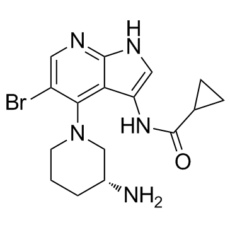
| SMILES |
C1C[C@H](CN(C1)C2=C3C(=CNC3=NC=C2Br)NC(=O)C4CC4)N
|
|
| InChi Key |
BAZRWWGASYWYGB-SNVBAGLBSA-N
|
|
| InChi Code |
InChI=1S/C16H20BrN5O/c17-11-6-19-15-13(14(11)22-5-1-2-10(18)8-22)12(7-20-15)21-16(23)9-3-4-9/h6-7,9-10H,1-5,8,18H2,(H,19,20)(H,21,23)/t10-/m1/s1
|
|
| 化学名 |
N-[4-[(3R)-3-aminopiperidin-1-yl]-5-bromo-1H-pyrrolo[2,3-b]pyridin-3-yl]cyclopropanecarboxamide
|
|
| 别名 |
|
|
| HS Tariff Code |
2934.99.9001
|
|
| 存储方式 |
Powder -20°C 3 years 4°C 2 years In solvent -80°C 6 months -20°C 1 month |
|
| 运输条件 |
Room temperature (This product is stable at ambient temperature for a few days during ordinary shipping and time spent in Customs)
|
| 溶解度 (体外实验) |
|
|||
|---|---|---|---|---|
| 溶解度 (体内实验) |
配方 1 中的溶解度: ≥ 2.5 mg/mL (6.61 mM) (饱和度未知) in 10% DMSO + 40% PEG300 + 5% Tween80 + 45% Saline (这些助溶剂从左到右依次添加,逐一添加), 澄清溶液。
例如,若需制备1 mL的工作液,可将100 μL 25.0 mg/mL澄清DMSO储备液加入到400 μL PEG300中,混匀;然后向上述溶液中加入50 μL Tween-80,混匀;加入450 μL生理盐水定容至1 mL。 *生理盐水的制备:将 0.9 g 氯化钠溶解在 100 mL ddH₂O中,得到澄清溶液。 配方 2 中的溶解度: ≥ 2.5 mg/mL (6.61 mM) (饱和度未知) in 10% DMSO + 90% (20% SBE-β-CD in Saline) (这些助溶剂从左到右依次添加,逐一添加), 澄清溶液。 例如,若需制备1 mL的工作液,可将 100 μL 25.0 mg/mL澄清DMSO储备液加入900 μL 20% SBE-β-CD生理盐水溶液中,混匀。 *20% SBE-β-CD 生理盐水溶液的制备(4°C,1 周):将 2 g SBE-β-CD 溶解于 10 mL 生理盐水中,得到澄清溶液。 请根据您的实验动物和给药方式选择适当的溶解配方/方案: 1、请先配制澄清的储备液(如:用DMSO配置50 或 100 mg/mL母液(储备液)); 2、取适量母液,按从左到右的顺序依次添加助溶剂,澄清后再加入下一助溶剂。以 下列配方为例说明 (注意此配方只用于说明,并不一定代表此产品 的实际溶解配方): 10% DMSO → 40% PEG300 → 5% Tween-80 → 45% ddH2O (或 saline); 假设最终工作液的体积为 1 mL, 浓度为5 mg/mL: 取 100 μL 50 mg/mL 的澄清 DMSO 储备液加到 400 μL PEG300 中,混合均匀/澄清;向上述体系中加入50 μL Tween-80,混合均匀/澄清;然后继续加入450 μL ddH2O (或 saline)定容至 1 mL; 3、溶剂前显示的百分比是指该溶剂在最终溶液/工作液中的体积所占比例; 4、 如产品在配制过程中出现沉淀/析出,可通过加热(≤50℃)或超声的方式助溶; 5、为保证最佳实验结果,工作液请现配现用! 6、如不确定怎么将母液配置成体内动物实验的工作液,请查看说明书或联系我们; 7、 以上所有助溶剂都可在 Invivochem.cn网站购买。 |
| 制备储备液 | 1 mg | 5 mg | 10 mg | |
| 1 mM | 2.6436 mL | 13.2181 mL | 26.4361 mL | |
| 5 mM | 0.5287 mL | 2.6436 mL | 5.2872 mL | |
| 10 mM | 0.2644 mL | 1.3218 mL | 2.6436 mL |
1、根据实验需要选择合适的溶剂配制储备液 (母液):对于大多数产品,InvivoChem推荐用DMSO配置母液 (比如:5、10、20mM或者10、20、50 mg/mL浓度),个别水溶性高的产品可直接溶于水。产品在DMSO 、水或其他溶剂中的具体溶解度详见上”溶解度 (体外)”部分;
2、如果您找不到您想要的溶解度信息,或者很难将产品溶解在溶液中,请联系我们;
3、建议使用下列计算器进行相关计算(摩尔浓度计算器、稀释计算器、分子量计算器、重组计算器等);
4、母液配好之后,将其分装到常规用量,并储存在-20°C或-80°C,尽量减少反复冻融循环。
计算结果:
工作液浓度: mg/mL;
DMSO母液配制方法: mg 药物溶于 μL DMSO溶液(母液浓度 mg/mL)。如该浓度超过该批次药物DMSO溶解度,请首先与我们联系。
体内配方配制方法:取 μL DMSO母液,加入 μL PEG300,混匀澄清后加入μL Tween 80,混匀澄清后加入 μL ddH2O,混匀澄清。
(1) 请确保溶液澄清之后,再加入下一种溶剂 (助溶剂) 。可利用涡旋、超声或水浴加热等方法助溶;
(2) 一定要按顺序加入溶剂 (助溶剂) 。
| NCT Number | Recruitment | interventions | Conditions | Sponsor/Collaborators | Start Date | Phases |
| NCT01564251 | Completed | Drug: GDC-0575 Drug: Gemcitabine |
Lymphoma, Solid Tumor | Genentech, Inc. | March 23, 2012 | Phase 1 |
|
InvivoChem的所有产品仅用于作科学研究,不面向患者销售
Copyright 2020 InvivoChem LLC | All Rights Reserved 粤ICP备20063088号-1


 COA
COA

 463611831
