| 规格 | 价格 | 库存 | 数量 |
|---|---|---|---|
| 10 mM * 1 mL in DMSO |
|
||
| 1mg |
|
||
| 2mg |
|
||
| 5mg |
|
||
| 10mg |
|
||
| 25mg |
|
||
| 50mg |
|
||
| 100mg |
|
||
| 250mg |
|
||
| Other Sizes |
|
| 靶点 |
1β-hydroxylase (CYP11B1) (IC50 = 35 nM)
|
|---|---|
| 体外研究 (In Vitro) |
皮质醇和醛固酮被奥西洛司他抑制(LCI699;0.01-10 μM;HAC15 细胞、17 种原代人肾上腺皮质细胞培养物、垂体腺瘤细胞)。奥西洛司他对肾上腺雄激素影响较小,并抑制皮质酮和 11-脱氧皮质醇的积聚 [2]。
体外酶抑制[1] LCI699/Osilodrostat剂量依赖性地抑制重组人醛固酮合酶的活性(IC50 = 0.7 nmol/L)的选择性是11β-羟化酶的3.6倍(IC50 = 2.5 nmol/L)(表1)。Lineweaver–Burk图(图1)显示,LCI699是重组人醛固酮合酶(Ki = 1.4 ± 0.2 nmol/L,平均值 ± SEM)和更高浓度的11β-羟化酶(Ki = 2.4 ± 0.3 nmol/L)。 使用大鼠重组酶的体外酶研究表明,LCI699抑制大鼠醛固酮合酶的效力比人酶低约230倍(表1)。然而,与重组人酶相比,LCI699对重组大鼠醛固酮合酶和11β-羟化酶具有相似的弱选择性(2.6倍的差异)。 在猴子肾上腺匀浆中,LCI699对醛固酮合酶的IC50比人重组酶高17倍,但比大鼠肾上腺匀浆中测量的IC50低67倍。在猴肾上腺匀浆中,醛固酮合酶的选择性是11β-羟化酶的5.2倍。 总之,LCI699/Osilodrostat抑制醛固酮合酶的相对物种排名顺序是人类 > 猴子 > 大鼠,而醛固酮合酶对11β-羟化酶的3至5倍选择性在这些物种中是相似的。 背景:美替卡松和酮康唑是治疗库欣综合征的常用类固醇生成抑制剂,可能与副作用和疗效有限有关。Osilodrostat是一种CYP11B1和CYP11B2抑制剂,对其他类固醇生成酶的影响尚不清楚。 目的:比较奥司他、美曲朋和酮康唑对体外肾上腺类固醇生成和垂体腺瘤细胞的影响。 方法:将HAC15细胞、17种原代人肾上腺皮质细胞培养物和垂体腺瘤细胞与Osilodrostat、Metrapone或酮康唑(0.01至10µM)一起孵育。皮质醇和促肾上腺皮质激素采用化学发光免疫测定法测定,类固醇谱采用液相色谱-质谱法测定。 结果:在HAC15细胞中,osilodrostat对皮质醇产生的抑制作用(IC50:0.035µM)明显强于美曲朋(0.068µM;P<0.0001)和酮康唑(0.621µM;P<0.001)。osilodrostat和metrapone对基础皮质醇产生的IC50值分别相差25倍和18倍,具有相当的效力。与美曲普酮和酮康唑相比,奥司他对醛固酮的抑制作用更强。Osilodrostat和metrapone治疗导致皮质酮和皮质醇的强烈抑制,11-脱氧皮质醇的积累,以及对肾上腺雄激素的适度影响。未观察到osilodrostat对垂体的直接影响。 结论:在我们的研究条件下,osilodrostat是人类肾上腺皮质细胞中一种强效的皮质醇生成抑制剂,与甲氧基吡咯烷酮相当。所有类固醇生成抑制剂在原代肾上腺皮质培养物之间的敏感性存在很大差异。Osilodrostat可能抑制CYP11B1和CYP11B2,在某些情况下抑制CYP17A1活性的程度较小,是类固醇生成的近端步骤。Osilodrostat是库欣综合征的一种有前景的治疗选择,与美曲普龙的体内差异可能是由药代动力学差异引起的[2]。 Osilodrostat、美曲普龙和酮康唑对体外基础和促肾上腺皮质激素刺激的皮质醇产生的影响[2] HAC15细胞系[2] 3天后,与美曲朋(0.0678µM;95%CI,0.0543至0.0848;P<0.0001)和酮康唑(0.621µM;95%CI,0.488至0.833;P<0.00001)相比,奥司他在显著较低的浓度下抑制了皮质醇的产生(IC50为0.0347µM;95%CI,0.0294至0.0410)(图1A)。在所有实验中,ACTH对皮质醇的平均刺激率为42%(±4%)(图1C)。对于osilodrostat,当HAC15细胞被ACTH刺激时,IC50值增加了1.7倍(与基础条件相比,P<0.0001),而甲氧基帕酮和酮康唑在ACTH刺激下的效力没有显著变化。比较三种化合物在ACTH刺激下的抑制作用,奥司洛司他抑制皮质醇产生的效果与美曲普酮一样强(IC50 0.0605µM;95%CI,0.0514至0.0714 vs IC50 0.0739µM;95%CI,0.0645至0.0847;P=0.0669),与酮康唑相比更有效(IC50 0.709µM;95%CI,0.523至0.962;P<0.0001)。添加类固醇生成抑制剂不会影响细胞数量。 原代肾上腺皮质培养[2] 在17种原代培养的人类肾上腺皮质组织中也评估了Osilodrostat、美曲普龙和酮康唑的影响:8种产生皮质醇的ACA,3种ACTH依赖性肾上腺增生,2种ACTH非依赖性肾上腺增殖,2种产生皮质醇和2种Conn综合征相关肾上腺增生。患者和组织特征如表1所示。表2列出了osilodrostat、metrapone和酮康唑对原代肾上腺皮质培养物中皮质醇产生的IC50值;剂量反应曲线如图2和(26)所示。在58个肾上腺培养板中的37个中进行了DNA测量,其中评估了化合物对皮质醇或醛固酮的剂量反应,结果显示任何药物对这些培养物中的细胞数量都没有影响。在原代肾上腺皮质培养物中,85pM ACTH诱导的皮质醇增加从48%到737%不等(表2)。 在未受刺激的原代ACA培养物中,Osilodrostat对皮质醇产生的IC50值差异为25倍(表2,图2A;0.0217,95%CI,0.0102至0.0461;0.534,95%CI,0.360至0.793),而美曲普酮的IC50值相差18倍,酮康唑的IC50值差别为84倍。与肾上腺增生的平均IC50(n=2;0.0269µM,95%CI,0.0210至0.0346;P<0.0001与ACA相比)相比,奥司罗司他在ACA中的平均IC50更高(n=7;0.104µM;95%CI,0.0716至0.151),尽管组数较小,但与ACC的IC50没有统计学上的显著差异。与ACC相比,osilodrostat在肾上腺增生中的平均IC50较低(P=0.0007)。在八种情况下(基础或促肾上腺皮质激素刺激),可以对osilodrostat和metrapone进行直接比较(表2)。与osilodrostat相比,Metyrapone在三种情况下更有效地抑制皮质醇的产生(P<0.05),而osilodrosta在ACTH依赖性肾上腺增生1号中更有效地控制皮质醇(P<0.0001)。在11种培养物中,有8种在基础条件下研究了Osilodrostat和酮康唑的疗效,与酮康唑相比,Osilodrosta对皮质醇的抑制作用更强(P<0.05至P<0.0001)。在促肾上腺皮质激素刺激下,与酮康唑相比,六种原代培养物中有两种对osilodrostat的IC50较低(P<0.01和P<0.001)。 在基础和ACTH模拟条件下比较疗效的三种培养物中,有两种培养物的疗效发生了变化,其中一种培养物在ACTH刺激条件下效力更高,另一种培养液效力更低(分别为P<0.01和P<0.05)。 Osilodrostat、美曲普龙和酮康唑对人肾上腺皮质细胞醛固酮生成的影响[2] 在血管紧张素II刺激的HAC15细胞中(图3D;醛固酮平均增加282%;P<0.0001),与美曲朋相比,Osilodrostat抑制醛固酮水平的浓度低10倍以上(图3A;IC50,0.0354μM;95%CI,0.0269至0.0465 vs 0.413μM;95%CI,0.306至0.557;P<0.00001)。未刺激的HAC15细胞中的醛固酮浓度太低,无法充分评估化合物的抑制作用。在导致Conn综合征的醛固酮生成性肾上腺增生中,Osilodrostat对醛固酮生成的抑制作用也比美曲普龙强得多(图3B;IC50,0.00281μM;95%CI,0.000910至0.00866 vs 0.822μM;95%CI,0.471至1.433;P<0.0001)。在第二例醛固酮生成性肾上腺增生中,在两种测试浓度(0.1和5μM,数据未显示)下,奥司罗司他和美曲朋对基础醛固酮浓度的抑制作用没有差异。在基础状态下的ACTH依赖性肾上腺增生1号中,奥司前列素对醛固酮的抑制作用明显强于美曲普酮(图3C;IC50,0.00469μM;95%CI,5.516E-5至0.398 vs 0.364μM;95%CI,0.05515至2.397;P<0.0001)和酮康唑(0.315μM;95%CI,0.05%至1.916;P<0.0011 vs奥司前列醇)。在这种原代培养中,与皮质醇抑制所需的浓度相比,osilodrostat在显著较低的浓度下抑制醛固酮的产生(IC50醛固酮为0.00469μM;95%CI为5.516E-5至0.398,而皮质醇为0.0311μM;95%CI为0.0242至0.0399;P=0.0164)。 |
| 体内研究 (In Vivo) |
在 Ang-II 和 ACTH 刺激的 Sprague Dawley 大鼠中,奥西洛司他(LCI699;0.1-100 mg/kg;口服;一次)抑制醛固酮和皮质酮的合成 [1]。 Osilodrostat(LCI699;3-100 mg/kg;口服;每天一次,持续 52 周)通过降低平均动脉压来延长 dTG 大鼠的生存期 [1]。
背景:醛固酮合酶的抑制有可能减弱醛固酮的盐皮质激素受体依赖性和非依赖性作用。重组人酶的体外研究表明,LCI699是一种强效、可逆、竞争性的醛固酮合酶抑制剂(人体内K i=1.4±0.2 nmol/L),对11β-羟化酶具有相对选择性。 方法:在促肾上腺皮质激素(ACTH)和血管紧张素II刺激醛固酮释放的大鼠和猴子体内模型中,研究了口服Osilodrostat/LC699的激素作用,并在一项随机、安慰剂对照研究中与盐皮质激素受体拮抗剂依普利酮进行了比较。在过表达人肾素和血管紧张素原的双转基因大鼠(dTG大鼠)模型中,研究了LCI699和依普利酮对醛固酮过量所致心脏和肾脏后遗症的影响。 结果:刺激醛固酮释放的大鼠和猴子体内模型预测了人类剂量和暴露反应关系,但高估了Osilodrostat在人类中的选择性。在dTG大鼠模型中,LCI699剂量依赖性地阻断醛固酮的增加,防止独立于血压变化的心肾功能异常的发展,并延长生存期。依普利酮在相似程度上延长了生存期,但在预防心脏和肾脏损伤方面效果较差。在健康人类受试者中,LCI699 0.5 mg选择性降低血浆和24小时尿醛固酮分别为49±3%和39±6%(第1天,平均值±SEM;与安慰剂相比P<0.001),这与钠尿症和血浆肾素活性增加有关。大于1mg的LCI699剂量抑制了基础皮质醇和ACTH刺激的皮质醇。100 mg依普利酮可增加血浆和24小时尿醛固酮,同时刺激钠分泌和增加肾素活性。与依普利酮相比,LCI699增加了醛固酮前体11-脱氧皮质酮和尿钾排泄。 结论:这些结果为实验模型中抑制醛固酮合酶的心脏和肾脏作用以及激素作用对人类的转化提供了新的见解。选择性抑制醛固酮合酶似乎是治疗醛固酮过量相关疾病的一种有前景的方法。[1] 生长抑素类似物帕瑞肽和11β-羟化酶抑制剂Osilodrostat(LCI699)通过不同的作用机制降低皮质醇水平。研究这两种药物联合使用的临床疗效是有科学依据的。这份手稿报告了一项大鼠毒理学研究的结果,评估了不同剂量的单独和联合使用的osilodrostat和pasireotide。将60只雄性和60只雌性大鼠随机分为单性别组,接受每日剂量的帕瑞肽(0.3mg/kg/天,皮下注射)、奥司罗他(20mg/kg/天,口服)、奥司罗他/帕瑞肽组合(低剂量,1.5/0.03mg/kg/天;中剂量,5/0.1mg/kg/天;或高剂量,20/0.3mg/kg/日)或赋形剂治疗13周。与对照组相比,帕瑞肽单独治疗组和联合治疗组从基线到第13周的平均体重增加明显较低,而接受奥司他单药治疗的雌性大鼠的平均体重增长明显较高。Osilodrostat和帕瑞肽单药治疗与垂体和肾上腺、肝脏和卵巢/输卵管的组织学和平均重量的显著变化有关。单独使用Osilodrostat与肾上腺皮质肥大和肝细胞肥大有关。联合使用,奥司他/帕瑞肽不会加剧任何靶器官的变化,并改善了单一疗法观察到的肝脏和肾上腺的变化。奥司他和帕瑞肽的Cmax和AUC0-24h以近似剂量成比例的方式增加。总之,与单一疗法相比,帕瑞肽和奥司他联合用药不会加剧靶器官重量或毒性的变化,并且具有可接受的安全性;在osilodrostat方案中加入帕瑞肽可能会减轻潜在的肾上腺过度活化和肝细胞肥大,这是osilodrosat单一疗法的潜在副作用[3]。 |
| 酶活实验 |
体外酶抑制[1]
实验设计[1] 细胞系和组织样本[1] 重组人细胞色素P450(CYP)11B2和CYP11B1酶分别由细胞系V79-4 CYP11B2肾上腺毒素-肾上腺毒素还原酶(AAR)#317和V79-4 CYP11B1-AAR#618制备。类似地制备重组大鼠CYP11B2和CYP11B1酶。所有细胞系均保存在添加了10%胎牛血清、0.5倍抗生素、800μg/mL遗传霉素和250μg/mL潮霉素的Dulbecco改良Eagle培养基中。 如前所述,从雄性Sprague-Dawley(S-D)大鼠的肾上腺制备大鼠肾上腺匀浆。从雌性食蟹猴的肾上腺制备猴CYB11B2和CYB11B1匀浆。将猴肾上腺组织在玻璃组织研磨机中在冰上切碎并均质化,每100mg组织加入1mL冰冷的均质缓冲液(每50mL缓冲液加入2.7mmol/L CaCl2和一片无乙二胺四乙酸(EDTA)的蛋白酶抑制剂片)。将均质材料在450℃下离心 g在4°C下放置5分钟,使上清液的最终甘油浓度达到5%,在液氮中快速冷冻,并在-80°C下储存直至分析。使用96孔板测定法定量醛固酮、皮质醇和皮质酮浓度(见附加文件1)。 CYP11B2和CYP11B1酶测定[1] 人CYP11B2和CYP11B1测定如前所述。使用11-DOC作为底物,对大鼠和猴子进行了类似的检测。 |
| 细胞实验 |
根据制造商的说明,将Osilodrostat、Metrapon和酮康唑的储备溶液分别溶解在0.01N盐酸、蒸馏水和无水乙醇中,并在-20°C下以10-2M的储备浓度储存。在每次实验开始时,将Osilodrostat、Metrapone和酮康唑在溶解的相同溶液中稀释至正确浓度。Synacten的储备浓度在4°C下储存,并在使用当天在培养基中稀释。血管紧张素II储备浓度储存在-20°C下,并在使用当天用蒸馏水稀释。使用的ACTH和血管紧张素II的浓度分别基于HAC15细胞中皮质醇和醛固酮产生的剂量反应曲线,并根据先前报道的研究。接种细胞一天后,开始孵育。对照细胞经过载体处理。对于HAC15,细胞以每孔100000个细胞的密度在0.5mL培养基中铺板。加入Osilodrostat、美曲普龙或酮康唑(0.01至5µM)3天,加入或不加入10 nM ACTH或100 nM血管紧张素II,分别评估对类固醇谱和醛固酮产生的影响。为了评估osilodrostat对小鼠促肾上腺皮质激素垂体细胞的影响,将AtT20细胞与osilodrosta一起孵育1、3和7天(0.01至10µM),以评估该药物在多种条件下(不同孵育时间和更高浓度)的潜在影响。对于7天的实验,培养基和化合物在3天后被刷新[2]。
原发性人类肾上腺和垂体腺瘤培养实验与HAC15和AtT20细胞的实验相似,但略有调整:ACTH的浓度为85 pM,血管紧张素II的浓度为10 nM,在细胞接种后3至4天开始治疗,然后进行培养基更新,在原发性促肾上腺皮质激素垂体腺瘤培养中,Osilodrostat的浓度仅为1µM。原代培养中使用的ACTH和血管紧张素II的浓度低于HAC15细胞中的浓度,因为原代培养对这些化合物的敏感性通常更高。由于从某些标本中获得的细胞数量有限,并非所有实验都可以在每个原代培养物中进行[2]。 |
| 动物实验 |
动物/疾病模型:雄性血管紧张素II (Ang-II) 和促肾上腺皮质激素 (ACTH) 刺激的Sprague Dawley大鼠[1]
剂量:0.1、0.3、1和3 mg/kg(Ang-II刺激的大鼠)和1、3、10、30和100 mg/kg(ACTH刺激的大鼠) 给药途径:口服;一次 实验结果:以剂量依赖的方式抑制Ang II或ACTH刺激引起的血浆醛固酮浓度升高。 动物/疾病模型:dTG大鼠[1] 剂量:3、10、30和100 mg/kg 给药途径:口服;每日一次,持续 52 周。 实验结果: 左心室(收缩期和舒张期)缩短分数增加,左心室等容舒张时间与 RR 间期比值 (IVRT/RR) 和心肌细胞体积正常化,左心室重量呈剂量依赖性增加。 LCI699/奥西洛司他制剂[1] LCI699/奥西洛司他溶液在每次实验前新鲜配制(由粉末配制)。在大鼠模型中,奥西洛司他/LCI699(游离碱)首先溶解于 1.5 倍摩尔当量的 1 N HCl 和 10 倍水的混合溶液中,然后用 3% 玉米淀粉溶液稀释(1 mL/kg 体积)。在猴模型中,LCI699(磷酸盐)溶解于水中(1 mL/kg 体积)。 LCI699 的给药途径为口服(大鼠和猴)或鼻胃管灌注(猴)。猴模型中化合物的剂量以游离碱当量表示。 大鼠模型实验方案[1] Ang-II 和 ACTH 刺激醛固酮合成的大鼠模型研究方案遵循已发表的方案。对于 Ang-II 输注模型,初始负荷剂量为 300 ng/kg 血管紧张素 II (Ang II),随后以 100 ng/kg/min 的速率静脉 (iv) 输注 9 小时。对于 ACTH 输注模型,ACTH 的负荷剂量和输注剂量分别为 100 ng/kg 和 30 ng/kg/min。在血管紧张素II (Ang II) 或促肾上腺皮质激素 (ACTH) 输注1小时后,采集血样,用于测定Ang II或ACTH输注后“基线”(即促分泌剂升高)血浆醛固酮和皮质酮浓度。在Ang II输注模型中,Osilodrostat/LCI699的给药剂量分别为0.1、0.3、1和3 mg/kg;在ACTH输注模型中,给药剂量分别为1、3、10、30和100 mg/kg。两种模型中,输注均持续8小时。分别于给药后15分钟、30分钟、1小时、2小时、3小时、4小时、5小时、6小时、7小时、8小时和24小时,从动脉插管中抽取含肝素(终浓度15 U/mL)的血样。采用放射免疫分析法测定血浆醛固酮和皮质醇,采用液相色谱-串联质谱法(LC-MS/MS)测定LCI699(见补充文件1)。 猴模型实验方案[1] 选取6只猴子(4.9–8.8 kg),随机分为两组,每组3只。导管/血管通路(VAP)手术后至少2周恢复期后方可开始实验。实验开始前30分钟,经皮将Huber针插入VAP,用于采集血样和注射ACTH。两次采样之间,用生理盐水冲洗导管/VAP,并用10 U/mL肝素保持通畅。在所有情况下,每周抽取的总血量不超过体重的1%,且连续实验之间至少间隔1周的恢复期。 在给药前0.5小时、0.25小时和给药前立即采集血样(0.3 mL,溶于15 U/mL肝素),用于基线药代动力学和药效学评估。给予Osilodrostat/LCI699(5、15、50或150 μg/kg)或赋形剂(水),3小时后静脉注射ACTH(1–24) 3000 ng/kg(0.1 mL/kg,约2分钟内注射完毕)。ACTH的3000 ng/kg剂量是根据一项预实验的剂量-反应结果确定的,该实验显示血浆醛固酮和皮质醇水平得到持续且最大的刺激。在注射ACTH后0.125、0.25、0.5、0.75和1小时采集血样,以评估血浆醛固酮和皮质醇刺激的时间进程。随后,在LCI699/载体给药后8小时、23.5小时和24小时再次采集血样。在8小时至23.5小时的两次采血之间,移除Huber针并将猴子放回笼中。在24小时最后一次采血后,移除所有仪器。采用放射免疫分析法测定血浆醛固酮和皮质醇水平,采用液相色谱-串联质谱法(LC-MS/MS)测定LCI699水平(见补充文件1)。将60只雄性和60只雌性大鼠随机分为单性别组,分别接受每日不同剂量的赋形剂、低剂量奥西洛司他/帕瑞肽、中剂量奥西洛司他/帕瑞肽、高剂量奥西洛司他/帕瑞肽、高剂量奥西洛司他单药治疗或高剂量帕瑞肽单药治疗(表1)。给药前,所有动物均进行称重,并按体重分层进行随机分组。大鼠每日接受其指定的治疗方案,持续13周;13周的治疗周期是根据欧洲药品管理局(EMA)的现行建议(ICH M3 [R2])选择的。三只雌性哨兵大鼠用于健康筛查程序。 使用塑料灌胃管口服给予奥西洛司他,随后,在适用情况下,于给予奥西洛司他后5分钟内,将帕瑞肽皮下注射至肩胛间区。除特定程序期间外,动物每天在大致相同的时间给药。选择口服和皮下注射途径分别作为奥西洛司他(osilodrostat)和帕瑞肽(pasireotide)的给药途径,因为这两种途径代表了在人体中的预期给药途径。 根据先前在大鼠中进行的单药治疗研究结果(奥西洛司他剂量范围:0.2–50 mg/kg/天,口服;帕瑞肽剂量范围:0.08–0.24 mg/kg/天,皮下注射;诺华制药公司,未发表数据),认为低、中、高剂量的奥西洛司他(1.5、5 和 20 mg/kg/天)和帕瑞肽(0.03、0.1 和 0.3 mg/kg/天)是合适的;预计这些剂量能够提供足够的暴露量,以达到治疗剂量下人体系统暴露量的倍数。在两项为期6个月的单药治疗研究中,奥西洛司他和帕瑞肽的剂量分别高达20 mg/kg/天和0.24 mg/kg/天,均耐受良好;奥西洛司他和帕瑞肽的未观察到不良反应剂量(NOAEL)分别为2 mg/kg/天和0.024 mg/kg/天。 化合物和制剂 [2] 奥西洛司他以超纯水配制,用于灌胃给药。帕瑞肽以醋酸盐缓冲溶液(pH 4.5)、醋酸和D-甘露醇配制于无菌水中,用于皮下注射。赋形剂对照组包括用于灌胃的超纯水和用于皮下注射的醋酸盐缓冲溶液(pH 4.5)、醋酸和D-甘露醇配制于无菌水中。口服灌胃给药(奥西洛司他和赋形剂)的剂量为 5 mL/kg,皮下注射给药(帕瑞肽和赋形剂)的剂量为 1 mL/kg。药物储存于 4 °C 并避光。奥西洛司他给药前,将给药溶液从冰箱取出,并在室温下搅拌至少 10 分钟。帕瑞肽给药前,将给药溶液从冰箱取出,并在室温下放置至少 30 分钟。 终点和评估[2] 本研究旨在确定每日单独或联合使用奥西洛司他和帕瑞肽对垂体-肾上腺轴的影响,并报告任何其他与治疗相关的毒性。同时,还测定了奥西洛司他和帕瑞肽的毒代动力学特征。 |
| 药代性质 (ADME/PK) |
吸收、分布和排泄
奥西洛司他口服吸收迅速,达峰时间(Tmax)约为1小时,且吸收基本完全。在标准剂量范围内,药物暴露量(即AUC和Cmax)的增加略高于剂量比例。奥西洛司他与食物同服对其药代动力学无显著影响。年龄和性别不影响其药代动力学,但亚裔患者的生物利用度和总暴露量较高(尽管无临床意义)。中重度肝功能损害患者接触奥西洛司他的风险更高——处方信息建议,中度肝功能损害(Child-Pugh B级)患者起始剂量为每日两次,每次1毫克;重度肝功能损害(Child-Pugh C级)患者起始剂量为每晚睡前1毫克。 口服放射性标记的奥西洛司他后,90.6%的放射性物质经尿液排出,仅有1.58%经粪便排出。仅有 5.2% 的给药剂量以原药形式经尿液排出,表明代谢后经尿液排泄是奥西洛司他的主要清除途径。 奥西洛司他的表观分布容积中位数为 100 L。 目前尚无奥西洛司他口服清除率的数据。 代谢/代谢物 奥西洛司他代谢广泛——口服给药后约 80% 的剂量以代谢物形式排出,这是药物清除的主要途径。血浆中最丰富的代谢物是 M35.4(双氧代奥西洛司他)、M16.5 和 M24.9,分别占给药剂量的 51%、9% 和 7%。代谢物 M34.5 和 M24.9 的半衰期比母体药物长,因此每日两次给药可能导致药物蓄积。在尿液中观察到的 13 种代谢物中,含量最高的是 M16.5(奥西洛司他葡萄糖醛酸苷)、M22(M34.5 的葡萄糖醛酸苷结合物)和 M24.9,分别占给药剂量的 17%、13% 和 11%。代谢物 M34.5 在尿液中的排泄量不到给药剂量的 1%,但其葡萄糖醛酸苷结合物 (M22) 约占 13%。奥西洛司他的生物转化由多种细胞色素 P450 (CYP) 和 UDP-葡萄糖醛酸转移酶 (UGT) 介导,但没有单一酶对总清除率的贡献超过 25%。在总清除率中,约 26% 由 CYP 介导,19% 由 UGT 介导,50% 由其他酶介导。奥西洛司他的主要代谢物 M34.5 的生成可能并非由 CYP 介导。其主要尿代谢物奥西洛司他葡萄糖醛酸苷 (M16.5) 的生成由 UGT1A4、UGT2B7 和 UGT2B10 催化。体外数据表明,这些代谢物均不影响奥西洛司他的治疗效果,但代谢物 M34.5 已被证实可抑制和/或诱导多种酶和转运蛋白。 生物半衰期 奥西洛司他的消除半衰期约为 4 小时。 |
| 毒性/毒理 (Toxicokinetics/TK) |
肝毒性
在预注册试验中,137 例接受 奥西洛司他 治疗库欣病的患者中有 37 例 (27%) 出现轻度、短暂的血清转氨酶升高,但仅有 8 例 (6%) 患者的转氨酶值超过正常值上限 (ULN) 的 3 倍,仅有 1 例超过 ULN 的 5 倍( 可能性评分:E(不太可能是临床上明显的肝损伤的原因))。 妊娠和哺乳期影响 ◉ 哺乳期用药概述 目前尚无关于哺乳期使用奥西洛司他的信息。由于哺乳婴儿可能出现严重不良反应,例如肾上腺功能不全,因此不建议在接受奥西洛司他治疗期间进行母乳喂养,并且应避免在末次给药后 1 周内进行母乳喂养。 ◉ 哺乳期影响婴儿 截至修订日期,未找到相关的已发表信息。 ◉ 对哺乳和母乳的影响 截至修订日期,未找到相关的已发表信息。 蛋白结合 奥西洛司他及其代谢物 M34.5 在血浆中的蛋白结合率均低于 40%。蛋白结合程度与药物浓度无关。奥西洛司他结合的具体血浆蛋白尚未明确。 毒代动力学[3] 在第 1 天,奥西洛司他剂量从 0.5 mg/kg/天增加到 20 mg/kg/天,并与帕瑞肽(剂量范围 0.03–0.3 mg/kg/天)联合使用,同时血浆最大浓度 (Cmax) 和 0 至 24 小时浓度-时间曲线下面积也呈近似剂量比例增加。表4显示了雄性和雌性大鼠中奥西洛司他(osilodrostat)的AUC0-24h。对于每个剂量组,多次给药(第77天)后奥西洛司他的AUC0-24h和Cmax与单次给药(第1天)后相似,且总体上雌雄之间也相似。 在第1天,将帕瑞肽剂量从0.03 mg/kg/天增加到0.3 mg/kg/天,并与奥西洛司他(剂量范围:0.5-20 mg/kg/天)联合使用,导致雄性和雌性大鼠中帕瑞肽的Cmax和AUC0-24h呈剂量比例增加(表4);与单次给药相比,多次给药后帕瑞肽的Cmax和AUC0-24h略有增加。在所有测试剂量下,帕瑞肽的暴露量均观察到轻微的性别差异(雄性/雌性AUC0-24h)。比例为 1.4–1.8)。未对单独给予奥西洛司他或帕瑞肽的大鼠进行毒代动力学参数评估。 |
| 参考文献 |
|
| 其他信息 |
奥西洛司他是一种11β-羟化酶(也称为CYP11B1)抑制剂,该酶催化内源性皮质醇生物合成的最后一步。它用于降低循环皮质醇水平,治疗库欣病,这是一种皮质醇水平长期且超生理水平升高的疾病。库欣病通常是由于垂体肿瘤引起的促肾上腺皮质激素(ACTH)分泌过多所致,而手术切除肿瘤通常是首选治疗方法。作为一种口服生物利用度高的药物,奥西洛司他为那些无法切除病因肿瘤或既往垂体手术未能治愈的患者提供了一种新的治疗选择。奥西洛司他由诺华公司生产,商品名为伊斯图里萨(Isturisa)。该药物曾进行过治疗实体瘤、高血压和心力衰竭的II期临床试验,但诺华公司于2013年1月终止了针对这些适应症的研发。奥西洛司他于2020年1月在欧盟获批用于治疗内源性库欣综合征(即库欣病),并于2020年3月获得美国FDA批准,获颁孤儿药资格,用于治疗同一适应症。
奥西洛司他是一种皮质醇合成抑制剂。奥西洛司他的作用机制是作为细胞色素P450 11B1抑制剂、细胞色素P450 1A2抑制剂、细胞色素P450 2C19抑制剂、细胞色素P450 2D6抑制剂、细胞色素P450 3A4抑制剂和细胞色素P450 3A5抑制剂。 奥西洛司他是一种皮质醇合成抑制剂,用于治疗标准疗法无法控制的库欣病。奥西洛司他治疗期间未发现血清转氨酶升高,也未发现临床上明显的肝损伤病例。 奥西洛司他是一种口服生物利用度高的抑制剂,可同时抑制类固醇11β-羟化酶(细胞色素P450 (CYP) 11B1)和醛固酮合成酶(CYP11B2;类固醇18-羟化酶),具有潜在的抗肾上腺活性,并可用于治疗库欣病 (CD)。给药后,奥西洛司他与CYP11B1(催化皮质醇由前体11-脱氧皮质醇合成的最后一步的酶)和CYP11B2(催化肾上腺中皮质酮和11-脱氧皮质酮合成醛固酮的酶)结合并抑制其活性。抑制 CYP11B1 可防止皮质醇过量产生,从而降低皮质醇水平并使其正常化。库欣氏病 (CD) 最常见的原因是分泌促肾上腺皮质激素 (ACTH) 的垂体肿瘤。 另见:磷酸奥西洛司他(活性成分)。 药物适应症 奥西洛司他适用于治疗不适合或无法通过垂体手术治愈的成年库欣氏病患者。 FDA 标签 伊斯图瑞沙适用于治疗成人内源性库欣氏综合征。 治疗肾上腺皮质功能亢进 作用机制 库欣氏综合征是一种内分泌疾病,由长期过量接触糖皮质激素引起,其症状可能包括皮肤和毛发变薄、体重增加、肌肉无力、骨质疏松以及一系列精神、心血管和免疫缺陷。库欣综合征最常见的诱发因素是外源性使用超生理剂量的糖皮质激素,例如鼻喷剂、乳膏和吸入剂中的糖皮质激素。库欣病——库欣综合征的另一种较少见的病因——通常是由于垂体腺瘤过度分泌促肾上腺皮质激素(ACTH)导致内源性皮质醇暴露增加所致。奥西洛司他是一种11β-羟化酶(CYP11B1)抑制剂,对醛固酮合成酶(CYP11B2)的抑制作用较弱。 CYP11B1酶负责催化皮质醇合成的最后一步——通过抑制该酶,奥西洛司他有助于使内源性皮质醇水平正常化,并缓解库欣病症状。 总之,我们发现,在药理浓度下,奥西洛司他能有效抑制人肾上腺皮质细胞体外皮质醇和醛固酮的分泌。我们发现,不同患者肾上腺组织对类固醇生成抑制剂的敏感性存在显著差异,这与药代动力学的差异一起,可能解释了接受同一种化合物治疗的患者之间临床观察到的差异。在本研究条件下,奥西洛司他和美替拉酮对类固醇谱的影响高度相似。奥西洛司他似乎能抑制CYP11B1和CYP11B2,在某些情况下,还能较轻地抑制CYP17A1裂解酶活性,并影响类固醇生成途径的近端步骤。体内奥西洛司他和美替拉酮的差异可能是药代动力学差异所致,而非对肾上腺皮质的药效学效应差异。这些数据表明,奥西洛司他有望成为库欣综合征患者的一种治疗选择。来自3期临床试验的更多信息将提供关于奥西洛司他疗效和安全性的重要数据。[2] 在大鼠中,帕瑞肽和奥西洛司他联合用药并未改变单独使用任一药物时观察到的毒性特征和血浆暴露量,而帕瑞肽加入奥西洛司他治疗方案中可能减轻潜在的肾上腺肥大和肝细胞肥大。因此,该研究表明帕瑞肽和奥西洛司他联合用药具有可接受的安全性。然而,在将帕瑞肽和奥西洛司他联合用药用于人体之前,还应考虑其他安全性问题,特别是对QT间期的影响。[3] |
| 分子式 |
C13H10FN3
|
|---|---|
| 分子量 |
227.24
|
| 精确质量 |
227.085
|
| 元素分析 |
C, 68.71; H, 4.44; F, 8.36; N, 18.49
|
| CAS号 |
928134-65-0
|
| 相关CAS号 |
Osilodrostat phosphate;1315449-72-9
|
| PubChem CID |
44139752
|
| 外观&性状 |
White to yellow solid powder
|
| 密度 |
1.3±0.1 g/cm3
|
| 沸点 |
433.8±45.0 °C at 760 mmHg
|
| 闪点 |
216.2±28.7 °C
|
| 蒸汽压 |
0.0±1.0 mmHg at 25°C
|
| 折射率 |
1.664
|
| LogP |
1.13
|
| tPSA |
41.61
|
| 氢键供体(HBD)数目 |
0
|
| 氢键受体(HBA)数目 |
3
|
| 可旋转键数目(RBC) |
1
|
| 重原子数目 |
17
|
| 分子复杂度/Complexity |
337
|
| 定义原子立体中心数目 |
1
|
| SMILES |
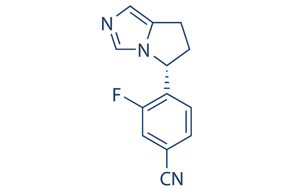
C1CC2=CN=CN2[C@H]1C3=C(C=C(C=C3)C#N)F
|
| InChi Key |
USUZGMWDZDXMDG-CYBMUJFWSA-N
|
| InChi Code |
InChI=1S/C13H10FN3/c14-12-5-9(6-15)1-3-11(12)13-4-2-10-7-16-8-17(10)13/h1,3,5,7-8,13H,2,4H2/t13-/m1/s1
|
| 化学名 |
4-[(5R)-6,7-dihydro-5H-pyrrolo[1,2-c]imidazol-5-yl]-3-fluorobenzonitrile
|
| 别名 |
Osilodrostat;LCI699; Isturisa; LCI 699; osilodrostat; (+)-Osilodrostat; LCI699-NX; LCI-699-NX; 5YL4IQ1078; UNII-5YL4IQ1078; ...; 928134-65-0; LCI-699
|
| HS Tariff Code |
2934.99.9001
|
| 存储方式 |
Powder -20°C 3 years 4°C 2 years In solvent -80°C 6 months -20°C 1 month |
| 运输条件 |
Room temperature (This product is stable at ambient temperature for a few days during ordinary shipping and time spent in Customs)
|
| 溶解度 (体外实验) |
|
|||
|---|---|---|---|---|
| 溶解度 (体内实验) |
配方 1 中的溶解度: ≥ 2.5 mg/mL (11.00 mM) (饱和度未知) in 10% DMSO + 40% PEG300 + 5% Tween80 + 45% Saline (这些助溶剂从左到右依次添加,逐一添加), 澄清溶液。
例如,若需制备1 mL的工作液,可将100 μL 25.0 mg/mL澄清DMSO储备液加入到400 μL PEG300中,混匀;然后向上述溶液中加入50 μL Tween-80,混匀;加入450 μL生理盐水定容至1 mL。 *生理盐水的制备:将 0.9 g 氯化钠溶解在 100 mL ddH₂O中,得到澄清溶液。 配方 2 中的溶解度: ≥ 2.5 mg/mL (11.00 mM) (饱和度未知) in 10% DMSO + 90% (20% SBE-β-CD in Saline) (这些助溶剂从左到右依次添加,逐一添加), 澄清溶液。 例如,若需制备1 mL的工作液,可将 100 μL 25.0 mg/mL澄清DMSO储备液加入900 μL 20% SBE-β-CD生理盐水溶液中,混匀。 *20% SBE-β-CD 生理盐水溶液的制备(4°C,1 周):将 2 g SBE-β-CD 溶解于 10 mL 生理盐水中,得到澄清溶液。 View More
配方 3 中的溶解度: ≥ 2.5 mg/mL (11.00 mM) (饱和度未知) in 10% DMSO + 90% Corn Oil (这些助溶剂从左到右依次添加,逐一添加), 澄清溶液。 配方 4 中的溶解度: ≥ 2.5 mg/mL (11.00 mM) (饱和度未知) in 10% EtOH + 40% PEG300 + 5% Tween80 + 45% Saline (这些助溶剂从左到右依次添加,逐一添加), 澄清溶液。 例如,若需制备1 mL的工作液,可将100 μL 25.0 mg/mL 澄清 EtOH 储备液加入400 μL PEG300 中,混匀;再向上述溶液中加入50 μL Tween-80,混匀;然后加入450 μL 生理盐水定容至1 mL。 *生理盐水的制备:将 0.9 g 氯化钠溶解在 100 mL ddH₂O中,得到澄清溶液。 配方 5 中的溶解度: ≥ 2.5 mg/mL (11.00 mM) (饱和度未知) in 10% EtOH + 90% (20% SBE-β-CD in Saline) (这些助溶剂从左到右依次添加,逐一添加), 澄清溶液。 例如,若需制备1 mL的工作液,可将100μL 25.0mg/mL澄清EtOH储备液加入到900μL 20%SBE-β-CD生理盐水中,混匀。 *20% SBE-β-CD 生理盐水溶液的制备(4°C,1 周):将 2 g SBE-β-CD 溶解于 10 mL 生理盐水中,得到澄清溶液。 配方 6 中的溶解度: ≥ 2.5 mg/mL (11.00 mM) (饱和度未知) in 10% EtOH + 90% Corn Oil (这些助溶剂从左到右依次添加,逐一添加), 澄清溶液。 例如,若需制备1 mL的工作液,可将 100 μL 25.0 mg/mL 澄清乙醇储备液加入到 900 μL 玉米油中并混合均匀。 1、请先配制澄清的储备液(如:用DMSO配置50 或 100 mg/mL母液(储备液)); 2、取适量母液,按从左到右的顺序依次添加助溶剂,澄清后再加入下一助溶剂。以 下列配方为例说明 (注意此配方只用于说明,并不一定代表此产品 的实际溶解配方): 10% DMSO → 40% PEG300 → 5% Tween-80 → 45% ddH2O (或 saline); 假设最终工作液的体积为 1 mL, 浓度为5 mg/mL: 取 100 μL 50 mg/mL 的澄清 DMSO 储备液加到 400 μL PEG300 中,混合均匀/澄清;向上述体系中加入50 μL Tween-80,混合均匀/澄清;然后继续加入450 μL ddH2O (或 saline)定容至 1 mL; 3、溶剂前显示的百分比是指该溶剂在最终溶液/工作液中的体积所占比例; 4、 如产品在配制过程中出现沉淀/析出,可通过加热(≤50℃)或超声的方式助溶; 5、为保证最佳实验结果,工作液请现配现用! 6、如不确定怎么将母液配置成体内动物实验的工作液,请查看说明书或联系我们; 7、 以上所有助溶剂都可在 Invivochem.cn网站购买。 |
| 制备储备液 | 1 mg | 5 mg | 10 mg | |
| 1 mM | 4.4006 mL | 22.0032 mL | 44.0063 mL | |
| 5 mM | 0.8801 mL | 4.4006 mL | 8.8013 mL | |
| 10 mM | 0.4401 mL | 2.2003 mL | 4.4006 mL |
1、根据实验需要选择合适的溶剂配制储备液 (母液):对于大多数产品,InvivoChem推荐用DMSO配置母液 (比如:5、10、20mM或者10、20、50 mg/mL浓度),个别水溶性高的产品可直接溶于水。产品在DMSO 、水或其他溶剂中的具体溶解度详见上”溶解度 (体外)”部分;
2、如果您找不到您想要的溶解度信息,或者很难将产品溶解在溶液中,请联系我们;
3、建议使用下列计算器进行相关计算(摩尔浓度计算器、稀释计算器、分子量计算器、重组计算器等);
4、母液配好之后,将其分装到常规用量,并储存在-20°C或-80°C,尽量减少反复冻融循环。
计算结果:
工作液浓度: mg/mL;
DMSO母液配制方法: mg 药物溶于 μL DMSO溶液(母液浓度 mg/mL)。如该浓度超过该批次药物DMSO溶解度,请首先与我们联系。
体内配方配制方法:取 μL DMSO母液,加入 μL PEG300,混匀澄清后加入μL Tween 80,混匀澄清后加入 μL ddH2O,混匀澄清。
(1) 请确保溶液澄清之后,再加入下一种溶剂 (助溶剂) 。可利用涡旋、超声或水浴加热等方法助溶;
(2) 一定要按顺序加入溶剂 (助溶剂) 。
InvivoChem的所有产品仅用于作科学研究,不面向患者销售
Copyright 2020 InvivoChem LLC | All Rights Reserved 粤ICP备20063088号-1


 COA
COA
 463611831
