| 规格 | 价格 | 库存 | 数量 |
|---|---|---|---|
| 10 mM * 1 mL in DMSO |
|
||
| 1mg |
|
||
| 2mg |
|
||
| 5mg |
|
||
| 50mg |
|
||
| 100mg |
|
||
| 250mg |
|
||
| 500mg |
|
||
| 1g |
|
||
| 2g |
|
||
| Other Sizes |
|

| 靶点 |
VEGFR1 (IC50 = 22 nM); VEGFR2 (IC50 = 4 nM); VEGFR3 (IC50 = 5.2 nM); FGFR1 (IC50 = 46 nM); PDGFRα (IC50 = 51 nM); PDGFRβ (IC50 = 39 nM); c-Kit (IC50 = 100 nM); FGFR2; FGFR3; FGFR4; RET
Vascular Endothelial Growth Factor Receptor (VEGFR) 1/2/3, Fibroblast Growth Factor Receptor (FGFR) 1/2/3/4, Platelet-Derived Growth Factor Receptor β (PDGFRβ), and Ret Proto-Oncogene (RET), tyrosine kinases involved in angiogenesis and cell proliferation. For Lenvatinib (E7080; ER-203492-00), literature [3] reported: VEGFR2 (IC50 = 4.0 nM), VEGFR3 (IC50 = 5.2 nM) via HTRF kinase assay [3] - Literature [4] supplemented: FGFR1 (IC50 = 46 nM), FGFR2 (IC50 = 51 nM), PDGFRβ (IC50 = 57 nM) via radioactive kinase assay [4] - Literature [2] confirmed: RET (IC50 = 21 nM), VEGFR1 (IC50 = 7.0 nM), FGFR3 (IC50 = 48 nM), FGFR4 (IC50 = 66 nM) via HTRF assay; no significant inhibition of EGFR (IC50 > 1 μM) [2] |
|---|---|
| 体外研究 (In Vitro) |
体外活性:E7080作为体外血管生成的有效抑制剂,对VEGF/KDR和SCF/Kit信号传导显示出显着的抑制作用。根据体外受体酪氨酸和丝氨酸/苏氨酸激酶测定,E7080 抑制 Flt-1、KDR、Flt-4,IC50 分别为 22、4.0 和 5.2 nM。除了这些激酶外,E7080 还抑制 FGFR1 和 PDGFR 酪氨酸激酶,对 FGFR1、PDGFRα 和 PDGFRβ 的 IC50 值分别为 46、51 和 100 nM。 E7080 有效抑制分别受 VEGF 和 VEGF-C 刺激的 HUVEC 中 VEGFR2(IC50,0.83 nM)和 VEGFR3(IC50,0.36 nM)的磷酸化。最近的一项研究表明,E7080 处理(1 μM 和 10 μM)可通过抑制 FGFR 和 PDGFR 信号传导显着抑制细胞迁移和侵袭。激酶测定:酪氨酸激酶测定通过 HTRF(KDR、VEGFR1、FGFR1、c-Met、EGFR)和 ELISA (PDGFRβ) 使用受体的重组激酶结构域进行。在这两种测定中,将 4 μL E7080 的连续稀释液与 10 μL 酶、16 μL 聚 (GT) 溶液 (250 ng) 和 10 μL ATP 溶液 (1 μM ATP) 混合在 96 孔圆板中( DMSO 的最终浓度为 0.1%)。在空白孔中,不添加酶。在对照孔中不添加测试物品。通过向每个孔中添加 ATP 溶液来启动激酶反应。 30°C 孵育 30 分钟后,通过向每孔的反应混合物中添加 0.5 M EDTA(10 μL/孔)来终止反应。将适合每种激酶测定的稀释缓冲液添加到反应混合物中。在 HTRF 测定中,将 50 μL 反应混合物转移至 96 孔 1/2 面积黑色 EIA/RIA 板,将 HTRF 溶液(50 μL/孔)添加到反应混合物中,然后通过以下方法测定激酶活性:使用时间分辨荧光检测器在 337 nm 激发波长和 620 和 665 nm 发射波长下测量荧光。在 ELISA 中,将 50 μL 反应混合物在亲和素包被的 96 孔聚苯乙烯板中室温孵育 30 分钟。用洗涤缓冲液洗涤后,加入PY20-HRP溶液(70μL/孔),并将反应混合物在室温下孵育30分钟。用洗涤缓冲液洗涤后,将TMB试剂(100μL/孔)添加到每个孔中。几分钟(10-30 分钟)后,向每个孔中添加 1 M H3PO4(100 μL/孔)。通过使用酶标仪测量 450 nm 处的吸光度来确定激酶活性。细胞测定:将 HUVEC(每孔 1,000 个细胞,在含有 2% 胎牛血清的无血清培养基中)和 L6 大鼠骨骼肌成肌细胞(每孔 5,000 个细胞,在无血清 DMEM 中)分配到 96 孔板中并孵育过夜。将 E7080 和含有 2% 胎牛血清的 VEGF (20 ng/mL) 或 FGF-2 (20 ng/mL) 以及 PDGFβ (40 ng/mL) 添加到每个孔中。将细胞孵育3天,然后用WST-1试剂测量存活细胞的比例。对于增殖测定,复制样品并进行三个单独的实验。
E7080的激酶抑制谱。[4] E7080的激酶抑制谱通过无细胞激酶试验确定(表1)。E7080有效抑制VEGF-R3激酶活性(IC50, 5.2 nmol/L;表1;补充图S1)和VEGF-R2激酶活性(IC50, 4.0 nmol/L)在相似程度上(表1)。E7080也抑制VEGF-R1, FGF-R1和PDGF-Rβ激酶,但抑制活性约为4至10倍(表1)。E7080不能有效抑制EGFR激酶。E7080分别对VEGF和VEGF- c刺激后HUVECs中VEGF- r2 (IC50, 0.83 nmol/L)和VEGF- r3 (IC50, 0.36 nmol/L)的磷酸化有很强的抑制作用(表1;图1)。这些数据表明E7080是VEGF-R3激酶和VEGF-R2激酶的有效抑制剂。E7080对vegf诱导的HUVEC增殖的抑制活性(IC50, 2.7 nmol/L)强于碱性FGF诱导的HUVEC增殖(IC50, 410 nmol/L)和pdgf诱导的L细胞增殖(IC50, 340 nmol/L;表1).我们无法确定VEGF-C诱导细胞增殖的IC50值,因为在我们的实验中VEGF-C没有刺激细胞增殖。 E7080抑制人乳腺癌细胞诱导的血管生成和淋巴管生成。[4] MDA-MB-231细胞是一种来源于胸腔积液的人乳腺腺癌细胞(25)。接种到mfp中的MDA-MB-231细胞转移在区域淋巴结和远端肺中发生的频率很高(表2),而MDA-MB-435细胞仅在远端肺中发生转移(数据未显示)。条件培养基的ELISA检测表明,两种肿瘤细胞都表达了大量的VEGF,但只有MDA-MB-231产生了大量的VEGF- c(表3),两种细胞系都没有产生可检测到的VEGF- d。这些数据表明,在MDA-MB-231 m.f.p.异种移植模型中,VEGF/VEGF- r2和VEGF- c /VEGF- r3信号可能被激活,导致肿瘤转移到局部淋巴结和远处肺,而在MDA-MB-435 m.f.p.异种移植模型中,只有VEGF/VEGF- r2信号可能被激活,导致肿瘤转移到远处肺。为了确定VEGF/VEGF- r2和VEGF- c /VEGF- r3信号在转移中的作用,我们检测了抗VEGF抗体贝伐单抗(VEGF信号的选择性抑制剂)和E7080 (VEGF- r2和VEGF- r3激酶的双重抑制剂)在两种m.f.p.异种移植模型中对血管生成和淋巴管生成的影响。用抗cd31抗体和抗lyve -1抗体分别对肿瘤组织进行染色,评价血管生成和淋巴管生成的程度。 小细胞肺癌(SCLC)细胞:在H146(SCLC,分泌干细胞因子)细胞中,Lenvatinib(0.01 μM–10 μM)抑制增殖,MTT法(72小时)IC50=0.2 μM。0.5 μM处理24小时后,ELISA显示VEGF分泌减少70%;0.3 μM处理24小时后,HUVEC管腔形成被抑制85% [3] - 乳腺癌细胞:在MDA-MB-231(三阴性乳腺癌,TNBC)细胞中,Lenvatinib(0.05 μM–10 μM)抑制增殖,CCK-8法(72小时)IC50=0.3 μM。Western blot显示0.5 μM处理2小时后p-VEGFR2/p-VEGFR3减少90% [4] - 肝细胞癌(HCC)细胞:在HepG2(HCC)细胞中,Lenvatinib(0.1 μM–10 μM)抑制增殖,MTT法(72小时)IC50=0.4 μM;0.5 μM处理24小时后,qRT-PCR显示FGFR驱动的cyclin D1表达减少65% [2] |
| 体内研究 (In Vivo) |
当在 H146 异种移植模型中口服给药时,E7080 在 30 和 100 mg/kg 剂量下以剂量依赖性方式抑制 H146 肿瘤的生长,并在 100 mg/kg 剂量下导致肿瘤消退。此外,100 mg/kg 的 E7080 比抗 VEGF 抗体和伊马替尼治疗更能降低微血管密度。 E7080 显着抑制 MDA-MB-231 乳腺脂肪垫 (mfp) 模型中的局部肿瘤生长,RTV(第 8 天计算的肿瘤体积/第 1 天的肿瘤体积)为 0.81,并减少已建立的转移结节的血管生成和淋巴管生成。淋巴结中的 MDA-MB-231 肿瘤。
E7080、伊马替尼和VEGF中和抗体对H146异种移植物模型的疗效[4] 为了研究SCF/KIT信号在肿瘤血管生成中的作用,研究人员利用H146异种移植物模型,评估了同时抑制KDR和KIT激酶的E7080、选择性抑制VEGF信号的VEGF中和抗体和单独抑制KIT激酶的伊马替尼的作用。口服E7080在剂量为30和100 mg/kg (BID, QDx21)时抑制H146肿瘤生长,呈剂量依赖性,在剂量为100 mg/kg时引起肿瘤消退(图6a)。伊马替尼剂量为160 mg/kg (BID, QDx21)或抗vegf抗体剂量为300和500 μg /只小鼠(每周两次)均可明显减缓肿瘤生长,但未引起肿瘤消退(图6a)。抗cd31抗体的免疫组化分析(图6b)显示,E7080在100 mg/kg时比抗vegf抗体和伊马替尼治疗更能降低微血管密度(图6c)。E7080可能通过抑制KIT和VEGF受体信号传导而具有强大的抗血管生成活性,从而实现肿瘤消退。 在MDA-MB-231 m.f.p.异种移植模型中,E7080抑制区域淋巴结和远端肺转移。[4] 接下来,研究人员评估了E7080和贝伐单抗对MDA-MB-231转移到区域淋巴结和远端肺的影响。MDA-MB-231发生转移的时间为~ 7周。我们在接种43天后用抑制剂治疗荷瘤小鼠,并给药56天(图4)。E7080和贝伐单抗在m.f.p均显著抑制局部肿瘤生长,治疗结束时,RTVs分别为0.81±1.00 (E7080)、5.11±6.54(贝伐单抗)和17.4±13.1(载药组);P < 0.05;图4). E7080还能显著抑制肿瘤向区域淋巴结和远端肺转移(P < 0.05;表2)。E7080治疗后,10只小鼠中有0只发生淋巴结转移,10只小鼠中有0只发生肺转移,而12只小鼠中有9只发生淋巴结和肺转移。贝伐单抗似乎也降低了转移到淋巴结(10例中有6例)和肺(10例中有3例)的发生率,但这种降低仅在肺中显著(表2)。这些结果表明贝伐单抗不能抑制VEGF-C/VEGF-R3信号。 E7080降低MDA-MB-231肿瘤淋巴结转移结节的血管生成和淋巴管生成。[4] 研究人员观察到,E7080治疗原发性MDA-MB-231肿瘤的淋巴管生成和血管生成均显著减少(图3)。因此,我们评估了E7080在切除原发肿瘤后,对转移结节生长、血管生成和淋巴结转移结节内淋巴管生成的影响(图5A)。在接种后90天切除原发肿瘤(图5A),并在肿瘤切除后2周开始给予E7080,持续4周(图5C)。E7080似乎能抑制转移性结节的生长(对照:11.8±10.8;E7080: 0.6±0.3;图5B和C),但由于rtv在载药组中变化较大,因此没有统计学差异,尽管抗cd31和抗lyve -1抗体的免疫组织化学分析(图6)表明E7080处理显著降低了MVD(载药组:94.3±12.6;E7080: 20.3±2.9/mm2;图6A和C)和LVD(载体:24.7±13.3;E7080: 1.0±0.9/mm2;图6B和C)在淋巴结转移结节内。这些结果表明,E7080抑制MDA-MB-231异种移植瘤模型中淋巴结转移结节内的血管生成和淋巴管生成。 HCC III期临床试验:在954例不可切除HCC患者中,Lenvatinib(体重<60 kg者8 mg/天;≥60 kg者12 mg/天,口服,连续给药)在总生存期(OS)上非劣于索拉非尼(400 mg每日两次):中位OS分别为13.6个月(仑伐替尼) vs. 12.3个月(索拉非尼)。客观缓解率(ORR):24.1%(仑伐替尼) vs. 9.2%(索拉非尼) [1] - SCLC异种移植模型:6周龄雄性裸鼠接种H146细胞,用Lenvatinib 10 mg/kg(口服每日一次)处理28天。肿瘤体积较溶媒组减少80%;肿瘤重量减少75% [3] - 乳腺癌转移模型:7周龄雌性裸鼠建立MDA-MB-231淋巴结/肺转移模型后,用Lenvatinib 15 mg/kg(口服每日一次)处理35天。淋巴结转移较溶媒组减少70%,肺转移结节减少65% [4] |
| 酶活实验 |
受体的重组激酶结构域用于 HTRF(KDR、VEGFR1、FGFR1、c-Met、EGFR)和 ELISA (PDGFRβ) 酪氨酸激酶测定。在这两种测定中,将 10 μL 酶、16 μL 聚 (GT) 溶液 (250 ng) 和 10 μL ATP 溶液 (1 μM ATP) 与 4 μL Lenvatinib (E7080)连续稀释液在 96 孔圆板中混合(DMSO终浓度为0.1%)。空白孔中不添加酶。对照孔中没有添加测试物品。每孔添加 ATP 溶液以启动激酶反应。在 30°C 孵育 30 分钟后,向每个孔中的反应混合物中添加 0.5 M EDTA(10 μL/孔)来终止反应。反应混合物中添加了适合每种激酶测定的稀释缓冲液。 HTRF 测定包括将 50 μL 反应混合物转移至 96 孔 1/2 面积黑色 EIA/RIA 板,每孔添加 50 μL HTRF 溶液,并使用时间分辨荧光检测器测量反应混合物的荧光发射波长为 620 和 665 nm,激发波长为 337 nm。这允许测定激酶活性。对于 ELISA,将涂有抗生物素蛋白的 96 孔聚苯乙烯板与 50 μL 反应混合物在室温下孵育 30 分钟。用洗涤缓冲液洗涤后,将反应混合物在室温下孵育 30 分钟,然后添加 PY20-HRP 溶液(70 μL/孔)。用洗涤缓冲液洗涤后,在每个孔中添加 100 μL TMB 试剂。几分钟(10-30 分钟)后,每个孔接收 100 μL 1 M H3PO4。通过使用酶标仪测量 450 nm 处的吸光度,可以鉴定激酶活性。
体外激酶测定[3] 酪氨酸激酶检测采用HTRF (KDR, VEGFR1, FGFR1, c-Met, EGFR)和ELISA (PDGFRβ),使用受体的重组激酶结构域。在两个实验中,将4 μL Lenvatinib (E7080)系列稀释剂与10 μL酶、16 μL聚(GT)溶液(250 ng)和10 μL ATP溶液(1 μmol/L ATP)(终浓度DMSO为0.1%)混合在96孔圆板中。在空白的孔中,没有添加酶。在对照井中不添加试验品。激酶反应是通过在每个孔中加入ATP溶液来启动的。30℃孵育30 min后,每孔加入0.5 mol/L EDTA (10 μL/孔)停止反应。在反应混合物中加入适合每个激酶测定的稀释缓冲液。 在HTRF实验中,将反应混合物的50 μL转移到96孔1/2面积的黑色EIA/RIA板上,在反应混合物中加入50 μL/孔的HTRF溶液,用时间分辨荧光检测器在激发波长为337 nm,发射波长为620和665 nm处测量荧光,测定激酶活性。 ELISA中,将反应液取50 μL于亲和素包被的96孔聚苯乙烯板中室温孵育30 min,用洗涤缓冲液洗涤后,加入PY20-HRP溶液(70 μL/孔),室温孵育30 min,用洗涤缓冲液洗涤后,每孔加入TMB试剂(100 μL/孔)。10 ~ 30min后,每孔加入1 mol/L H3PO4 (100 μL/孔)。激酶活性用酶标仪测定450 nm处吸光度。 ProQinase公司检测了除KDR、VEGFR1、FGFR1、c-Met、EGFR和PDGFRβ外,Lenvatinib (E7080)的激酶抑制活性。 无细胞激酶试验/细胞磷酸化试验。[4] 酪氨酸激酶活性通过均匀时间分辨荧光法(VEGF-R2、VEGF-R1、成纤维细胞生长因子受体1 (FGF-R1)和表皮生长因子受体)和ELISA法(血小板衍生生长因子(PDGF)受体β)测定,利用这些受体的重组激酶结构域。使用ProQinase公司的技术平台检测Lenvatinib (E7080)对VEGF-R3的激酶抑制活性。对于无细胞激酶试验,重复样品并进行两到三个单独的实验。HUVECs在含0.5%胎牛血清的无血清培养基中培养24小时,细胞用Lenvatinib (E7080)处理,用VEGF (20 ng/mL)或VEGF- c (100 ng/mL)刺激10分钟,然后收集在裂解缓冲液中。为了检测VEGF-R2和磷酸化的VEGF-R2,电泳10 ~ 20 μg的细胞裂解物。为了检测VEGF-R3和磷酸化的VEGF-R3,用抗VEGF-R3免疫沉淀400 ~ 1000 μg的细胞裂解物。免疫复合物在60 μL样品缓冲液中溶解,电泳。通过Western blot分析分离的蛋白与指定抗体:VEGF-R2和磷酸化VEGF-R2, VEGF-R3和抗磷酸酪氨酸IgG。使用Image Master VDS-CL化学发光显示免疫反应条带。使用1D图像分析软件测量每个波段的强度。在细胞磷酸化实验中,我们分别做了三个独立的实验。 VEGFR2/3 HTRF激酶实验(文献[3]):将重组人VEGFR2(786–1356位氨基酸)或VEGFR3(803–1363位氨基酸)与生物素化肽底物(Ac-EAIYAAPFAKKK-NH2,20 μM)、Eu标记抗磷酸酪氨酸抗体及ATP(10 μM)共同孵育于激酶缓冲液(25 mM Tris-HCl pH 7.5、10 mM MgCl₂、1 mM DTT)中。加入系列稀释的Lenvatinib(0.001 nM–100 nM),30°C孵育60分钟。检测时间分辨荧光(激发光340 nm,发射光620 nm),计算IC50 [3] - FGFR/PDGFRβ放射性实验(文献[4]):重组FGFR1/2或PDGFRβ与[γ-³²P]-ATP(10 μM,3000 Ci/mmol)、肽底物(FGFR:KKKSPGEYVNIEFG,PDGFRβ:KEAELTVEEVRK,20 μM)共同孵育于缓冲液(25 mM Tris-HCl pH 7.5、10 mM MgCl₂、1 mM DTT)中。加入Lenvatinib(0.01 nM–100 nM),30°C孵育30分钟。用30% TCA终止反应,将沉淀的底物转移至P81滤膜,液体闪烁计数仪检测放射性 [4] |
| 细胞实验 |
H146(1.2×10³ 细胞/50 μL/孔)在 96 孔多孔板中用含有 0.5% BSA 的 SFM 培养。在 37°C 培养过夜后,添加含有 0.5% FBS 和各种 SCF 浓度的 SFM(150 μL/孔),可添加或不添加各种化合物浓度。 WST-1 用于测量 72 小时培养后存活细胞的比例。
流式细胞术分析[3] 先用胰蛋白酶分离细胞,离心后用PBS或1 μg的一抗(抗kit抗体)在4°C下孵育30分钟,然后用50 μL的抗pe偶联二抗在PBS中稀释1:50孵育。用流式细胞术分析染色细胞,用FACS Calibur仪器量化染色强度,结果显示为直方图。 增殖试验[3] 将含0.5% BSA的SFM中的H146 (1.2 × 10³个细胞/50 μL/孔)培养于96孔多孔板中。37°C培养过夜后,加入含有0.5% FBS和几种SCF浓度的SFM (150 μL/孔),添加或不添加几种浓度的化合物。培养72h后,用WST-1法测定存活细胞比例。 生长因子刺激增殖试验。将HUVECs(每孔1000个细胞在含2%胎牛血清的无血清培养基中)和L6大鼠骨骼肌成肌细胞(每孔5000个细胞在无血清的DMEM中)分配在96孔板中孵育过夜。每孔加入Lenvatinib (E7080)和VEGF (20 ng/mL)或FGF-2 (20 ng/mL),其中含有2%胎牛血清和PDGFβ (40 ng/mL)。细胞孵育3 d,用WST-1试剂测定细胞存活率。增殖实验,重复样品,分别做3次实验。 SCLC与HUVEC实验(文献[3]):H146细胞以5×10³个细胞/孔接种于96孔板,用Lenvatinib(0.01 μM–10 μM)处理72小时。MTT法检测活力;0.5 μM处理24小时后ELISA分析VEGF分泌。HUVECs接种于Matrigel进行管腔形成实验(0.3 μM,24小时) [3] - 乳腺癌细胞实验(文献[4]):MDA-MB-231细胞以5×10³个细胞/孔接种于96孔板,用Lenvatinib(0.05 μM–10 μM)处理72小时。CCK-8法检测活力;0.5 μM处理2小时后Western blot检测p-VEGFR2/p-VEGFR3 [4] - HCC细胞qRT-PCR实验(文献[2]):HepG2细胞以3×10⁵个细胞/孔接种于6孔板,用Lenvatinib(0.5 μM)处理24小时。提取总RNA,qRT-PCR定量cyclin D1 mRNA [2] |
| 动物实验 |
实验在洁净室条件下进行,饲养8-12周龄、体重20-25克的雌性BALB/c裸鼠。将6.5×10⁶个H146肿瘤细胞皮下植入小鼠侧腹。注射后12天为实验第1天,此时将小鼠随机分为治疗组(n=6或n=5)和对照组(n=12)。从第1天到第21天,分别口服乐伐替尼、STI571和VEGF中和抗体。乐伐替尼和STI571每日两次口服,VEGF中和抗体每周两次口服。这些药物分别悬浮于0.5%甲基纤维素溶液和生理盐水中。在指定日期,测量并计算肿瘤体积。相对肿瘤体积 (RTV) 是抗肿瘤活性的指标,计算方法为第 1 天的肿瘤体积除以指定日期的肿瘤体积。
肿瘤异种移植模型 [3] 本研究使用购自 Charles River 公司(日本神奈川县)的雌性 BALB/c 裸鼠(8-12 周龄,20-25 克)。动物饲养于洁净室条件下。将 H146 肿瘤细胞(6.5 × 10⁶)皮下注射到小鼠侧腹部。接种后12天,将小鼠随机分为对照组(n = 12)和治疗组(n = 6或n = 5),并将此时间点定义为第1天。将乐伐替尼(E7080)、伊马替尼和VEGF中和抗体分别悬浮于0.5%甲基纤维素和生理盐水中,从第1天到第21天,每天两次口服乐伐替尼和伊马替尼,每周两次口服VEGF中和抗体。在指定日期测量肿瘤体积,并根据以下公式计算:肿瘤体积(mm³)= 长度 × 宽度²/2。抗肿瘤活性以相对肿瘤体积(RTV = 指定日期计算的肿瘤体积/第1天的肿瘤体积)表示。对mfp异种移植模型中的血管生成和淋巴管生成进行免疫组织化学分析。 [4]从接受乐伐替尼(E7080)(n = 5)或贝伐珠单抗(n = 5)治疗1周(第8天)的小鼠以及未接受治疗的小鼠(n = 5)中取出MDA-MB-231和MDA-MB-435肿瘤,包埋于OCT化合物中,干冰冷冻,并进行双重染色,分别检测内皮细胞标志物CD31(使用大鼠单克隆抗小鼠CD31抗体,克隆号MEC13.3)和淋巴内皮细胞标志物(使用兔多克隆抗LYVE-1抗体)。CD31和LYVE-1分别用品红和3,3′-二氨基联苯胺染色显色。通过计数肿瘤微血管和淋巴管成分(每个肿瘤4-5个视野)并计算肿瘤微血管或淋巴管密度(即每个视野中的血管成分数量)来评估微血管密度(MVD)和淋巴管密度(LVD)。实验重复进行,并使用Dunnett型多重比较法进行统计分析。 Lenvatinib (E7080) 对mfp中原发肿瘤生长和转移的影响。[4] 将高表达rsGFP的MDA-MB-231细胞皮下植入裸鼠侧腹。从皮下培养的100至200 mm³肿瘤中制备肿瘤碎片(17 ± 2 mg),然后接种到小鼠体内。接种后约2周,于第1天将小鼠随机分为对照组(n = 12)和治疗组(n = 10)。从第1天到第56天,分别每日口服一次乐伐替尼(E7080)(溶于水)或每周静脉注射两次贝伐珠单抗(溶于生理盐水)。抗肿瘤活性以相对肿瘤体积(RTV = 计算肿瘤体积/第1天肿瘤体积)表示。治疗56天后,使用荧光成像检测系统检测淋巴结和肺中表达rsGFP的肿瘤。数据包括RTV的平均值±标准差以及携带转移结节的小鼠数量比例。实验重复进行,并采用Dunnett型多重比较法进行统计分析。 Lenvatinib (E7080)对原发肿瘤切除后淋巴结转移结节肿瘤生长的影响。[4] 将rsGFP MDA-MB-231肿瘤组织块移植到淋巴结,使其生长至出现转移(约90天),转移灶通过荧光成像检测系统进行检测,然后切除原发肿瘤。将8只小鼠分为两组。在切除原发肿瘤2周后(第1天)开始给予Lenvatinib (E7080)。 乐伐替尼 (E7080) 从第 1 天到第 28 天每天口服一次。统计分析采用 Dunnett 型多重比较法。 H146 小细胞肺癌异种移植方案(文献 [3]):将 5×10⁶ 个 H146 细胞皮下植入 6 周龄雄性裸鼠体内。当肿瘤体积达到约 100 mm³ 时,将乐伐替尼溶解于 0.5% 甲基纤维素 + 0.1% Tween 80 溶液中,每天口服一次(10 mg/kg),持续 28 天。每 3 天测量一次肿瘤体积(长×宽²/2);小鼠于第28天处死,称量肿瘤重量[3] - MDA-MB-231转移方案(文献[4]):将2×10⁶个MDA-MB-231细胞经尾静脉注射到7周龄雌性裸鼠体内以诱导转移。7天后,每日一次口服乐伐替尼(15 mg/kg,溶于0.5%羟丙基甲基纤维素),持续35天。收集淋巴结和肺组织以计数转移结节[4] - HCC III期方案(文献[1]):符合条件的不可切除HCC患者(ECOG PS 0-1)接受持续口服乐伐替尼(体重<60 kg者每日8 mg;体重≥60 kg者每日12 mg)或索拉非尼400 mg,每日两次。每6周通过影像学评估疗效;不良事件根据 CTCAE v4.0 进行分级 [1] |
| 药代性质 (ADME/PK) |
吸收、分布和排泄
给药后 1 至 4 小时达到血浆峰值浓度。与食物同服不影响吸收程度,但会降低吸收速率,并将中位达峰时间 (Tmax) 从 2 小时延迟至 4 小时。 服用放射性标记剂量后,约 64% 和 25% 的放射性标记物分别经粪便和尿液排出。 代谢/代谢物 乐伐替尼由 CYP3A 和醛氧化酶代谢。 生物半衰期 乐伐替尼的末端消除半衰期约为 28 小时。 人体药代动力学(文献 [2]):在治疗剂量(8–12 mg/天)下,患者的 Cmax = 32.5–50.3 ng/mL,Tmax = 1–4 小时,末端半衰期 (t₁/₂) = 27–35 小时,口服生物利用度 = 80%–90%。稳态血浆浓度在7-10天内达到[2] - 人血浆蛋白结合率:98%-99%(平衡透析[2]) - 代谢(文献[2]):在人肝微粒体中,乐伐替尼主要通过CYP3A4(60%)和醛氧化酶(30%)代谢;尿液中原形药物的排泄量<5%[2] |
| 毒性/毒理 (Toxicokinetics/TK) |
肝毒性
在乐伐替尼的大型临床试验中,血清转氨酶水平升高较为常见,发生率达52%。然而,仅有3%至5%的患者出现转氨酶水平超过正常值上限(ULN)5倍的情况。血清碱性磷酸酶升高也较为常见,发生率达28%,其中2%的患者碱性磷酸酶水平超过ULN 3倍。此外,在预注册临床试验中,1160例接受治疗的患者中有3例报告发生致命性肝衰竭,另有1例患者出现症状性但自限性急性肝炎伴黄疸。然而,这些事件与乐伐替尼治疗的相关性尚不明确。乐伐替尼的产品说明书中,血清ALT、AST和碱性磷酸酶升高被列为不良反应,而急性肝炎则被描述为罕见事件。建议在治疗前、治疗期间每 2 周监测一次血清酶,持续 2 个月,之后每月监测一次,并根据异常的程度和持续时间酌情减少剂量或停药。 可能性评分:D(可能导致临床上明显的肝损伤)。 妊娠和哺乳期影响 ◉ 哺乳期用药概述 目前尚无关于乐伐替尼在哺乳期临床应用的信息。由于乐伐替尼与血浆蛋白的结合率超过 98%,因此其在乳汁中的含量可能较低。然而,其半衰期约为 28 小时,因此可能在婴儿体内蓄积。制造商建议在乐伐替尼治疗期间以及末次给药后至少1周内停止母乳喂养。 ◉ 对母乳喂养婴儿的影响 截至修订日期,未找到相关的已发表信息。 ◉ 对泌乳和母乳的影响 截至修订日期,未找到相关的已发表信息。 蛋白结合 乐伐替尼与人血浆蛋白的体外结合率在98%至99%之间。 III期临床毒性(文献[1]):乐伐替尼最常见的治疗相关不良事件(TRAE):高血压(42.1%)、腹泻(39.5%)、疲乏(30.4%)、食欲下降(28.8%)。 3-4级治疗相关不良事件:高血压(16.9%)、蛋白尿(6.6%)、肝性脑病(3.7%)[1] - 体外细胞毒性:在正常人肝细胞(NHH)和外周血单核细胞(PBMC)中,乐伐替尼(浓度高达10 μM,作用72小时)的细胞活力>80%,表明其非特异性毒性较低[2] - 体内急性毒性:大鼠口服乐伐替尼10 mg/kg(28天)后出现轻度高血压(15%的动物),但未出现严重的肝肾损伤(ALT/AST/肌酐水平正常)[2] |
| 参考文献 |
|
| 其他信息 |
药效学
基于X射线晶体衍射和动力学相互作用研究,乐伐替尼通过环丙烷环与VEGFR2的腺苷5'-三磷酸结合位点及其邻近区域结合,从而抑制酪氨酸激酶活性及相关信号通路。 乐伐替尼(E7080;ER-203492-00)是一种多靶点酪氨酸激酶抑制剂,已获批用于治疗不可切除的肝细胞癌、分化型甲状腺癌和肾细胞癌(与依维莫司联合用药)[2] - 其作用机制包括与VEGFR、FGFR、PDGFRβ和RET的ATP结合口袋结合,抑制酪氨酸激酶活化和下游信号通路(ERK/AKT),从而抑制血管生成、肿瘤生长和转移[3][4] - 在III期REFLECT试验显示,在不可切除的肝细胞癌(HCC)患者中,其总生存期不劣于索拉非尼,且客观缓解率更高,支持其作为一线治疗方案[1] - 2018年获得FDA批准用于不可切除HCC的一线治疗;建议肝肾功能不全患者调整剂量[2] |
| 分子式 |
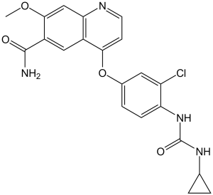
C21H19CLN4O4
|
|---|---|
| 分子量 |
426.85
|
| 精确质量 |
426.109
|
| 元素分析 |
C, 59.09; H, 4.49; Cl, 8.30; N, 13.13; O, 14.99
|
| CAS号 |
417716-92-8
|
| 相关CAS号 |
Lenvatinib mesylate;857890-39-2;Lenvatinib-d4;Lenvatinib-d5
|
| PubChem CID |
9823820
|
| 外观&性状 |
Off-white to light yellow solid powder
|
| 密度 |
1.5±0.1 g/cm3
|
| 沸点 |
627.2±55.0 °C at 760 mmHg
|
| 闪点 |
333.1±31.5 °C
|
| 蒸汽压 |
0.0±1.8 mmHg at 25°C
|
| 折射率 |
1.697
|
| LogP |
3.39
|
| tPSA |
115.57
|
| 氢键供体(HBD)数目 |
3
|
| 氢键受体(HBA)数目 |
5
|
| 可旋转键数目(RBC) |
6
|
| 重原子数目 |
30
|
| 分子复杂度/Complexity |
634
|
| 定义原子立体中心数目 |
0
|
| SMILES |
ClC1C([H])=C(C([H])=C([H])C=1N([H])C(N([H])C1([H])C([H])([H])C1([H])[H])=O)OC1C([H])=C([H])N=C2C([H])=C(C(C(N([H])[H])=O)=C([H])C=12)OC([H])([H])[H]
|
| InChi Key |
WOSKHXYHFSIKNG-UHFFFAOYSA-N
|
| InChi Code |
InChI=1S/C21H19ClN4O4/c1-29-19-10-17-13(9-14(19)20(23)27)18(6-7-24-17)30-12-4-5-16(15(22)8-12)26-21(28)25-11-2-3-11/h4-11H,2-3H2,1H3,(H2,23,27)(H2,25,26,28)
|
| 化学名 |
4-[3-chloro-4-(cyclopropylcarbamoylamino)phenoxy]-7-methoxyquinoline-6-carboxamide
|
| 别名 |
E-7080; E7080; E 7080; ER-203492-00; Lenvatinib; Brand name: Lenvima
|
| HS Tariff Code |
2934.99.9001
|
| 存储方式 |
Powder -20°C 3 years 4°C 2 years In solvent -80°C 6 months -20°C 1 month |
| 运输条件 |
Room temperature (This product is stable at ambient temperature for a few days during ordinary shipping and time spent in Customs)
|
| 溶解度 (体外实验) |
|
|||
|---|---|---|---|---|
| 溶解度 (体内实验) |
配方 1 中的溶解度: ≥ 0.64 mg/mL (1.50 mM) (饱和度未知) in 5% DMSO + 95% (20% SBE-β-CD in Saline) (这些助溶剂从左到右依次添加,逐一添加), 澄清溶液。
*20% SBE-β-CD 生理盐水溶液的制备(4°C,1 周):将 2 g SBE-β-CD 溶解于 10 mL 生理盐水中,得到澄清溶液。 配方 2 中的溶解度: 0.5% methylcellulose: 30 mg/kg View More
配方 3 中的溶解度: 6.67 mg/mL (15.63 mM) in 0.5% Methylcellulose/saline water (这些助溶剂从左到右依次添加,逐一添加), 悬浊液; 超声助溶。 1、请先配制澄清的储备液(如:用DMSO配置50 或 100 mg/mL母液(储备液)); 2、取适量母液,按从左到右的顺序依次添加助溶剂,澄清后再加入下一助溶剂。以 下列配方为例说明 (注意此配方只用于说明,并不一定代表此产品 的实际溶解配方): 10% DMSO → 40% PEG300 → 5% Tween-80 → 45% ddH2O (或 saline); 假设最终工作液的体积为 1 mL, 浓度为5 mg/mL: 取 100 μL 50 mg/mL 的澄清 DMSO 储备液加到 400 μL PEG300 中,混合均匀/澄清;向上述体系中加入50 μL Tween-80,混合均匀/澄清;然后继续加入450 μL ddH2O (或 saline)定容至 1 mL; 3、溶剂前显示的百分比是指该溶剂在最终溶液/工作液中的体积所占比例; 4、 如产品在配制过程中出现沉淀/析出,可通过加热(≤50℃)或超声的方式助溶; 5、为保证最佳实验结果,工作液请现配现用! 6、如不确定怎么将母液配置成体内动物实验的工作液,请查看说明书或联系我们; 7、 以上所有助溶剂都可在 Invivochem.cn网站购买。 |
| 制备储备液 | 1 mg | 5 mg | 10 mg | |
| 1 mM | 2.3427 mL | 11.7137 mL | 23.4274 mL | |
| 5 mM | 0.4685 mL | 2.3427 mL | 4.6855 mL | |
| 10 mM | 0.2343 mL | 1.1714 mL | 2.3427 mL |
1、根据实验需要选择合适的溶剂配制储备液 (母液):对于大多数产品,InvivoChem推荐用DMSO配置母液 (比如:5、10、20mM或者10、20、50 mg/mL浓度),个别水溶性高的产品可直接溶于水。产品在DMSO 、水或其他溶剂中的具体溶解度详见上”溶解度 (体外)”部分;
2、如果您找不到您想要的溶解度信息,或者很难将产品溶解在溶液中,请联系我们;
3、建议使用下列计算器进行相关计算(摩尔浓度计算器、稀释计算器、分子量计算器、重组计算器等);
4、母液配好之后,将其分装到常规用量,并储存在-20°C或-80°C,尽量减少反复冻融循环。
计算结果:
工作液浓度: mg/mL;
DMSO母液配制方法: mg 药物溶于 μL DMSO溶液(母液浓度 mg/mL)。如该浓度超过该批次药物DMSO溶解度,请首先与我们联系。
体内配方配制方法:取 μL DMSO母液,加入 μL PEG300,混匀澄清后加入μL Tween 80,混匀澄清后加入 μL ddH2O,混匀澄清。
(1) 请确保溶液澄清之后,再加入下一种溶剂 (助溶剂) 。可利用涡旋、超声或水浴加热等方法助溶;
(2) 一定要按顺序加入溶剂 (助溶剂) 。
Safety and Efficacy Study of Pembrolizumab (MK-3475) Combined With Lenvatinib (MK-7902/E7080) as First-line Intervention in Adults With Advance Melanoma (MK-7902-003/E7080-G000-312/LEAP-003)
CTID: NCT03820986
Phase: Phase 3 Status: Completed
Date: 2024-12-02
|
|
|
|
 VEGFR2-IN-7
 SYHA1813
 VEGFR-2-IN-38
 BHEP
BHEP
InvivoChem的所有产品仅用于作科学研究,不面向患者销售
Copyright 2020 InvivoChem LLC | All Rights Reserved 粤ICP备20063088号-1


 COA
COA


 463611831
