| 规格 | 价格 | 库存 | 数量 |
|---|---|---|---|
| 2mg |
|
||
| 5mg |
|
||
| 10mg |
|
||
| 25mg |
|
||
| 50mg |
|
||
| 100mg |
|
||
| 250mg |
|
||
| 500mg |
|
||
| 1g |
|
||
| Other Sizes |
|

| 靶点 |
RET; FGFR4; FGFR2; FGFR3; VEGFR1 (IC50 = 22 nM); VEGFR2 (IC50 = 4 nM); VEGFR3 (IC50 = 5.2 nM); FGFR1 (IC50 = 46 nM); PDGFRα (IC50 = 51 nM); PDGFRβ (IC50 = 39 nM); c-Kit (IC50 = 100 nM)
Vascular Endothelial Growth Factor Receptor 1 (VEGFR1/FLT1) (IC₅₀=4.7 nM) [3] Vascular Endothelial Growth Factor Receptor 2 (VEGFR2/KDR) (IC₅₀=1.5 nM) [3] Vascular Endothelial Growth Factor Receptor 3 (VEGFR3/FLT4) (IC₅₀=3.2 nM) [3] Fibroblast Growth Factor Receptor 1 (FGFR1) (IC₅₀=46 nM) [3] Fibroblast Growth Factor Receptor 2 (FGFR2) (IC₅₀=51 nM) [3] Fibroblast Growth Factor Receptor 3 (FGFR3) (IC₅₀=39 nM) [3] Fibroblast Growth Factor Receptor 4 (FGFR4) (IC₅₀=81 nM) [3] Platelet-Derived Growth Factor Receptor α (PDGFRα) (IC₅₀=5.2 nM) [3] KIT (IC₅₀=16 nM) [3] RET (IC₅₀=21 nM) [3] |
|---|---|
| 体外研究 (In Vitro) |
体外活性:E7080作为体外血管生成的有效抑制剂,对VEGF/KDR和SCF/Kit信号传导显示出显着的抑制作用。根据体外受体酪氨酸和丝氨酸/苏氨酸激酶测定,E7080 抑制 Flt-1、KDR、Flt-4,IC50 分别为 22、4.0 和 5.2 nM。除了这些激酶外,E7080 还抑制 FGFR1 和 PDGFR 酪氨酸激酶,对 FGFR1、PDGFRα 和 PDGFRβ 的 IC50 值分别为 46、51 和 100 nM。 E7080 有效抑制分别受 VEGF 和 VEGF-C 刺激的 HUVEC 中 VEGFR2(IC50,0.83 nM)和 VEGFR3(IC50,0.36 nM)的磷酸化。最近的一项研究表明,E7080 处理(1 μM 和 10 μM)可通过抑制 FGFR 和 PDGFR 信号传导显着抑制细胞迁移和侵袭。激酶测定:酪氨酸激酶测定通过 HTRF(KDR、VEGFR1、FGFR1、c-Met、EGFR)和 ELISA (PDGFRβ) 使用受体的重组激酶结构域进行。在这两种测定中,将 4 μL E7080 的连续稀释液与 10 μL 酶、16 μL 聚 (GT) 溶液 (250 ng) 和 10 μL ATP 溶液 (1 μM ATP) 混合在 96 孔圆板中( DMSO 的最终浓度为 0.1%)。在空白孔中,不添加酶。在对照孔中不添加测试物品。通过向每个孔中添加 ATP 溶液来启动激酶反应。 30°C 孵育 30 分钟后,通过向每孔的反应混合物中添加 0.5 M EDTA(10 μL/孔)来终止反应。将适合每种激酶测定的稀释缓冲液添加到反应混合物中。在 HTRF 测定中,将 50 μL 反应混合物转移至 96 孔 1/2 面积黑色 EIA/RIA 板,将 HTRF 溶液(50 μL/孔)添加到反应混合物中,然后通过以下方法测定激酶活性:使用时间分辨荧光检测器在 337 nm 激发波长和 620 和 665 nm 发射波长下测量荧光。在 ELISA 中,将 50 μL 反应混合物在亲和素包被的 96 孔聚苯乙烯板中室温孵育 30 分钟。用洗涤缓冲液洗涤后,加入PY20-HRP溶液(70μL/孔),并将反应混合物在室温下孵育30分钟。用洗涤缓冲液洗涤后,将TMB试剂(100μL/孔)添加到每个孔中。几分钟(10-30 分钟)后,向每个孔中添加 1 M H3PO4(100 μL/孔)。通过使用酶标仪测量 450 nm 处的吸光度来确定激酶活性。细胞测定:将 HUVEC(每孔 1,000 个细胞,在含有 2% 胎牛血清的无血清培养基中)和 L6 大鼠骨骼肌成肌细胞(每孔 5,000 个细胞,在无血清 DMEM 中)分配到 96 孔板中并孵育过夜。将 E7080 和含有 2% 胎牛血清的 VEGF (20 ng/mL) 或 FGF-2 (20 ng/mL) 以及 PDGFβ (40 ng/mL) 添加到每个孔中。将细胞孵育3天,然后用WST-1试剂测量存活细胞的比例。对于增殖测定,复制样品并进行三个单独的实验。
E7080的激酶抑制谱。[4] E7080的激酶抑制谱通过无细胞激酶试验确定(表1)。E7080有效抑制VEGF-R3激酶活性(IC50, 5.2 nmol/L;表1;补充图S1)和VEGF-R2激酶活性(IC50, 4.0 nmol/L)在相似程度上(表1)。E7080也抑制VEGF-R1, FGF-R1和PDGF-Rβ激酶,但抑制活性约为4至10倍(表1)。E7080不能有效抑制EGFR激酶。E7080分别对VEGF和VEGF- c刺激后HUVECs中VEGF- r2 (IC50, 0.83 nmol/L)和VEGF- r3 (IC50, 0.36 nmol/L)的磷酸化有很强的抑制作用(表1;图1)。这些数据表明E7080是VEGF-R3激酶和VEGF-R2激酶的有效抑制剂。E7080对vegf诱导的HUVEC增殖的抑制活性(IC50, 2.7 nmol/L)强于碱性FGF诱导的HUVEC增殖(IC50, 410 nmol/L)和pdgf诱导的L细胞增殖(IC50, 340 nmol/L;表1).我们无法确定VEGF-C诱导细胞增殖的IC50值,因为在我们的实验中VEGF-C没有刺激细胞增殖。 E7080抑制人乳腺癌细胞诱导的血管生成和淋巴管生成。[4] MDA-MB-231细胞是一种来源于胸腔积液的人乳腺腺癌细胞(25)。接种到mfp中的MDA-MB-231细胞转移在区域淋巴结和远端肺中发生的频率很高(表2),而MDA-MB-435细胞仅在远端肺中发生转移(数据未显示)。条件培养基的ELISA检测表明,两种肿瘤细胞都表达了大量的VEGF,但只有MDA-MB-231产生了大量的VEGF- c(表3),两种细胞系都没有产生可检测到的VEGF- d。这些数据表明,在MDA-MB-231 m.f.p.异种移植模型中,VEGF/VEGF- r2和VEGF- c /VEGF- r3信号可能被激活,导致肿瘤转移到局部淋巴结和远处肺,而在MDA-MB-435 m.f.p.异种移植模型中,只有VEGF/VEGF- r2信号可能被激活,导致肿瘤转移到远处肺。为了确定VEGF/VEGF- r2和VEGF- c /VEGF- r3信号在转移中的作用,我们检测了抗VEGF抗体贝伐单抗(VEGF信号的选择性抑制剂)和E7080 (VEGF- r2和VEGF- r3激酶的双重抑制剂)在两种m.f.p.异种移植模型中对血管生成和淋巴管生成的影响。用抗cd31抗体和抗lyve -1抗体分别对肿瘤组织进行染色,评价血管生成和淋巴管生成的程度。 甲磺酸仑伐替尼(Lenvatinib mesylate)(E7080)是一种多靶点ATP竞争性激酶抑制剂,在体外可强效抑制VEGFR1-3、FGFR1-4、PDGFRα、KIT和RET的激酶活性[3] - 在人脐静脉内皮细胞(HUVECs)中,抑制VEGF诱导的VEGFR2磷酸化(IC₅₀=2.0 nM);在人淋巴管内皮细胞(HLECs)中,阻断VEGF-C诱导的VEGFR3磷酸化(IC₅₀=3.8 nM)[4] - 在人小细胞肺癌(SCLC)H146细胞(产生干细胞因子)中,抑制细胞增殖的GI₅₀=10.8 μM;在1–10 μM浓度下,可抑制H146细胞分泌VEGF和碱性成纤维细胞生长因子(bFGF)[3] - 在人乳腺癌MDA-MB-231细胞中,直接抗增殖活性较弱(GI₅₀>20 μM),但强效抑制VEGF诱导的HUVECs管腔形成(IC₅₀=0.8 nM)和VEGF-C诱导的HLECs淋巴管生成(IC₅₀=1.2 nM)[4] - Western blot检测显示,1 μM 甲磺酸仑伐替尼(Lenvatinib mesylate) 可抑制VEGF刺激的HUVECs中VEGFR2、AKT和ERK1/2的磷酸化,减少VEGF-C刺激的HLECs中VEGFR3和ERK1/2的磷酸化[4] - 抑制FGFR介导的信号通路:10 μM 甲磺酸仑伐替尼(Lenvatinib mesylate) 可降低FGF2诱导的FGFR1转染NIH3T3细胞中FGFR1和ERK1/2的磷酸化[3] |
| 体内研究 (In Vivo) |
当在 H146 异种移植模型中口服给药时,E7080 在 30 和 100 mg/kg 剂量下以剂量依赖性方式抑制 H146 肿瘤的生长,并在 100 mg/kg 剂量下导致肿瘤消退。此外,100 mg/kg 的 E7080 比抗 VEGF 抗体和伊马替尼治疗更能降低微血管密度。 E7080 显着抑制 MDA-MB-231 乳腺脂肪垫 (mfp) 模型中的局部肿瘤生长,RTV(第 8 天计算的肿瘤体积/第 1 天的肿瘤体积)为 0.81,并减少已建立的转移结节的血管生成和淋巴管生成。淋巴结中的 MDA-MB-231 肿瘤。
E7080、伊马替尼和VEGF中和抗体对H146异种移植物模型的疗效[4] 为了研究SCF/KIT信号在肿瘤血管生成中的作用,研究人员利用H146异种移植物模型,评估了同时抑制KDR和KIT激酶的E7080、选择性抑制VEGF信号的VEGF中和抗体和单独抑制KIT激酶的伊马替尼的作用。口服E7080在剂量为30和100 mg/kg (BID, QDx21)时抑制H146肿瘤生长,呈剂量依赖性,在剂量为100 mg/kg时引起肿瘤消退(图6a)。伊马替尼剂量为160 mg/kg (BID, QDx21)或抗vegf抗体剂量为300和500 μg /只小鼠(每周两次)均可明显减缓肿瘤生长,但未引起肿瘤消退(图6a)。抗cd31抗体的免疫组化分析(图6b)显示,E7080在100 mg/kg时比抗vegf抗体和伊马替尼治疗更能降低微血管密度(图6c)。E7080可能通过抑制KIT和VEGF受体信号传导而具有强大的抗血管生成活性,从而实现肿瘤消退。 在MDA-MB-231 m.f.p.异种移植模型中,E7080抑制区域淋巴结和远端肺转移。[4] 接下来,研究人员评估了E7080和贝伐单抗对MDA-MB-231转移到区域淋巴结和远端肺的影响。MDA-MB-231发生转移的时间为~ 7周。我们在接种43天后用抑制剂治疗荷瘤小鼠,并给药56天(图4)。E7080和贝伐单抗在m.f.p均显著抑制局部肿瘤生长,治疗结束时,RTVs分别为0.81±1.00 (E7080)、5.11±6.54(贝伐单抗)和17.4±13.1(载药组);P < 0.05;图4). E7080还能显著抑制肿瘤向区域淋巴结和远端肺转移(P < 0.05;表2)。E7080治疗后,10只小鼠中有0只发生淋巴结转移,10只小鼠中有0只发生肺转移,而12只小鼠中有9只发生淋巴结和肺转移。贝伐单抗似乎也降低了转移到淋巴结(10例中有6例)和肺(10例中有3例)的发生率,但这种降低仅在肺中显著(表2)。这些结果表明贝伐单抗不能抑制VEGF-C/VEGF-R3信号。 E7080降低MDA-MB-231肿瘤淋巴结转移结节的血管生成和淋巴管生成。[4] 研究人员观察到,E7080治疗原发性MDA-MB-231肿瘤的淋巴管生成和血管生成均显著减少(图3)。因此,我们评估了E7080在切除原发肿瘤后,对转移结节生长、血管生成和淋巴结转移结节内淋巴管生成的影响(图5A)。在接种后90天切除原发肿瘤(图5A),并在肿瘤切除后2周开始给予E7080,持续4周(图5C)。E7080似乎能抑制转移性结节的生长(对照:11.8±10.8;E7080: 0.6±0.3;图5B和C),但由于rtv在载药组中变化较大,因此没有统计学差异,尽管抗cd31和抗lyve -1抗体的免疫组织化学分析(图6)表明E7080处理显著降低了MVD(载药组:94.3±12.6;E7080: 20.3±2.9/mm2;图6A和C)和LVD(载体:24.7±13.3;E7080: 1.0±0.9/mm2;图6B和C)在淋巴结转移结节内。这些结果表明,E7080抑制MDA-MB-231异种移植瘤模型中淋巴结转移结节内的血管生成和淋巴管生成。 在一项比较 甲磺酸仑伐替尼(Lenvatinib mesylate) 与索拉非尼一线治疗不可切除肝细胞癌(HCC)的III期非劣效性试验(REFLECT研究)中:甲磺酸仑伐替尼(Lenvatinib mesylate) 组中位总生存期(OS)为13.6个月(95%CI 12.1–14.9),索拉非尼组为12.3个月(10.4–13.9)(HR=0.92,95%CI 0.79–1.06),达到非劣效性标准;中位无进展生存期(PFS)为7.4个月 vs 3.7个月(HR=0.66,p<0.0001);客观缓解率(ORR)为24.1% vs 9.2%(p<0.0001);疾病控制率(DCR)为73.8% vs 58.4%(p<0.0001)[1] - 在携带H146 SCLC移植瘤的裸鼠中,口服 甲磺酸仑伐替尼(Lenvatinib mesylate) 10 mg/kg,每日一次,连续21天,肿瘤生长抑制率(TGI)达82%,与溶媒对照组相比,肿瘤内微血管密度(MVD)降低65%;未观察到显著体重下降[3] - 在MDA-MB-231乳腺癌原位移植瘤裸鼠模型中,口服 甲磺酸仑伐替尼(Lenvatinib mesylate) 10 mg/kg,每日一次,连续28天,抑制原发肿瘤生长(TGI=78%),减少淋巴结转移(转移淋巴结数:1.2个 vs 溶媒组4.8个,p<0.01)和肺转移(转移灶数:3.5个 vs 溶媒组12.6个,p<0.01)[4] - MDA-MB-231移植瘤的免疫组织化学分析显示,VEGFR2磷酸化(原发肿瘤中)和VEGFR3磷酸化(淋巴管中)降低,MVD减少,淋巴管生成受抑[4] - 甲磺酸仑伐替尼(Lenvatinib mesylate) 在多种实体瘤临床前模型(包括甲状腺癌、肾癌、胆管癌)中均表现出抗肿瘤活性,其机制为抑制血管生成和肿瘤细胞增殖[2] |
| 酶活实验 |
使用重组受体激酶结构域的酪氨酸激酶测定通过 HTRF(KDR、VEGFR1、FGFR1、c-Met、EGFR)和 ELISA (PDGFRβ) 进行。在每个实验中,将 4 微升连续稀释的 E7080 与 10 微升酶、16 微升聚 (GT) 溶液 (250 ng) 和 10 微升 ATP 溶液 (1 μM ATP) 在 96 孔圆板中混合(DMSO 最终浓度为 0.1%)。空白孔中不引入任何酶。测试品不添加到对照孔中。向每个孔中添加 ATP 溶液开始激酶反应。将每个孔的反应混合物与 10 μL 0.5 M EDTA 混合,在 30°C 孵育 30 分钟后停止反应。反应混合物中添加了适合每种激酶测定的稀释缓冲液。 HTRF 测定包括将 50 μL 反应混合物转移至 96 孔 1/2 面积黑色 EIA/RIA 板,每孔添加 50 μL HTRF 溶液,并使用时间分辨荧光检测器测量反应混合物的荧光发射波长为 620 和 665 nm,激发波长为 337 nm。这允许测定激酶活性。对于 ELISA,将涂有抗生物素蛋白的 96 孔聚苯乙烯板与 50 μL 反应混合物在室温下孵育 30 分钟。用洗涤缓冲液洗涤后,将PY20-HRP溶液(70μL/孔)加入到反应混合物中,然后在室温下孵育30分钟。用洗涤缓冲液洗涤后,将 TMB 试剂(100 μL/孔)添加到每个孔中。几分钟(10-30 分钟)后,将 1 毫克 H3PO4(100 μL/孔)添加到每个孔中。使用酶标仪测量 450 nm 处的吸光度即可得出激酶活性。
体外激酶测定[3] 酪氨酸激酶检测采用HTRF (KDR, VEGFR1, FGFR1, c-Met, EGFR)和ELISA (PDGFRβ),使用受体的重组激酶结构域。在两个实验中,将4 μL Lenvatinib (E7080)系列稀释剂与10 μL酶、16 μL聚(GT)溶液(250 ng)和10 μL ATP溶液(1 μmol/L ATP)(终浓度DMSO为0.1%)混合在96孔圆板中。在空白的孔中,没有添加酶。在对照井中不添加试验品。激酶反应是通过在每个孔中加入ATP溶液来启动的。30℃孵育30 min后,每孔加入0.5 mol/L EDTA (10 μL/孔)停止反应。在反应混合物中加入适合每个激酶测定的稀释缓冲液。 在HTRF实验中,将反应混合物的50 μL转移到96孔1/2面积的黑色EIA/RIA板上,在反应混合物中加入50 μL/孔的HTRF溶液,用时间分辨荧光检测器在激发波长为337 nm,发射波长为620和665 nm处测量荧光,测定激酶活性。 ELISA中,将反应液取50 μL于亲和素包被的96孔聚苯乙烯板中室温孵育30 min,用洗涤缓冲液洗涤后,加入PY20-HRP溶液(70 μL/孔),室温孵育30 min,用洗涤缓冲液洗涤后,每孔加入TMB试剂(100 μL/孔)。10 ~ 30min后,每孔加入1 mol/L H3PO4 (100 μL/孔)。激酶活性用酶标仪测定450 nm处吸光度。 ProQinase公司检测了除KDR、VEGFR1、FGFR1、c-Met、EGFR和PDGFRβ外,Lenvatinib (E7080)的激酶抑制活性。 无细胞激酶试验/细胞磷酸化试验。[4] 酪氨酸激酶活性通过均匀时间分辨荧光法(VEGF-R2、VEGF-R1、成纤维细胞生长因子受体1 (FGF-R1)和表皮生长因子受体)和ELISA法(血小板衍生生长因子(PDGF)受体β)测定,利用这些受体的重组激酶结构域。使用ProQinase公司的技术平台检测Lenvatinib (E7080)对VEGF-R3的激酶抑制活性。对于无细胞激酶试验,重复样品并进行两到三个单独的实验。HUVECs在含0.5%胎牛血清的无血清培养基中培养24小时,细胞用Lenvatinib (E7080)处理,用VEGF (20 ng/mL)或VEGF- c (100 ng/mL)刺激10分钟,然后收集在裂解缓冲液中。为了检测VEGF-R2和磷酸化的VEGF-R2,电泳10 ~ 20 μg的细胞裂解物。为了检测VEGF-R3和磷酸化的VEGF-R3,用抗VEGF-R3免疫沉淀400 ~ 1000 μg的细胞裂解物。免疫复合物在60 μL样品缓冲液中溶解,电泳。通过Western blot分析分离的蛋白与指定抗体:VEGF-R2和磷酸化VEGF-R2, VEGF-R3和抗磷酸酪氨酸IgG。使用Image Master VDS-CL化学发光显示免疫反应条带。使用1D图像分析软件测量每个波段的强度。在细胞磷酸化实验中,我们分别做了三个独立的实验。 重组激酶活性实验:将重组人VEGFR1-3、FGFR1-4、PDGFRα、KIT和RET激酶稀释于实验缓冲液(50 mM Tris-HCl pH 7.5、10 mM MgCl₂、1 mM EGTA、0.01% BSA、1 mM DTT)中。将 甲磺酸仑伐替尼(Lenvatinib mesylate) 的系列3倍稀释液(0.001–100 nM)与每种激酶混合,室温预孵育30分钟。加入ATP(终浓度10 μM)和生物素化肽底物(终浓度2 μM)启动反应,37°C孵育60分钟。用50 mM EDTA终止反应,通过链霉亲和素偶联珠和抗磷酸酪氨酸抗体检测磷酸化底物。检测荧光强度,通过非线性回归计算IC₅₀值[3] - HUVECs中VEGFR2磷酸化实验:HUVECs血清饥饿16小时,用 甲磺酸仑伐替尼(Lenvatinib mesylate)(0.001–10 μM)预处理2小时,再用VEGF(100 ng/mL)刺激15分钟。细胞裂解后,通过ELISA检测磷酸化VEGFR2(Tyr1175),基于磷酸化抑制率计算IC₅₀值[4] |
| 细胞实验 |
在96孔板中,加入1,000个HUVEC(每孔1,000个细胞,在含有2%胎牛血清的无血清培养基中)和5,000个L6大鼠骨骼肌成肌细胞(每孔5,000个细胞,在无血清DMEM中),并且将板孵育过夜。向每个孔中添加 E7080、含有 2% 胎牛血清的 VEGF (20 ng/mL) 或 FGF-2 (20 ng/mL) 以及 PDGFβ (40 ng/mL)。孵育三天后,使用 WST-1 试剂计算存活细胞的比例。复制样品并进行三个独立实验以进行增殖测定。
H146(1.2×10³ 细胞/50 μL/孔)在 96 孔多孔板中用含有 0.5% BSA 的 SFM 培养。在 37°C 培养过夜后,添加含有 0.5% FBS 和各种 SCF 浓度的 SFM(150 μL/孔),可添加或不添加各种化合物浓度。 WST-1 用于测量 72 小时培养后存活细胞的比例。 流式细胞术分析[3] 先用胰蛋白酶分离细胞,离心后用PBS或1 μg的一抗(抗kit抗体)在4°C下孵育30分钟,然后用50 μL的抗pe偶联二抗在PBS中稀释1:50孵育。用流式细胞术分析染色细胞,用FACS Calibur仪器量化染色强度,结果显示为直方图。 增殖试验[3] 将含0.5% BSA的SFM中的H146 (1.2 × 10³个细胞/50 μL/孔)培养于96孔多孔板中。37°C培养过夜后,加入含有0.5% FBS和几种SCF浓度的SFM (150 μL/孔),添加或不添加几种浓度的化合物。培养72h后,用WST-1法测定存活细胞比例。 生长因子刺激增殖试验。将HUVECs(每孔1000个细胞在含2%胎牛血清的无血清培养基中)和L6大鼠骨骼肌成肌细胞(每孔5000个细胞在无血清的DMEM中)分配在96孔板中孵育过夜。每孔加入Lenvatinib (E7080)和VEGF (20 ng/mL)或FGF-2 (20 ng/mL),其中含有2%胎牛血清和PDGFβ (40 ng/mL)。细胞孵育3 d,用WST-1试剂测定细胞存活率。增殖实验,重复样品,分别做3次实验。 肿瘤细胞增殖实验:将H146(SCLC)和MDA-MB-231(乳腺癌)细胞以5×10³个/孔接种到96孔板,过夜孵育。加入 甲磺酸仑伐替尼(Lenvatinib mesylate) 的系列3倍稀释液(0.01–100 μM),培养72小时。通过MTT实验检测细胞活力,计算GI₅₀值[3][4] - 内皮细胞管腔形成实验:将HUVECs以1×10⁴个/孔接种到基质胶包被的96孔板,加入 甲磺酸仑伐替尼(Lenvatinib mesylate)(0.001–10 μM)和VEGF(50 ng/mL)。孵育18小时后,显微镜下观察管腔形成情况,通过图像分析软件量化管腔长度,基于管腔形成抑制率确定IC₅₀值[4] - 淋巴管生成实验:将HLECs以1×10⁴个/孔接种到基质胶包被的96孔板,加入 甲磺酸仑伐替尼(Lenvatinib mesylate)(0.001–10 μM)和VEGF-C(50 ng/mL)。24小时后,拍摄淋巴管样管腔形成图像并计数管腔数量,计算IC₅₀值[4] - 信号通路Western blot实验:VEGF刺激的HUVECs或VEGF-C刺激的HLECs用 甲磺酸仑伐替尼(Lenvatinib mesylate)(0.1–10 μM)处理2小时后裂解,蛋白经SDS-PAGE分离,膜上用抗p-VEGFR2(Tyr1175)、p-VEGFR3(Tyr1230/1231)、VEGFR2、VEGFR3、p-AKT(Ser473)、AKT、p-ERK1/2(Thr202/Tyr204)、ERK1/2和β-肌动蛋白抗体进行免疫印迹[4] - 细胞因子分泌实验:H146细胞用 甲磺酸仑伐替尼(Lenvatinib mesylate)(1–10 μM)处理48小时,收集培养上清液,通过ELISA检测VEGF和bFGF水平[3] |
| 动物实验 |
雌性BALB/c裸鼠
30和100 mg/kg 口服. 在洁净室条件下饲养8-12周龄、20-25 g的雌性BALB/c裸鼠。将6.5×106个H146肿瘤细胞皮下植入小鼠侧腹。注射后12天为实验第1天,此时将小鼠随机分为治疗组(n = 6或n = 5)和对照组(n = 12)。从第1天到第21天,分别口服乐伐替尼、STI571和VEGF中和抗体,乐伐替尼和STI571每日两次,VEGF中和抗体每周两次。这些药物分别悬浮于0.5%甲基纤维素溶液和生理盐水中。在指定日期,测量并计算肿瘤体积。相对肿瘤体积 (RTV) 是抗肿瘤活性的指标,计算方法为第 1 天的肿瘤体积除以指定日期的肿瘤体积。 肿瘤异种移植模型 [3] 本研究使用购自 Charles River 公司(日本神奈川县)的雌性 BALB/c 裸鼠(8-12 周龄,20-25 克)。动物饲养于洁净室条件下。将 H146 肿瘤细胞(6.5 × 10⁶)皮下注射到小鼠侧腹部。接种后12天,将小鼠随机分为对照组(n = 12)和治疗组(n = 6或n = 5),并将此时间点定义为第1天。将乐伐替尼(E7080)、伊马替尼和VEGF中和抗体分别悬浮于0.5%甲基纤维素和生理盐水中,从第1天到第21天,每天两次口服乐伐替尼和伊马替尼,每周两次口服VEGF中和抗体。在指定日期测量肿瘤体积,并根据以下公式计算:肿瘤体积(mm³)= 长度 × 宽度²/2。抗肿瘤活性以相对肿瘤体积(RTV = 指定日期计算的肿瘤体积/第1天的肿瘤体积)表示。对mfp异种移植模型中的血管生成和淋巴管生成进行免疫组织化学分析。 [4] 从接受乐伐替尼 (E7080) (n = 5) 或贝伐珠单抗 (n = 5) 治疗 1 周(第 8 天)的小鼠以及未接受治疗的小鼠 (n = 5) 中取出 MDA-MB-231 和 MDA-MB-435 肿瘤,包埋于 OCT 化合物中,干冰冷冻,并进行双重染色,分别检测内皮细胞标志物 CD31(使用大鼠单克隆抗小鼠 CD31 抗体,克隆 MEC13.3)和淋巴内皮细胞标志物(使用兔多克隆抗 LYVE-1 抗体)。CD31 和 LYVE-1 分别用品红和 3,3′-二氨基联苯胺染色显色。通过计数肿瘤微血管和淋巴管成分(每个肿瘤4-5个视野)并计算肿瘤微血管或淋巴管密度(即每个视野中的血管成分数量)来评估微血管密度(MVD)和淋巴管密度(LVD)。实验重复进行,并使用Dunnett型多重比较法进行统计分析。 Lenvatinib (E7080) 对mfp中原发肿瘤生长和转移的影响。[4] 将高表达rsGFP的MDA-MB-231细胞皮下植入裸鼠侧腹。从皮下培养的100至200 mm³肿瘤中制备肿瘤碎片(17 ± 2 mg),然后接种到小鼠体内。接种后约2周,于第1天将小鼠随机分为对照组(n = 12)和治疗组(n = 10)。从第1天到第56天,分别每日口服一次乐伐替尼(E7080)(溶于水)或每周静脉注射两次贝伐珠单抗(溶于生理盐水)。抗肿瘤活性以相对肿瘤体积(RTV = 计算肿瘤体积/第1天肿瘤体积)表示。治疗56天后,使用荧光成像检测系统检测淋巴结和肺中表达rsGFP的肿瘤。数据包括RTV的平均值±标准差以及携带转移结节的小鼠数量比例。实验重复进行,并采用Dunnett型多重比较法进行统计分析。 Lenvatinib (E7080)对原发肿瘤切除后淋巴结转移结节肿瘤生长的影响。[4] 将rsGFP MDA-MB-231肿瘤组织块移植到淋巴结,使其生长至出现转移(约90天),转移灶通过荧光成像检测系统进行检测,然后切除原发肿瘤。将8只小鼠分为两组。在切除原发肿瘤2周后(第1天)开始给予Lenvatinib (E7080)。 乐伐替尼 (E7080) 从第 1 天至第 28 天每日口服一次。统计分析采用 Dunnett 型多重比较法。 III 期临床试验(REFLECT 研究):不可切除的肝细胞癌 (HCC) 患者被随机分配接受乐伐替尼甲磺酸盐(体重≥60 kg 的患者每日一次,每次 12 mg;体重<60 kg 的患者每日一次,每次 8 mg)或索拉非尼(每日两次,每次 400 mg)口服,直至疾病进展或出现不可耐受的毒性。疗效终点包括总生存期 (OS)、无进展生存期 (PFS)、客观缓解率 (ORR) 和疾病控制率 (DCR);安全性通过不良事件报告和实验室检测进行监测[1] - H146 小细胞肺癌异种移植模型:将 5×10⁶ 个 H146 细胞皮下植入 6-8 周龄的裸鼠体内。当肿瘤体积达到 100–150 mm³ 时,将小鼠随机分为载体对照组和治疗组(每组 n=6)。Lenvatinib mesylate 以 0.5% 羧甲基纤维素钠 + 0.1% Tween 80 配制,每日一次口服 10 mg/kg,持续 21 天。每 3 天测量一次肿瘤大小;在研究结束时,通过免疫组织化学(CD31 染色)分析微血管密度 (MVD) [3] - MDA-MB-231 乳腺癌原位移植模型:将 2×10⁶ 个 MDA-MB-231 细胞植入裸鼠乳腺脂肪垫。植入 7 天后,小鼠每日一次口服 10 mg/kg Lenvatinib mesylate,持续 28 天。每 4 天测量一次原发肿瘤体积;研究结束时,通过组织病理学分析评估淋巴结和肺转移情况[4] |
| 药代性质 (ADME/PK) |
吸收、分布和排泄
给药后 1 至 4 小时达到血浆峰浓度。与食物同服不影响吸收程度,但会降低吸收速率,并将中位 Tmax 从 2 小时延迟至 4 小时。 服用放射性标记剂量后,约 64% 和 25% 的放射性标记物分别经粪便和尿液排出。 代谢/代谢物 乐伐替尼由 CYP3A 和醛氧化酶代谢。 生物半衰期 乐伐替尼的末端消除半衰期约为 28 小时。 在人体内:口服生物利用度约为 85%;给药后 1-4 小时达到血浆峰浓度 (Cmax);7 天内达到稳态;中位末端半衰期 (t₁/₂) 为 28 小时;AUC₀–24h 随剂量 2–12 mg 呈比例增加 [2] - 人血浆蛋白结合率为 98–99%(平衡透析,0.1–10 μg/mL)[2] - 代谢:主要在肝脏中通过细胞色素 P450 3A4 (CYP3A4) 代谢;主要代谢产物包括 M3、M1 和 M7,无显著激酶抑制活性 [2] - 排泄:64% 的剂量经粪便排泄,25% 经尿液排泄(主要以代谢产物的形式)[2] |
| 毒性/毒理 (Toxicokinetics/TK) |
妊娠期和哺乳期影响
◉ 哺乳期用药概述 目前尚无关于乐伐替尼在哺乳期临床应用的信息。由于乐伐替尼与血浆蛋白的结合率超过98%,因此其在乳汁中的含量可能很低。然而,其半衰期约为28小时,因此可能会在婴儿体内蓄积。制造商建议在乐伐替尼治疗期间以及末次给药后至少 1 周内停止母乳喂养。 ◉ 对母乳喂养婴儿的影响 截至修订日期,未找到相关的已发表信息。 ◉ 对哺乳和母乳的影响 截至修订日期,未找到相关的已发表信息。 III 期临床试验(REFLECT 研究):乐伐替尼甲磺酸盐组最常见的治疗相关不良事件 (TRAE) 为高血压 (42%)、腹泻 (39%)、疲乏 (37%)、食欲下降 (34%) 和体重减轻 (31%);3-4 级 TRAE 包括高血压 (16%)、蛋白尿 (6%)、疲乏 (5%) 和腹泻 (4%);治疗相关死亡发生率为 2.4%(索拉非尼组为 4.1%)[1] - 大鼠临床前毒性(28 天口服给药):剂量高达 30 mg/kg/天时未见明显死亡;在 20–30 mg/kg/天剂量下观察到轻度至中度胃肠道毒性(腹泻、呕吐)和血液学变化(轻度贫血);未报告严重毒性[2] - 在推荐剂量下的临床试验中未观察到明显的 QT 间期延长[2] - 药物相互作用:体外抑制 CYP3A4;与强效 CYP3A4 抑制剂合用可能增加乐伐替尼甲磺酸盐的血浆浓度[2] |
| 参考文献 |
|
| 其他信息 |
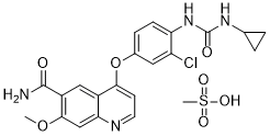
乐伐替尼甲磺酸盐是乐伐替尼与等摩尔量的甲磺酸反应制得的甲磺酸盐。它是一种多激酶抑制剂和孤儿药,以甲磺酸盐的形式用于治疗对放射性碘治疗无效的各种甲状腺癌。它具有多种功能,包括作为EC 2.7.10.1(受体蛋白酪氨酸激酶)抑制剂、成纤维细胞生长因子受体拮抗剂、孤儿药、血管内皮生长因子受体拮抗剂和抗肿瘤药物。它含有乐伐替尼(1+)分子。
乐伐替尼甲磺酸盐是一种合成的、口服有效的血管内皮生长因子受体2(VEGFR2,也称为KDR/FLK-1)酪氨酸激酶抑制剂,具有潜在的抗肿瘤活性。 E7080 可阻断 VEGF 对 VEGFR2 的激活,从而抑制 VEGF 受体信号转导通路,减少血管内皮细胞的迁移和增殖,并诱导血管内皮细胞凋亡。 另见:乐伐替尼(具有活性成分)。 药物适应症 Kisplyx 适用于治疗成人晚期肾细胞癌 (RCC):与帕博利珠单抗联合用于一线治疗(参见 5.1 节);与依维莫司联合用于既往接受过一种血管内皮生长因子 (VEGF) 靶向治疗的患者。 Lenvima® 适用于治疗对放射性碘 (RAI) 治疗无效的进展性、局部晚期或转移性分化型(乳头状/滤泡状/Hürthle 细胞)甲状腺癌 (DTC) 成人患者,作为单药治疗。乐伐替尼®适用于既往未接受过全身治疗的晚期或不可切除肝细胞癌(HCC)成人患者的单药治疗。乐伐替尼属于喹啉类药物,是4-{3-氯-4-[(环丙基氨基甲酰基)氨基]苯氧基}-7-甲氧基喹啉-6-羧酸的羧酰胺衍生物。它是一种多激酶抑制剂和孤儿药,(以其甲磺酸盐形式)用于治疗对放射性碘治疗无效的各种甲状腺癌。它具有多种功能,包括血管内皮生长因子受体拮抗剂、孤儿药、抗肿瘤药、EC 2.7.10.1(受体蛋白酪氨酸激酶)抑制剂和成纤维细胞生长因子受体拮抗剂。它属于喹啉类、芳香醚类、单羧酸酰胺类、芳香酰胺类、单氯苯类、环丙烷类和苯脲类化合物。它是乐伐替尼(1+)的共轭碱。 乐伐替尼是一种受体酪氨酸激酶 (RTK) 抑制剂,可抑制血管内皮生长因子 (VEGF) 受体 VEGFR1 (FLT1)、VEGFR2 (KDR) 和 VEGFR3 (FLT4) 的激酶活性。乐伐替尼除了抑制其正常的细胞功能外,还抑制其他与致病性血管生成、肿瘤生长和癌症进展相关的 RTK,包括成纤维细胞生长因子 (FGF) 受体 FGFR1、2、3 和 4;血小板衍生生长因子受体 α (PDGFRα)、KIT 和 RET。这些位于细胞膜上的受体酪氨酸激酶 (RTK) 在激活参与细胞增殖、迁移、凋亡和分化等正常细胞过程以及病理性血管生成、淋巴生成、肿瘤生长和癌症进展的信号转导通路中发挥着核心作用。特别是,VEGF 已被确定为生理性和病理性血管生成的关键调节因子,VEGF 表达增加与多种癌症的不良预后相关。乐伐替尼适用于治疗局部复发或转移性、进展性、放射性碘 (RAI) 难治性分化型甲状腺癌患者。大多数甲状腺癌患者通过手术和激素治疗的治疗预后良好(5 年生存率达 98%)。然而,对于放射性碘难治性甲状腺癌患者,治疗选择有限,预后较差,因此迫切需要开发更具靶向性的疗法,例如乐伐替尼。 乐伐替尼是一种激酶抑制剂。其作用机制是作为受体酪氨酸激酶抑制剂。 乐伐替尼是一种口服多激酶抑制剂和抗肿瘤药物,用于治疗晚期、转移性甲状腺髓样癌和难治性肾细胞癌。乐伐替尼治疗期间血清酶升高发生率较低,并且与罕见的临床表现明显的急性肝损伤病例有关,其中一些病例甚至导致死亡。 乐伐替尼是一种合成的口服血管内皮生长因子受体2(VEGFR2,也称为KDR/FLK-1)酪氨酸激酶抑制剂,具有潜在的抗肿瘤活性。乐伐替尼通过阻断 VEGF 对 VEGFR2 的激活,抑制 VEGF 受体信号转导通路,从而减少血管内皮细胞的迁移和增殖,并诱导血管内皮细胞凋亡。 另见:甲磺酸乐伐替尼(有盐形式)。 药物适应症 乐伐替尼适用于治疗以下癌症:分化型甲状腺癌 (DTC) - 用于治疗局部复发或转移、进展性、放射性碘难治性分化型甲状腺癌;肾细胞癌 (RCC) - 与帕博利珠单抗联合用于一线治疗,适用于晚期肾细胞癌成人患者;肝细胞癌 (HCC) - 与依维莫司联合用于治疗既往接受过 ≥1 种抗血管生成疗法的晚期肾细胞癌成人患者;肝细胞癌 (HCC) - 一线治疗不可切除的肝细胞癌患者 子宫内膜癌 - 用于治疗非微卫星不稳定性高 (MSI-H) 或错配修复缺陷 (dMMR) 的晚期子宫内膜癌,与帕博利珠单抗联合使用,适用于既往接受过全身治疗后出现疾病进展且不适合根治性手术或放疗的患者。 FDA 标签 Kisplyx 适用于治疗成人晚期肾细胞癌 (RCC):与帕博利珠单抗联合使用,作为一线治疗(参见 5.1 节);与依维莫司联合使用,用于既往接受过一种血管内皮生长因子 (VEGF) 靶向治疗的患者。 Lenvima® 适用于单药治疗对放射性碘 (RAI) 耐药的进展性、局部晚期或转移性分化型(乳头状/滤泡状/Hürthle 细胞)甲状腺癌 (DTC) 成人患者。乐伐替尼®适用于治疗既往未接受过全身治疗的晚期或不可切除的肝细胞癌 (HCC) 成年患者。 治疗除造血和淋巴组织肿瘤、乳头状甲状腺癌、滤泡状甲状腺癌和骨肉瘤以外的所有恶性肿瘤。 治疗滤泡状甲状腺癌、治疗骨肉瘤、治疗乳头状甲状腺癌。 作用机制 乐伐替尼是一种受体酪氨酸激酶 (RTK) 抑制剂,可抑制血管内皮生长因子 (VEGF) 受体 VEGFR1 (FLT1)、VEGFR2 (KDR) 和 VEGFR3 (FLT4) 的激酶活性。除了正常的细胞功能外,乐伐替尼还能抑制其他与致病性血管生成、肿瘤生长和癌症进展有关的RTK,包括成纤维细胞生长因子(FGF)受体FGFR1、2、3和4;血小板衍生生长因子受体α(PDGFRα)、KIT和RET。 背景:在一项II期试验中,作为VEGF受体1-3、FGF受体1-4、PDGF受体α、RET和KIT抑制剂的乐伐替尼,在肝细胞癌中显示出活性。本研究旨在比较接受乐伐替尼与索拉非尼一线治疗的不可切除肝细胞癌患者的总生存期。方法:这是一项开放标签、多中心、非劣效性III期临床试验,纳入了来自亚太、欧洲和北美地区20个国家154个研究中心的、既往未接受过晚期疾病治疗的不可切除肝细胞癌患者。患者通过交互式语音应答系统按1:1的比例随机分组,分组因素包括所在地区、是否存在肉眼可见的门静脉侵犯、肝外转移或两者兼有、美国东部肿瘤协作组(ECOG)体能状态评分以及体重。患者分别接受口服乐伐替尼(体重≥60 kg者每日12 mg,体重<60 kg者每日8 mg)或索拉非尼(400 mg,每日两次),28天为一个疗程。主要终点为总生存期,定义为从随机分组之日起至因任何原因死亡之日止的时间。疗效分析遵循意向性治疗原则,安全性分析仅纳入接受治疗的患者。非劣效性界值设定为1.08。该试验已在ClinicalTrials.gov注册,注册号为NCT01761266。结果:2013年3月1日至2015年7月30日期间,共招募了1492例患者。其中954例符合条件的患者被随机分配至乐伐替尼组(n=478)或索拉非尼组(n=476)。乐伐替尼的中位生存时间为 13.6 个月(95% CI 12.1-14.9),不劣于索拉非尼(12.3 个月,10.4-13.9;风险比 0.92,95% CI 0.79-1.06),符合非劣效性标准。乐伐替尼组最常见的任何级别不良事件为高血压(201例[42%])、腹泻(184例[39%])、食欲减退(162例[34%])和体重下降(147例[31%]);索拉非尼组最常见的任何级别不良事件为手足综合征(249例[52%])、腹泻(220例[46%])、高血压(144例[30%])和食欲减退(127例[27%])。结论:在未经治疗的晚期肝细胞癌患者中,乐伐替尼在总生存期方面不劣于索拉非尼。乐伐替尼的安全性和耐受性与既往观察结果一致。[1] 乐伐替尼是一种小分子酪氨酸激酶抑制剂,可抑制血管内皮生长因子受体 (VEGFR1-3)、成纤维细胞生长因子受体 (FGFR1-4)、血小板衍生生长因子受体α (PDGFRα)、干细胞因子受体 (KIT) 和转染重排受体 (RET)。这些受体对肿瘤血管生成至关重要,乐伐替尼通过抑制这些受体的功能来抑制肿瘤血管生成。乐伐替尼的 I 期临床试验在日本、欧洲和美国同时进行,在甲状腺癌、子宫内膜癌、黑色素瘤、肾细胞癌、肉瘤和结肠癌中均观察到肿瘤缩小效果。乐伐替尼是一种很有前景的药物,已显示出对多种实体瘤的治疗效果。乐伐替尼治疗可能出现高血压、蛋白尿、腹泻和伤口愈合延迟等不良反应。控制这些不良反应对于乐伐替尼的使用至关重要。本文概述了乐伐替尼在甲状腺癌、肝细胞癌、肾细胞癌和肺癌治疗中的现状、毒性和未来前景。[2] E7080 是一种口服有效的多种受体酪氨酸激酶抑制剂,包括 VEGF、FGF 和 SCF 受体。本研究表明,E7080 在体外可抑制 SCF 诱导的血管生成,在体内可抑制产生 SCF 的人小细胞肺癌 H146 细胞的肿瘤生长。 E7080 能抑制 SCF 驱动的 HUVEC 细胞管状结构形成,该细胞表达 SCF 受体 KIT,其 IC50 值为 5.2 nM,对 VEGF 驱动的管状结构形成抑制作用几乎相同(IC50 = 5.1 nM)。为了评估 SCF/KIT 信号通路在肿瘤血管生成中的作用,我们研究了选择性 KIT 激酶抑制剂伊马替尼对裸鼠体内 H146 细胞肿瘤生长的影响。由于 H146 细胞不表达 KIT,伊马替尼在体外未显示出显著的抗肿瘤活性(IC50 = 2200 nM)。然而,口服 160 mg/kg 伊马替尼可显著减缓裸鼠体内 H146 细胞肿瘤的生长,并伴有微血管密度的降低。口服 E7080 可剂量依赖性地抑制 H146 细胞的肿瘤生长,剂量分别为 30 和 100 mg/kg,并在 100 mg/kg 剂量下引起肿瘤消退。抗 VEGF 抗体虽然也能减缓肿瘤生长,但并未引起肿瘤消退。这些结果表明,KIT 信号通路在产生 SCF 的 H146 细胞的肿瘤血管生成中发挥作用,而 E7080 通过抑制 KIT 和 VEGF 受体信号通路发挥抗血管生成活性,从而导致 H146 肿瘤消退。E7080 可能对治疗产生 SCF 的肿瘤具有潜在的治疗价值。[3] 目的:血管内皮生长因子 (VEGF)-C/VEGF 受体 3 (VEGF-R3) 信号通路通过影响淋巴管,在淋巴管生成和肿瘤转移中发挥重要作用。然而,关于使用小分子激酶抑制剂抑制 VEGF-R3 对淋巴管生成和淋巴结转移的影响知之甚少。实验设计:我们利用表达过量 VEGF-C 的 MDA-MB-231 细胞,在乳腺脂肪垫异种移植人乳腺癌模型中,评估了 VEGF-R2 和 VEGF-R3 激酶的强效抑制剂 E7080 以及贝伐珠单抗对淋巴管生成和血管生成的影响。淋巴管生成通过淋巴管密度 (LVD) 测定,血管生成通过微血管密度 (MVD) 测定。结果:与表达与 MDA-MB-231 细胞相似但 VEGF-C 含量无法检测的 MDA-MB-435 细胞相比,只有 MDA-MB-231 细胞在原发肿瘤中表现出淋巴管生成。 E7080(而非贝伐单抗)显著降低了MDA-MB-231肿瘤内的淋巴管密度(LVD)。E7080和贝伐单抗均降低了MDA-MB-231和MDA-MB-435模型中的微血管密度(MVD)。E7080显著抑制了MDA-MB-231的区域淋巴结转移和远处肺转移,而贝伐单抗仅显著抑制了肺转移。E7080还降低了原发肿瘤切除后淋巴结转移结节内的MVD和LVD。结论:E7080通过抑制VEGF-R3激酶,有效降低了表达VEGF-C的MDA-MB-231肿瘤内的LVD。 E7080 同时抑制 VEGF-R2 和 VEGF-R3 激酶可能是一种有前景的新策略,可用于控制区域淋巴结和远处肺转移。[4] 甲磺酸仑伐替尼是一种口服多靶点酪氨酸激酶抑制剂 (TKI),靶向血管生成(VEGFR1-3、FGFR1-4、PDGFRα)和肿瘤细胞增殖(KIT、RET),与其他抗血管生成药物相比,具有独特的激酶抑制谱。[2] - 它已获得 FDA 和 EMA 批准,用于不可切除肝细胞癌 (HCC) 的一线治疗,以及与依维莫司联合用于治疗放射性碘难治性分化型甲状腺癌 (DTC) 和晚期肾细胞癌 (RCC)。[2] - REFLECT 研究证实,其在总生存期 (OS) 和中期生存期 (CTR) 方面不劣于索拉非尼。该药物在不可切除肝细胞癌的无进展生存期(PFS)和客观缓解率(ORR)方面具有优势,使其成为一线标准疗法[1] - 其抗肿瘤机制涉及双重抑制肿瘤血管生成(通过VEGFR/FGFR/PDGFR)和肿瘤细胞存活/增殖(通过KIT/RET),使其对多种激酶信号通路失调的癌症类型有效[2] - 在乳腺癌模型中,该药物通过靶向VEGFR2(血管生成)和VEGFR3(淋巴管生成)抑制血行和淋巴转移,为转移性乳腺癌提供了一种潜在的治疗策略[4] - 体重<60 kg、肝功能不全或肾功能不全的患者需要调整剂量[2] |
| 分子式 |
C22H23CLN4O7S
|
|---|---|
| 分子量 |
522.96
|
| 精确质量 |
522.097
|
| 元素分析 |
C, 50.53; H, 4.43; Cl, 6.78; N, 10.71; O, 21.42; S, 6.13
|
| CAS号 |
857890-39-2
|
| 相关CAS号 |
Lenvatinib;417716-92-8
|
| PubChem CID |
11237762
|
| 外观&性状 |
White to off-white solid powder
|
| LogP |
5.818
|
| tPSA |
182.8
|
| 氢键供体(HBD)数目 |
4
|
| 氢键受体(HBA)数目 |
8
|
| 可旋转键数目(RBC) |
6
|
| 重原子数目 |
35
|
| 分子复杂度/Complexity |
727
|
| 定义原子立体中心数目 |
0
|
| SMILES |
ClC1C([H])=C(C([H])=C([H])C=1N([H])C(N([H])C1([H])C([H])([H])C1([H])[H])=O)OC1C([H])=C([H])N=C2C([H])=C(C(C(N([H])[H])=O)=C([H])C=12)OC([H])([H])[H].S(C([H])([H])[H])(=O)(=O)O[H]
|
| InChi Key |
HWLFIUUAYLEFCT-UHFFFAOYSA-N
|
| InChi Code |
InChI=1S/C21H19ClN4O4.CH4O3S/c1-29-19-10-17-13(9-14(19)20(23)27)18(6-7-24-17)30-12-4-5-16(15(22)8-12)26-21(28)25-11-2-3-11;1-5(2,3)4/h4-11H,2-3H2,1H3,(H2,23,27)(H2,25,26,28);1H3,(H,2,3,4)
|
| 化学名 |
4-[3-chloro-4-(cyclopropylcarbamoylamino)phenoxy]-7-methoxyquinoline-6-carboxamide;methanesulfonic acid
|
| 别名 |
E-7080 mesylate; E7080; E 7080; LENVATINIB MESYLATE; 857890-39-2; lenvatinibMesylate; Lenvima; Lenvatinib mesilate; E7080 MESYLATE; Lenvatinib mesylate [USAN]; UNII-3J78384F61; ER-203492-00 mesylate; Lenvatinib mesylate; Brand name Lenvima
|
| HS Tariff Code |
2934.99.9001
|
| 存储方式 |
Powder -20°C 3 years 4°C 2 years In solvent -80°C 6 months -20°C 1 month 注意: 请将本产品存放在密封且受保护的环境中(例如氮气保护),避免吸湿/受潮和光照。 |
| 运输条件 |
Room temperature (This product is stable at ambient temperature for a few days during ordinary shipping and time spent in Customs)
|
| 溶解度 (体外实验) |
|
|||
|---|---|---|---|---|
| 溶解度 (体内实验) |
配方 1 中的溶解度: ≥ 2.08 mg/mL (3.98 mM) (饱和度未知) in 10% DMSO + 40% PEG300 + 5% Tween80 + 45% Saline (这些助溶剂从左到右依次添加,逐一添加), 澄清溶液。
例如,若需制备1 mL的工作液,可将100 μL 20.8 mg/mL澄清DMSO储备液加入400 μL PEG300中,混匀;然后向上述溶液中加入50 μL Tween-80,混匀;加入450 μL生理盐水定容至1 mL。 *生理盐水的制备:将 0.9 g 氯化钠溶解在 100 mL ddH₂O中,得到澄清溶液。 配方 2 中的溶解度: ≥ 2.08 mg/mL (3.98 mM) (饱和度未知) in 10% DMSO + 90% (20% SBE-β-CD in Saline) (这些助溶剂从左到右依次添加,逐一添加), 澄清溶液。 例如,若需制备1 mL的工作液,可将 100 μL 20.8 mg/mL澄清DMSO储备液加入900 μL 20% SBE-β-CD生理盐水溶液中,混匀。 *20% SBE-β-CD 生理盐水溶液的制备(4°C,1 周):将 2 g SBE-β-CD 溶解于 10 mL 生理盐水中,得到澄清溶液。 View More
配方 3 中的溶解度: ≥ 2.08 mg/mL (3.98 mM) (饱和度未知) in 10% DMSO + 90% Corn Oil (这些助溶剂从左到右依次添加,逐一添加), 澄清溶液。 配方 4 中的溶解度: 0.5% methylcellulose: 30 mg/kg 1、请先配制澄清的储备液(如:用DMSO配置50 或 100 mg/mL母液(储备液)); 2、取适量母液,按从左到右的顺序依次添加助溶剂,澄清后再加入下一助溶剂。以 下列配方为例说明 (注意此配方只用于说明,并不一定代表此产品 的实际溶解配方): 10% DMSO → 40% PEG300 → 5% Tween-80 → 45% ddH2O (或 saline); 假设最终工作液的体积为 1 mL, 浓度为5 mg/mL: 取 100 μL 50 mg/mL 的澄清 DMSO 储备液加到 400 μL PEG300 中,混合均匀/澄清;向上述体系中加入50 μL Tween-80,混合均匀/澄清;然后继续加入450 μL ddH2O (或 saline)定容至 1 mL; 3、溶剂前显示的百分比是指该溶剂在最终溶液/工作液中的体积所占比例; 4、 如产品在配制过程中出现沉淀/析出,可通过加热(≤50℃)或超声的方式助溶; 5、为保证最佳实验结果,工作液请现配现用! 6、如不确定怎么将母液配置成体内动物实验的工作液,请查看说明书或联系我们; 7、 以上所有助溶剂都可在 Invivochem.cn网站购买。 |
| 制备储备液 | 1 mg | 5 mg | 10 mg | |
| 1 mM | 1.9122 mL | 9.5610 mL | 19.1219 mL | |
| 5 mM | 0.3824 mL | 1.9122 mL | 3.8244 mL | |
| 10 mM | 0.1912 mL | 0.9561 mL | 1.9122 mL |
1、根据实验需要选择合适的溶剂配制储备液 (母液):对于大多数产品,InvivoChem推荐用DMSO配置母液 (比如:5、10、20mM或者10、20、50 mg/mL浓度),个别水溶性高的产品可直接溶于水。产品在DMSO 、水或其他溶剂中的具体溶解度详见上”溶解度 (体外)”部分;
2、如果您找不到您想要的溶解度信息,或者很难将产品溶解在溶液中,请联系我们;
3、建议使用下列计算器进行相关计算(摩尔浓度计算器、稀释计算器、分子量计算器、重组计算器等);
4、母液配好之后,将其分装到常规用量,并储存在-20°C或-80°C,尽量减少反复冻融循环。
计算结果:
工作液浓度: mg/mL;
DMSO母液配制方法: mg 药物溶于 μL DMSO溶液(母液浓度 mg/mL)。如该浓度超过该批次药物DMSO溶解度,请首先与我们联系。
体内配方配制方法:取 μL DMSO母液,加入 μL PEG300,混匀澄清后加入μL Tween 80,混匀澄清后加入 μL ddH2O,混匀澄清。
(1) 请确保溶液澄清之后,再加入下一种溶剂 (助溶剂) 。可利用涡旋、超声或水浴加热等方法助溶;
(2) 一定要按顺序加入溶剂 (助溶剂) 。
| NCT Number | Recruitment | interventions | Conditions | Sponsor/Collaborators | Start Date | Phases |
| NCT03477175 | Active Recruiting |
Drug: E7080 Drug: Comparator Drug |
Solid Tumors | Eisai Inc. | August 16, 2018 | Phase 2 |
| NCT05339581 | Not yet recruiting | Drug: Sintilimab Drug: Tislelizumab |
Liver Cancer Portal Vein Thrombosis |
RenJi Hospital | May 20, 2022 | Not Applicable |
| NCT05617859 | Recruiting | Drug: Lenvatinib mesylate capsule |
Effectiveness Sexuality |
Henan Cancer Hospital | April 30, 2023 | Phase 2 |
| NCT05296512 | Recruiting | Drug: Lenvatinib Drug: Pembrolizumab |
Ovarian Clear Cell Carcinoma Gynecologic Cancer |
Elizabeth K. Lee MD | September 23, 2022 | Phase 2 |
| NCT05342194 | Not yet recruiting | Drug: Toripalimab Drug: Placebo IV |
Intrahepatic Cholangiocarcinoma | Shanghai Junshi Bioscience Co., Ltd. |
October 1, 2022 | Phase 3 |
|
|
 VEGFR2-IN-7
 SYHA1813
 VEGFR-2-IN-38
 BHEP
BHEP
InvivoChem的所有产品仅用于作科学研究,不面向患者销售
Copyright 2020 InvivoChem LLC | All Rights Reserved 粤ICP备20063088号-1


 COA
COA


 463611831
