| 规格 | 价格 | 库存 | 数量 |
|---|---|---|---|
| 1mg |
|
||
| 5mg |
|
||
| 10mg |
|
||
| Other Sizes |
|

| 靶点 |
sphingomyelin synthase (SMS)(IC50 = 1.5~3 μM)
|
|---|---|
| 体外研究 (In Vitro) |
研究了malabarine C(mal C)对人乳腺癌症MCF-7细胞系的细胞毒性机制。Mal C剂量依赖性地增加了亚G1细胞群,与细胞质寡核小体形成和染色质凝缩有关。mal C诱导的凋亡导致线粒体损伤,如JC-1染色细胞的荧光显微镜和流式细胞术所示,以及线粒体特异性核酸酶蛋白AIF和内切G的释放。mal C也从MCF-7细胞中释放细胞内Ca(2+),但Ca(2-)调节剂BAPTA-AM和Ru360仅部分消除了凋亡。malC激活钙蛋白酶对其细胞毒性没有任何影响。另一方面,在malC处理后,比线粒体损伤更早地观察到明显的溶酶体膜透性(LMP),以及组织蛋白酶B的释放、Bid切割及其向线粒体的易位。这表明mal C对人MCF-7人癌症细胞系的细胞毒性可能通过LMP作为触发胱天蛋白酶非依赖性但组织蛋白酶B和t-Bid依赖性内在线粒体凋亡途径的初始事件进行。S期或G2-M期细胞的显著积累,以及由于mal C暴露引起的细胞周期蛋白E和A的上调,使其有望成为潜在的抗癌剂。[3]
malabarine C(mal C)抑制T细胞活化、增殖和细胞因子产生。 malabarine C(mal C)抑制丝裂原诱导的细胞表面标志物MAPK和NF-κB的激活。 malabarine C(mal C)调节淋巴细胞中的细胞氧化还原状态[2]。 |
| 体内研究 (In Vivo) |
天然存在的抑制剂和靶向膜蛋白之间的相互作用可能是治疗代谢综合征,特别是肥胖的替代药物策略。在这项研究中,我们鉴定了从肉豆蔻果实中分离出的马拉巴锥A-C和E(1-4)作为鞘磷脂合酶(SMS)的天然抑制剂,SMS是一种负责鞘脂生物合成的膜蛋白。在高脂饮食诱导的肥胖小鼠模型中,口服化合物3/malabaricone C(mal C)具有最有前景的抑制作用,在减少体重增加、改善葡萄糖耐量和减少肝脂肪变性方面表现出多种功效。肝脏脂质分析揭示了化合物3的SMS活性与其体内外脂质代谢之间的关键联系。化合物3/malabaricone C(mal C)的无毒性使其成为寻找治疗和预防肥胖药物的合适候选者。[1]
malabarine C(mal C)给药抑制了离体T细胞活化[2] 为了确定malabarine C(mal C)作为体内免疫抑制剂的能力,小鼠腹腔注射载体(DMSO)或mal C,24小时后分离淋巴细胞。然后,用CFSE染色细胞并用Con A刺激。与载体对照组相比,mal C给药显著降低了增殖细胞的百分比(图4A和B)。Mal C给药导致T细胞离体分泌丝裂原诱导的细胞因子(IL-2、IFN-γ和IL-6)显著减少 为了研究malabarine C(mal C)预防急性GvHD的效用,将C57BL/6小鼠的淋巴细胞用mal C(10μM)处理4小时,并用作同种异体供体细胞。受体BALB/c小鼠通过暴露于6 Gy剂量的WBI而出现淋巴细胞减少。供体细胞通过侧尾静脉注射(每只小鼠1000万个细胞)过继转移,并监测GvHD宿主的发病率和死亡率。用载体处理的同种异体细胞重组的小鼠(GvHD组)在30天内死亡。然而,供体细胞的Mal C治疗完全抑制了GvHD相关的死亡率和发病率(图4D和E)。与未受辐射的对照组相比,GvHD相关的体重减轻在第10天很明显,这在用Mal C处理的细胞重建的小鼠中得到了显著预防(图4E)。与未受照射的对照组相比,接受WBI(6 Gy)的小鼠IL6水平显著升高(图4F)。GvHD相关IFN-γ水平显著高于WBI 6Gy组。此外,与接受载体处理的供体细胞的宿主相比,用Mal C处理的供体电池重构的宿主中IL6和IFN-γ的血清水平降低(图4F)。图4G显示了Mal C治疗对淋巴细胞减少宿主中同基因CD4+T细胞稳态增殖的影响。Mal C治疗不影响CD4+T细胞的稳态增殖[2]。 |
| 细胞实验 |
表面染色和细胞内染色[2]
如前所述,用荧光染料(PE或FITC)偶联的单克隆抗体对细胞进行染色(Sharma等人,2012)。简而言之,将淋巴细胞(2.5×10⁶)与malabaricone C (mal C)(10μM,2小时)一起孵育,然后用Con A刺激(2.5μg/mL,24小时用于表面染色,2.5μg/mL,2小时用于细胞内染色),随后用荧光染料偶联的CD69或CD25单克隆抗体染色。使用未染色和同种型染色的细胞作为对照。然后,在BD FACS Melody流式细胞仪上采集细胞,并使用FlowJo软件进行分析。 ROS和GSH的估算[2] 对于基于DCF荧光(485/535nm处的激发/发射)的ROS估计,用H2DCF-DA对malabaricone C (mal C)或载体处理的细胞进行染色,并使用光谱荧光计 进行监测。为了定量GSH(还原/氧化)水平,用Mal C或载体对照处理细胞,收获并裂解,并如前所述探测细胞提取物的GSH定量(Patwardhan等人,2015)。为了评估细胞巯基,细胞用MCB染色(在390/478 nm处激发/发射),然后进行流式细胞术采集。 高效液相色谱法分析malabaricone C (mal C)和NAC相互作用[2] 我们采用多步梯度流动相,乙腈为溶剂a,甲酸水溶液(0.1%)为溶剂B,使用C-18柱进行色谱分离,并采用优化的方法。梯度设置为:40%溶剂A 2分钟;80%溶剂A处理22分钟;100%溶剂A浸泡2分钟;40%溶剂A 4分钟,流速保持在1 mL/min。注射用样品体积为20μL,总运行时间为30分钟,在15.773分钟的保留时间(Rt)下检测Mal C。使用二极管阵列检测器在274 nm下进行检测。制备100 mM浓度的NAC储备溶液,并在1X PBS中稀释。将Mal C加入NAC中(Mal C 100μM+NAC 100µM),在37°C下孵育1小时,并在HPLC上运行(Dionex Ultima 3000系列HPLC系统,带Chromeleon软件(版本6.8))。 malabaricone C (mal C)和NAC相互作用的光谱分析[2] Mal C(100μM)与NAC(100μM)在37°C的水中孵育1小时,使用Jasco分光光度计记录250至700 nm的吸收光谱。使用Jasco Spectra Manager Ver.2软件绘制了显示吸光度与波长(nm)的图。 蛋白质印迹[2] 收获用malabaricone C (mal C)或Con A(2.5μg/mL)或两者处理的细胞(10×106/mL),并使用1X RIPA裂解缓冲液进行裂解,该缓冲液由50 mM Tris-HCl、150 mM NaCl、1.0%(v/v)NP-40、0.5%(w/v)脱氧胆酸钠、1.0 mM EDTA、0.1%(w/v。使用Bradford测定法进行蛋白质估算,然后在SDS-PAGE(10%)上进行电泳分离。将蛋白质转移到PVDF膜上,使用单克隆抗体和HRP偶联的二抗探测pERK、pJNK、ERK和JNK。使用增强化学发光试剂盒(POD)在SynGene:GBox凝胶记录系统中可视化条带。 转录因子凝胶转移分析[2] 细胞(10×106/mL)用载体或指定浓度的malabaricone C (mal C)或Con A(2.5μg/mL)或两者处理。如前所述(Patwardhan等人,20152016),从细胞中制备核提取物,并与来自人类免疫缺陷病毒长末端重复序列的32P末端标记的45-mer双链NF-κB寡核苷酸(5′-TTGTTACAGGGACTTTCCGTGGGGACTTCGGGAGGCGTG-3′)一起孵育。样品在天然聚丙烯酰胺凝胶上运行,然后干燥凝胶,随后将其暴露于荧光图像板上,使用荧光图像板扫描仪观察放射性带。 |
| 动物实验 |
淋巴细胞体外刺激[2]
马拉巴酮C (mal C) 的高亲脂性和低水溶性导致其口服生物利用度差,因此,小鼠腹腔注射马拉巴酮C (mal C)(10 mg/kg 体重)或载体(每组3只小鼠),并在注射后24小时处死。用Con A (2.5 μg/mL) 刺激淋巴细胞24小时,以使用ELISA法测定分泌的细胞因子。另一组淋巴细胞用CFSE染色,并用Con A (2.5 μg/mL) 刺激72小时,以通过流式细胞术评估细胞增殖。 移植物抗宿主病[2] 受体BALB/c小鼠在血液照射器中接受6 Gy的全身照射(WBI),剂量率为1 Gy/min,以诱导淋巴细胞减少症(每组6只小鼠)。将来自同种异体C57BL/6供体的淋巴细胞在体外用载体对照或马拉巴酮C(mal C)处理,并在WBI后48小时,将1000万个细胞经尾静脉注射到淋巴细胞减少的受体小鼠体内。移植后,监测淋巴细胞减少的受体小鼠,评估其体重变化和存活率,持续30天。移植后第5天,采集淋巴细胞减少的受体小鼠的血液,分离血清,并使用ELISA检测细胞因子水平。 体内稳态增殖监测[2] 使用磁珠从BALB/c小鼠的淋巴细胞中分选出CD4+ T细胞,并用CFSE染色。将细胞与马拉巴酮C(mal C)(10 μM)或溶剂在37°C、5% CO2条件下孵育4小时。洗涤细胞后,将100万个CFSE+ CD4+ T细胞静脉注射到淋巴细胞减少的同基因BALB/c小鼠体内(每组3只小鼠)。72小时后,分离淋巴细胞减少受体小鼠的淋巴细胞,并通过流式细胞术监测供体CFSE+细胞的频率和增殖情况。 |
| 参考文献 |
|
| 其他信息 |
马拉巴林C(mal C)是一种丁酮类化合物,具有代谢产物的作用。据报道,马拉巴林C存在于肉豆蔻科植物、香肉豆蔻以及其他有相关数据的生物体中。总之,从肉桂肉豆蔻(M. cinnamomea)果实中分离得到的酰基酚类化合物马拉巴林C(mal C)已被鉴定为一种天然的鞘磷脂合成酶抑制剂。马拉巴林C的作用机制与先前报道的SMS基因敲除研究相同,它能高效地阻止油酸跨膜吸收,从而减少体外脂滴的形成。15马拉巴林C还被发现能够减少体重增加、改善葡萄糖耐量并降低体内肝脏脂质积累,因此,这是首个报道植物来源的SMS抑制剂对抗高脂饮食诱导肥胖的案例。马拉巴酮C无毒的特性使其成为进一步开发用于治疗和预防肥胖症的新药或保健品的理想候选药物。[1]
T细胞是适应性免疫反应的重要介质。然而,在自身免疫、过敏、慢性炎症、COVID-19和急性移植物抗宿主病(GvHD)等特定病理条件下,需要抑制T细胞活化(Maurice等,1997;Griffiths等,2011;Weyand等,2018)。过去几十年,在开发用于抑制临床上不必要的T细胞活化的预防性和治疗性药物方面,取得了前所未有的研究成果。包括我们课题组在内的许多研究人员此前已强调了细胞氧化还原状态和周围氧化还原环境对T细胞活化、增殖和分化的调控作用(Checker等,2009;Kesarwani等,2013;Gambhir等,2014)。许多氧化还原活性物质,包括抗氧化剂和促氧化剂,已被证实能够调节T细胞活化。我们之前已证明,诸如铅丹素、1,4-萘醌和甲萘醌等促氧化剂可通过调节细胞内谷胱甘肽(GSH)水平,在亚微摩尔浓度下抑制T细胞活化(Checker等,2010,2011)。抗氧化剂,如GSH、N-乙酰半胱氨酸(NAC)、叶绿素、黄芩苷和维生素C,已被证实能够调节T细胞的活化和分化(Sharma等,2007;Patwardhan等,2016)。GSH失调也与早期实验性移植物抗宿主病(GvHD)的严重程度有关(Suh等,2014)。T细胞活化还依赖于线粒体中活性氧(ROS)的内在生成和代谢重编程(Sena等,2013)。孟买肉豆蔻,又称假肉豆蔻,是一种香料,提取自印度药用植物肉豆蔻(Myristica malabarica,肉豆蔻科;阿育吠陀名称:Rampatri)的果实假种皮(Patro et al. 2005)。基于其化学结构和活性成分已知的生物学特性,我们假设马拉巴林C(mal C)可能影响T细胞活化。研究发现,马拉巴林C(mal C)处理显著抑制了小鼠T细胞的活化、增殖和细胞因子分泌。此外,Mal C处理抑制了Con A诱导的活性氧(ROS)生成。我们观察到,T细胞活化的抑制伴随着总ROS水平的降低。淋巴细胞与Mal C孵育导致还原型谷胱甘肽(GSH)水平显著降低。因此,一个问题随之而来:Mal C 作为免疫调节剂的功效究竟是源于其抗氧化特性,还是源于其硫醇结合能力?有趣的是,硫醇抗氧化剂能够消除 Mal C 的抗增殖作用,这提示 Mal C 可能与细胞硫醇存在相互作用。事实上,生物物理学研究表明,Mal C 可以在水溶液环境中与 NAC 发生物理相互作用。更有趣的是,补充 NAC 可以恢复 Mal C 处理细胞中的硫醇水平,这可能是硫醇抗氧化剂存在下 Mal C 对 T 细胞抗增殖作用被消除的原因。这些研究表明,Mal C 可能与细胞蛋白和非蛋白硫醇相互作用,从而破坏细胞氧化还原平衡。细胞氧化还原变化通过氧化还原传感信号分子影响增殖、分化、存活和凋亡等生物学反应(Chiu 和 Dawes 2012;Hancock 和 Whiteman 2018)。 MAP激酶可通过氧化还原敏感性磷酸酶的作用,响应细胞氧化还原状态的变化而改变其磷酸化状态(Kamata和Hirata 1999;Seth和Rudolph 2006)。T细胞受体配体结合可诱导不同的MAPK信号通路,从而调节T细胞反应(Adachi和Davis 2011)。T细胞的促分裂原刺激可导致MAP激酶磷酸化,进而激活免疫调节转录因子NF-κB。Mal C处理可抑制促分裂原刺激后ERK和JNK的磷酸化以及NF-κB的后续DNA结合。T细胞在T细胞受体和共刺激分子传递的信号激活后,会上调CD25(IL2Rα)和CD69(C型凝集素)的表达。 CD25表达决定IL-2反应性,而CD69激活则刺激钙离子内流和细胞外激酶ERK1/2的激活,从而促进T细胞增殖(Chen et al. 2020)。Mal C介导的T细胞早期激活标志物(CD69)和晚期激活标志物(CD25)的抑制表明,Mal C干扰T细胞的早期激活,使其对有丝分裂原刺激无反应。基于这些结果,我们假设T细胞短暂暴露于Mal C可能有利于预防急性移植物抗宿主病(GvHD)。我们使用完全同种异体淋巴细胞移植的小鼠模型,发现Mal C完全消除了淋巴细胞减少宿主的急性GvHD相关发病率和死亡率。Mal C处理供体T细胞也显著降低了宿主血清中GvHD相关细胞因子的水平。体内T细胞增殖抑制也会导致免疫稳态紊乱。为了研究Mal C对同基因T细胞行为的影响,我们评估了同基因淋巴细胞减少宿主中纯化的CD4+ T细胞的稳态增殖。Mal C并未抑制T细胞的稳态增殖。Mal C处理通过同时清除活性氧(ROS)和硫醇,导致非经典的细胞氧化还原紊乱。然而,谷胱甘肽对于抗原诱导的T细胞增殖至关重要,但对于稳态增殖并非必需(Sena和Chandel 2012;Sena等 2013)。因此,可以预期Mal C处理不会影响T细胞的稳态增殖。[2] 急性移植物抗宿主病(GvHD)期间T细胞抗原特异性增殖的信号传导需求与淋巴细胞减少症反应中的稳态增殖信号传导需求不同。我们的研究结果表明,马拉巴林C(mal C)对T细胞抗原/有丝分裂原诱导的增殖具有差异性和特异性的抑制作用,但对T细胞的稳态增殖没有抑制作用。[2] 总之,这是首篇报道马拉巴林C(mal C)通过调节细胞氧化还原平衡(图5)抑制T细胞活化、增殖和细胞因子产生的研究。由于Mal C仅抑制有丝分裂原/同种异体抗原诱导的增殖,而不抑制稳态增殖,因此它可能有助于预防急性移植物抗宿主病(GvHD),且不会破坏宿主的免疫重建。目前的研究结果提示,有必要开展进一步的机制研究,以阐明Mal C在T细胞中的分子机制和生化靶点。[2] |
| 分子式 |
C21H26O5
|
|---|---|
| 分子量 |
358.42814
|
| 精确质量 |
358.178
|
| 元素分析 |
C, 70.37; H, 7.31; O, 22.32
|
| CAS号 |
63335-25-1
|
| PubChem CID |
100313
|
| 外观&性状 |
White to off-white solid powder
|
| 熔点 |
119 - 121 °C
|
| LogP |
4.665
|
| tPSA |
97.99
|
| 氢键供体(HBD)数目 |
4
|
| 氢键受体(HBA)数目 |
5
|
| 可旋转键数目(RBC) |
10
|
| 重原子数目 |
26
|
| 分子复杂度/Complexity |
403
|
| 定义原子立体中心数目 |
0
|
| SMILES |
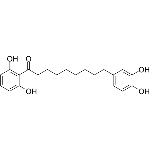
C1=CC(=C(C(=C1)O)C(=O)CCCCCCCCC2=CC(=C(C=C2)O)O)O
|
| InChi Key |
HCOZRFYGIFMIEX-UHFFFAOYSA-N
|
| InChi Code |
InChI=1S/C21H26O5/c22-16-13-12-15(14-20(16)26)8-5-3-1-2-4-6-9-17(23)21-18(24)10-7-11-19(21)25/h7,10-14,22,24-26H,1-6,8-9H2
|
| 化学名 |
1-(2,6-dihydroxyphenyl)-9-(3,4-dihydroxyphenyl)nonan-1-one
|
| 别名 |
Malabaricone C; 63335-25-1; 1-(2,6-dihydroxyphenyl)-9-(3,4-dihydroxyphenyl)nonan-1-one; CHEBI:69015; C9K53R3PRN; DTXSID40212721; NSC 287968; NSC-287968;
|
| HS Tariff Code |
2934.99.9001
|
| 存储方式 |
Powder -20°C 3 years 4°C 2 years In solvent -80°C 6 months -20°C 1 month 注意: 请将本产品存放在密封且受保护的环境中(例如氮气保护),避免吸湿/受潮和光照。 |
| 运输条件 |
Room temperature (This product is stable at ambient temperature for a few days during ordinary shipping and time spent in Customs)
|
| 溶解度 (体外实验) |
DMSO : ~100 mg/mL (~278.99 mM)
|
|---|---|
| 溶解度 (体内实验) |
配方 1 中的溶解度: ≥ 2.5 mg/mL (6.97 mM) (饱和度未知) in 10% DMSO + 40% PEG300 + 5% Tween80 + 45% Saline (这些助溶剂从左到右依次添加,逐一添加), 澄清溶液。
例如,若需制备1 mL的工作液,可将100 μL 25.0 mg/mL澄清DMSO储备液加入到400 μL PEG300中,混匀;然后向上述溶液中加入50 μL Tween-80,混匀;加入450 μL生理盐水定容至1 mL。 *生理盐水的制备:将 0.9 g 氯化钠溶解在 100 mL ddH₂O中,得到澄清溶液。 配方 2 中的溶解度: ≥ 2.5 mg/mL (6.97 mM) (饱和度未知) in 10% DMSO + 90% (20% SBE-β-CD in Saline) (这些助溶剂从左到右依次添加,逐一添加), 澄清溶液。 例如,若需制备1 mL的工作液,可将 100 μL 25.0 mg/mL澄清DMSO储备液加入900 μL 20% SBE-β-CD生理盐水溶液中,混匀。 *20% SBE-β-CD 生理盐水溶液的制备(4°C,1 周):将 2 g SBE-β-CD 溶解于 10 mL 生理盐水中,得到澄清溶液。 View More
配方 3 中的溶解度: ≥ 2.5 mg/mL (6.97 mM) (饱和度未知) in 10% DMSO + 90% Corn Oil (这些助溶剂从左到右依次添加,逐一添加), 澄清溶液。 1、请先配制澄清的储备液(如:用DMSO配置50 或 100 mg/mL母液(储备液)); 2、取适量母液,按从左到右的顺序依次添加助溶剂,澄清后再加入下一助溶剂。以 下列配方为例说明 (注意此配方只用于说明,并不一定代表此产品 的实际溶解配方): 10% DMSO → 40% PEG300 → 5% Tween-80 → 45% ddH2O (或 saline); 假设最终工作液的体积为 1 mL, 浓度为5 mg/mL: 取 100 μL 50 mg/mL 的澄清 DMSO 储备液加到 400 μL PEG300 中,混合均匀/澄清;向上述体系中加入50 μL Tween-80,混合均匀/澄清;然后继续加入450 μL ddH2O (或 saline)定容至 1 mL; 3、溶剂前显示的百分比是指该溶剂在最终溶液/工作液中的体积所占比例; 4、 如产品在配制过程中出现沉淀/析出,可通过加热(≤50℃)或超声的方式助溶; 5、为保证最佳实验结果,工作液请现配现用! 6、如不确定怎么将母液配置成体内动物实验的工作液,请查看说明书或联系我们; 7、 以上所有助溶剂都可在 Invivochem.cn网站购买。 |
| 制备储备液 | 1 mg | 5 mg | 10 mg | |
| 1 mM | 2.7899 mL | 13.9497 mL | 27.8995 mL | |
| 5 mM | 0.5580 mL | 2.7899 mL | 5.5799 mL | |
| 10 mM | 0.2790 mL | 1.3950 mL | 2.7899 mL |
1、根据实验需要选择合适的溶剂配制储备液 (母液):对于大多数产品,InvivoChem推荐用DMSO配置母液 (比如:5、10、20mM或者10、20、50 mg/mL浓度),个别水溶性高的产品可直接溶于水。产品在DMSO 、水或其他溶剂中的具体溶解度详见上”溶解度 (体外)”部分;
2、如果您找不到您想要的溶解度信息,或者很难将产品溶解在溶液中,请联系我们;
3、建议使用下列计算器进行相关计算(摩尔浓度计算器、稀释计算器、分子量计算器、重组计算器等);
4、母液配好之后,将其分装到常规用量,并储存在-20°C或-80°C,尽量减少反复冻融循环。
计算结果:
工作液浓度: mg/mL;
DMSO母液配制方法: mg 药物溶于 μL DMSO溶液(母液浓度 mg/mL)。如该浓度超过该批次药物DMSO溶解度,请首先与我们联系。
体内配方配制方法:取 μL DMSO母液,加入 μL PEG300,混匀澄清后加入μL Tween 80,混匀澄清后加入 μL ddH2O,混匀澄清。
(1) 请确保溶液澄清之后,再加入下一种溶剂 (助溶剂) 。可利用涡旋、超声或水浴加热等方法助溶;
(2) 一定要按顺序加入溶剂 (助溶剂) 。
 AOH-1996
 hMAO-B-IN-3
 Flupyrimin
 阿曲库铵
阿曲库铵
InvivoChem的所有产品仅用于作科学研究,不面向患者销售
Copyright 2020 InvivoChem LLC | All Rights Reserved 粤ICP备20063088号-1


 COA
COA
 463611831
