| 规格 | 价格 | 库存 | 数量 |
|---|---|---|---|
| 1mg |
|
||
| 5mg |
|
||
| 10mg |
|
||
| 25mg |
|
||
| 100mg |
|
||
| 250mg |
|
||
| Other Sizes |
|

| 靶点 |
The target of Maslinic acid is the nuclear factor kappa B (NF-κB) signaling pathway. It inhibits the activation of the NF-κB pathway [1]
|
|---|---|
| 体外研究 (In Vitro) |
山楂酸已被证明可以控制 IκB-α 的磷酸化和 LPS 诱导的 NF-κB 从细胞到细胞核的易位。据报道,山楂酸可降低 TNF-α 诱导的 NF-κB 活性及其下游基因在胰腺中的表达,并调节支架单核细胞中 NF-κB 调节的破骨细胞生成。需要确定山楂酸有效浓度为 10 -20 μM 的实验剂量,以确认橄榄果渣提取物 (OPE) 在 RAW264.7 细胞中的抗炎活性是否能够最终消除山楂酸。在 RAW 264.7 细胞中,20 μM 山楂酸显着降低 COX-2、IL-1 和 IL-6 mRNA 的表达以及 TNF-α 的产生。在 RAW 264.7 细胞中,LPS 引起的乳腺酸(10 和 20 μM)显着降低 NF-κB p65 的 DNA 结合活性。山楂酸能够显着降低 LPS 诱导的 IκB-α 磷酸化 [1]。
1. 在脂多糖(LPS)激活的RAW264.7巨噬细胞中,山楂酸呈剂量依赖性抑制促炎介质的产生。浓度为25 μM和50 μM时,与仅LPS处理组相比,分别使肿瘤坏死因子-α(TNF-α)mRNA水平降低约45%和70%、白细胞介素-6(IL-6)mRNA水平降低约40%和65%、诱导型一氧化氮合酶(iNOS)mRNA水平降低约35%和60%;对应的TNF-α、IL-6、iNOS蛋白水平也在这些浓度下降低30%-65% [1] 2. 山楂酸可抑制LPS刺激的RAW264.7细胞中NF-κB通路的活化。25 μM和50 μM 山楂酸处理分别使NF-κB抑制蛋白α(IκBα)的磷酸化水平降低约40%和65%,并抑制NF-κB p65亚基的核转位。免疫荧光结果显示,药物处理组细胞中p65的核积累量减少50%-70% [1] 3. MTT实验表明,山楂酸浓度在25 μM至100 μM范围内,对RAW264.7细胞无显著细胞毒性,细胞活力保持在90%以上 [1] |
| 体内研究 (In Vivo) |
当动物在注射 λ-角叉菜胶四小时后给予 200 mg/kg 山楂酸时,与角叉菜胶诱导的支架相比,它们的爪子肿胀减少(分别为 0.91 ± 0.51 mm 和 1.79 ± 0.4 mm [1]) 。
1. 在胶原诱导关节炎(CIA)小鼠模型中,口服给予山楂酸(20 mg/kg、40 mg/kg,每日一次,连续21天)可显著缓解关节炎症状。与CIA模型组相比,该剂量分别使关节炎评分(基于爪肿胀和发红程度)降低约35%(20 mg/kg)和55%(40 mg/kg),爪体积分别减少30%和50% [1] 2. 山楂酸可改善CIA小鼠的关节组织病理状态。组织学分析显示,40 mg/kg 山楂酸处理后,与模型组相比,滑膜增生、炎症细胞浸润和软骨破坏程度分别减少约60%、55%和50% [1] 3. 在CIA小鼠血清中,山楂酸(20 mg/kg、40 mg/kg)可降低促炎细胞因子水平:TNF-α分别降低35%和60%、IL-6分别降低30%和55%、IL-1β分别降低25%和50%。此外,关节组织中NF-κB通路活化受抑制,表现为p-IκBα和核内p65水平降低 [1] |
| 酶活实验 |
NF-κB报告基因检测实验:将HEK293细胞接种于24孔板,培养至融合度70%。使用转染试剂将NF-κB-荧光素酶报告质粒与海肾荧光素酶质粒(内参)共转染至细胞。转染24小时后,用山楂酸(12.5 μM、25 μM、50 μM)或二甲基亚砜(DMSO,对照)预处理细胞1小时,再用肿瘤坏死因子-α(TNF-α,10 ng/mL)刺激6小时。裂解细胞后,采用双荧光素酶报告基因检测系统测定荧光素酶活性,通过计算相对荧光素酶活性(NF-κB荧光素酶/海肾荧光素酶)评估山楂酸对NF-κB活化的抑制作用 [1]
|
| 细胞实验 |
1. RAW264.7巨噬细胞培养与处理:将RAW264.7细胞置于含10%胎牛血清和1%抗生素的完全培养基中,在37°C、5% CO2培养箱中培养。将细胞接种于6孔板(用于mRNA/蛋白检测)或96孔板(用于MTT实验),贴壁24小时后,用山楂酸(12.5 μM、25 μM、50 μM)或DMSO预处理1小时,再用LPS(1 μg/mL)刺激24小时(用于细胞因子检测)或15分钟(用于NF-κB通路蛋白检测) [1]
2. MTT细胞活力实验:处理结束后,向96孔板每孔加入20 μL MTT溶液(5 mg/mL),37°C孵育4小时。弃去上清液,加入150 μL DMSO溶解甲臜结晶,用酶标仪测定570 nm处吸光度,以对照组为基准计算细胞活力百分比 [1] 3. 定量实时PCR(qPCR):用RNA提取试剂盒提取RAW264.7细胞总RNA,通过逆转录合成cDNA,使用TNF-α、IL-6、iNOS及内参基因GAPDH的特异性引物进行qPCR,采用2^(-ΔΔCt)法计算相对mRNA表达量 [1] 4. Western blot检测:提取细胞总蛋白,用蛋白定量试剂盒测定蛋白浓度。将等量蛋白通过SDS-PAGE电泳分离,转移至PVDF膜,用5%脱脂牛奶封闭1小时。膜与抗TNF-α、IL-6、iNOS、IκBα、p-IκBα、NF-κB p65及内参GAPDH的一抗在4°C孵育过夜,再与二抗室温孵育1小时。用增强化学发光试剂盒显影条带,通过图像分析软件定量条带灰度值 [1] 5. NF-κB p65免疫荧光实验:将RAW264.7细胞接种于盖玻片,按上述方法处理后,用4%多聚甲醛固定、0.1% Triton X-100透化,1%牛血清白蛋白(BSA)封闭。加入抗NF-κB p65一抗4°C孵育过夜,再加入荧光二抗和DAPI(细胞核染色)室温孵育1小时。封片后,在荧光显微镜下观察p65的定位情况 [1] |
| 动物实验 |
1. 胶原诱导性关节炎 (CIA) 模型建立:将 100 μg 牛 II 型胶原蛋白乳化于弗氏完全佐剂中,皮内注射至 DBA/1 小鼠(雄性,6-8 周龄)尾根部进行免疫。在首次免疫后第 21 天,注射 50 μg II 型胶原蛋白(溶于弗氏不完全佐剂)进行加强免疫[1]
2. 分组和给药:将小鼠分为四组(每组 n=6):正常对照组(未免疫,未给药)、CIA 模型组(免疫,口服 0.5% 羧甲基纤维素钠 (CMC-Na))、低剂量马斯林酸组(免疫,口服 20 mg/kg 马斯林酸(溶于 0.5% CMC-Na))和高剂量马斯林酸组(免疫,口服 40 mg/kg 马斯林酸(溶于 0.5% CMC-Na))。首次免疫后第22天开始给药,每日一次,持续21天[1] 3. 样本采集与分析:实验期间,使用体积描记器测量爪体积,并每3天评估一次关节炎评分。第43天(治疗结束),对小鼠进行麻醉,从心脏采集血液分离血清用于细胞因子检测。取出后爪关节,用4%多聚甲醛固定,脱钙,石蜡包埋,切片,并用苏木精-伊红(H&E)染色进行组织学分析。关节组织也用于Western blot检测NF-κB通路相关蛋白[1] |
| 毒性/毒理 (Toxicokinetics/TK) |
1. 体内毒性:在接受马斯林酸(20 mg/kg 和 40 mg/kg,口服,21 天)治疗的 CIA 小鼠中,与正常对照组相比,未观察到体重的显著变化。血清丙氨酸氨基转移酶 (ALT)、天冬氨酸氨基转移酶 (AST)、血尿素氮 (BUN) 和肌酐 (Cr) 水平在正常范围内,表明未观察到明显的肝肾毒性 [1]
2. 体外毒性:MTT 实验表明,浓度高达 100 μM 的马斯林酸对 RAW264.7 细胞无显著细胞毒性,细胞活力保持在 90% 以上 [1] |
| 参考文献 | |
| 其他信息 |
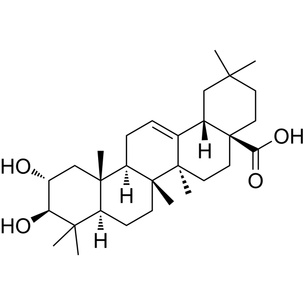
马斯林酸是一种五环三萜类化合物,其结构为齐墩果-12-烯,在2位和3位被羟基取代,在28位被羧基取代(2α,3β立体异构体)。它从油橄榄(Olea europaea)和加那利鼠尾草(Salvia canariensis)中分离得到,具有抗炎、抗氧化和抗肿瘤活性。它是一种植物代谢产物,具有抗氧化、抗肿瘤、抗炎等多种功能。它是一种五环三萜类化合物,也是一种二羟基单羧酸。它来源于齐墩果烷的氢化物。
据报道,丹参、白花铁线莲以及其他有相关数据的生物体中均含有马斯林酸。 另见:红花矢车菊(Centaurium erythraea)全株(部分)。 作用机制 马斯林酸是一种五环三萜,存在于油橄榄(Olea europaea L.)叶片和果实的蜡状保护层中,是一种有前景的结肠癌预防药物。研究人员此前已证实,马斯林酸能显著抑制结肠癌细胞增殖并激活线粒体凋亡。本研究旨在探究该化合物的凋亡分子机制。研究人员使用了HT29腺癌细胞。通过单细胞凝胶电泳(彗星试验)分析了其遗传毒性的变化。采用流式细胞术测定细胞周期。最后,通过蛋白质印迹法检测蛋白质表达的变化。采用学生t检验进行统计学比较。用马斯林酸处理的HT29细胞在处理72小时后,基因毒性显著增加,G0/G1期细胞周期阻滞显著,96小时后出现凋亡亚G0/G1期峰值……马斯林酸的抗肿瘤活性可能通过p53介导的细胞凋亡发挥作用,其机制是通过作用于导致p53活性增加的主要信号通路成分,并诱导参与凋亡通路的其他因子。研究人员发现,在HT29细胞中,马斯林酸激活了c-Jun氨基末端激酶(JNK)的表达,从而诱导p53的表达。用马斯林酸处理肿瘤细胞后,Bid 和 Bax 的表达增加,Bcl-2 的表达受到抑制,细胞色素 c 释放,以及 caspase-9、-3 和 -7 的表达增加。此外,马斯林酸还能诱导 caspase-8 的延迟激活,从而增强初始的线粒体凋亡信号。所有这些结果表明,马斯林酸通过激活 p53,经由 JNK-Bid 介导的线粒体凋亡途径诱导人 HT29 结肠癌细胞凋亡…… 马斯林酸(2-α,3-β-二羟基齐墩果-12-烯-28-酸)是一种来自油橄榄的天然三萜类化合物。该化合物在体外可抑制氧化应激和促炎细胞因子的生成。本研究利用脂多糖(LPS)刺激的大鼠星形胶质细胞培养物,探讨了马斯林酸在中枢神经系统中的抗炎作用。研究采用蛋白质印迹法和定量实时PCR技术,评估了核因子κB(NF-κB)信号转导通路中涉及的不同蛋白。结果表明,马斯林酸通过抑制一氧化氮和肿瘤坏死因子α(TNF-α)的产生,发挥了显著的抗炎作用。蛋白质印迹分析显示,马斯林酸以浓度依赖的方式减弱了LPS诱导的NF-κB p65亚基向细胞核的转位,并抑制了LPS诱导的IκBα磷酸化。此外,马斯林酸显著抑制了环氧合酶2(COX-2)和诱导型一氧化氮合酶(iNOS)在蛋白和mRNA水平的表达。这些结果表明,马斯林酸可能通过抑制培养的皮质星形胶质细胞中的NF-κB信号转导通路来减轻神经炎症。 受体激活因子NF-κB配体(RANKL)激活NF-κB和MAPK/激活蛋白1(AP-1)信号通路对于破骨细胞活性至关重要。靶向NF-κB和MAPK/AP-1信号通路来调节破骨细胞活性一直是治疗破骨细胞相关疾病的一种有前景的策略。本研究通过体外和体内实验系统,探讨了广泛存在于食用植物中的五环三萜酸——马斯林酸(MA)对RANKL诱导的破骨细胞生成、破骨细胞功能和信号通路的影响。在小鼠骨髓单核细胞 (BMM) 和 RAW264.7 细胞中,MA 在非生长抑制浓度范围内以剂量依赖的方式抑制 RANKL 诱导的破骨细胞生成,并降低破骨细胞生成相关标志基因的表达,包括 TRACP、MMP9、c-Src、CTR 和组织蛋白酶 K。具体而言,MA 在早期阶段抑制破骨细胞生成和肌动蛋白环的形成。在卵巢切除小鼠中,给予 MA 可通过抑制破骨细胞活性来预防卵巢切除引起的骨丢失。在分子水平上,MA 可消除 MAPK 的磷酸化和 AP-1 的活性,抑制 IκBα 的磷酸化和降解,并通过下调 RANK 的表达和阻断 RANK 与 TRAF6 的相互作用来抑制 NF-κB/p65 的磷酸化、核转位和 DNA 结合活性。这些数据共同表明,MA通过NF-κB和MAPK/AP-1信号通路抑制RANKL诱导的破骨细胞生成,并且MA有望成为治疗骨质疏松症等破骨细胞相关疾病的有效药物。 1. 马斯林酸(Maslinic acid)是一种五环三萜类化合物,主要从橄榄皮提取物中分离得到。其抗炎和抗关节炎作用主要通过抑制NF-κB信号通路实现——具体而言,是通过降低IκBα磷酸化水平和阻止NF-κB p65核转位,从而抑制促炎细胞因子和介质的产生[1] 2. 研究表明,马斯林酸能够缓解小鼠胶原诱导性关节炎,且无明显毒性,提示其具有开发抗炎或抗关节炎药物的潜力[1] |
| 分子式 |
C30H48O4
|
|---|---|
| 分子量 |
472.6997
|
| 精确质量 |
472.355
|
| CAS号 |
4373-41-5
|
| PubChem CID |
73659
|
| 外观&性状 |
White to off-white solid powder
|
| 密度 |
1.1±0.1 g/cm3
|
| 沸点 |
570.0±50.0 °C at 760 mmHg
|
| 熔点 |
249 - 250 °C
|
| 闪点 |
312.6±26.6 °C
|
| 蒸汽压 |
0.0±3.6 mmHg at 25°C
|
| 折射率 |
1.568
|
| LogP |
7.87
|
| tPSA |
77.76
|
| 氢键供体(HBD)数目 |
3
|
| 氢键受体(HBA)数目 |
4
|
| 可旋转键数目(RBC) |
1
|
| 重原子数目 |
34
|
| 分子复杂度/Complexity |
919
|
| 定义原子立体中心数目 |
9
|
| SMILES |
C[C@@]12CC[C@@H]3[C@@]([C@H]1CC=C4[C@]2(CC[C@@]5([C@H]4CC(CC5)(C)C)C(=O)O)C)(C[C@H]([C@@H](C3(C)C)O)O)C
|
| InChi Key |
MDZKJHQSJHYOHJ-LLICELPBSA-N
|
| InChi Code |
InChI=1S/C30H48O4/c1-25(2)12-14-30(24(33)34)15-13-28(6)18(19(30)16-25)8-9-22-27(5)17-20(31)23(32)26(3,4)21(27)10-11-29(22,28)7/h8,19-23,31-32H,9-17H2,1-7H3,(H,33,34)/t19-,20+,21-,22+,23-,27-,28+,29+,30-/m0/s1
|
| 化学名 |
(4aS,6aR,6aS,6bR,8aR,10R,11R,12aR,14bS)-10,11-dihydroxy-2,2,6a,6b,9,9,12a-heptamethyl-1,3,4,5,6,6a,7,8,8a,10,11,12,13,14b-tetradecahydropicene-4a-carboxylic acid
|
| HS Tariff Code |
2934.99.9001
|
| 存储方式 |
Powder -20°C 3 years 4°C 2 years In solvent -80°C 6 months -20°C 1 month |
| 运输条件 |
Room temperature (This product is stable at ambient temperature for a few days during ordinary shipping and time spent in Customs)
|
| 溶解度 (体外实验) |
DMSO : ~100 mg/mL (~211.55 mM)
|
|---|---|
| 溶解度 (体内实验) |
配方 1 中的溶解度: 2.5 mg/mL (5.29 mM) in 10% DMSO + 90% (20% SBE-β-CD in Saline) (这些助溶剂从左到右依次添加,逐一添加), 悬浮液;超声助溶。
例如,若需制备1 mL的工作液,可将100 μL 25.0 mg/mL澄清DMSO储备液加入900 μL 20% SBE-β-CD生理盐水溶液中,混匀。 *20% SBE-β-CD 生理盐水溶液的制备(4°C,1 周):将 2 g SBE-β-CD 溶解于 10 mL 生理盐水中,得到澄清溶液。 配方 2 中的溶解度: ≥ 2.5 mg/mL (5.29 mM) (饱和度未知) in 10% DMSO + 90% Corn Oil (这些助溶剂从左到右依次添加,逐一添加), 澄清溶液。 例如,若需制备1 mL的工作液,可将 100 μL 25.0 mg/mL 澄清 DMSO 储备液添加到 900 μL 玉米油中并混合均匀。 请根据您的实验动物和给药方式选择适当的溶解配方/方案: 1、请先配制澄清的储备液(如:用DMSO配置50 或 100 mg/mL母液(储备液)); 2、取适量母液,按从左到右的顺序依次添加助溶剂,澄清后再加入下一助溶剂。以 下列配方为例说明 (注意此配方只用于说明,并不一定代表此产品 的实际溶解配方): 10% DMSO → 40% PEG300 → 5% Tween-80 → 45% ddH2O (或 saline); 假设最终工作液的体积为 1 mL, 浓度为5 mg/mL: 取 100 μL 50 mg/mL 的澄清 DMSO 储备液加到 400 μL PEG300 中,混合均匀/澄清;向上述体系中加入50 μL Tween-80,混合均匀/澄清;然后继续加入450 μL ddH2O (或 saline)定容至 1 mL; 3、溶剂前显示的百分比是指该溶剂在最终溶液/工作液中的体积所占比例; 4、 如产品在配制过程中出现沉淀/析出,可通过加热(≤50℃)或超声的方式助溶; 5、为保证最佳实验结果,工作液请现配现用! 6、如不确定怎么将母液配置成体内动物实验的工作液,请查看说明书或联系我们; 7、 以上所有助溶剂都可在 Invivochem.cn网站购买。 |
| 制备储备液 | 1 mg | 5 mg | 10 mg | |
| 1 mM | 2.1155 mL | 10.5775 mL | 21.1551 mL | |
| 5 mM | 0.4231 mL | 2.1155 mL | 4.2310 mL | |
| 10 mM | 0.2116 mL | 1.0578 mL | 2.1155 mL |
1、根据实验需要选择合适的溶剂配制储备液 (母液):对于大多数产品,InvivoChem推荐用DMSO配置母液 (比如:5、10、20mM或者10、20、50 mg/mL浓度),个别水溶性高的产品可直接溶于水。产品在DMSO 、水或其他溶剂中的具体溶解度详见上”溶解度 (体外)”部分;
2、如果您找不到您想要的溶解度信息,或者很难将产品溶解在溶液中,请联系我们;
3、建议使用下列计算器进行相关计算(摩尔浓度计算器、稀释计算器、分子量计算器、重组计算器等);
4、母液配好之后,将其分装到常规用量,并储存在-20°C或-80°C,尽量减少反复冻融循环。
计算结果:
工作液浓度: mg/mL;
DMSO母液配制方法: mg 药物溶于 μL DMSO溶液(母液浓度 mg/mL)。如该浓度超过该批次药物DMSO溶解度,请首先与我们联系。
体内配方配制方法:取 μL DMSO母液,加入 μL PEG300,混匀澄清后加入μL Tween 80,混匀澄清后加入 μL ddH2O,混匀澄清。
(1) 请确保溶液澄清之后,再加入下一种溶剂 (助溶剂) 。可利用涡旋、超声或水浴加热等方法助溶;
(2) 一定要按顺序加入溶剂 (助溶剂) 。
InvivoChem的所有产品仅用于作科学研究,不面向患者销售
Copyright 2020 InvivoChem LLC | All Rights Reserved 粤ICP备20063088号-1


 463611831
