| 规格 | 价格 | 库存 | 数量 |
|---|---|---|---|
| 1mg |
|
||
| 2mg |
|
||
| 5mg |
|
||
| 10mg |
|
||
| 25mg |
|
||
| 50mg |
|
||
| 100mg |
|
||
| 250mg |
|
||
| Other Sizes |
|
| 靶点 |
Tubulin; microtubule
|
|---|---|
| 体外研究 (In Vitro) |
MMAF 可预防间变性大细胞淋巴瘤的生长。体外细胞毒性测定对 Karpas 299、乳腺癌 H3396、肾细胞癌 786-O 和 Caki-1 细胞的 IC50 值分别为 119、105、257 和 200 nM[4]。
体外生长抑制。cAC10−L1−MMAF4在CD30阳性和阴性细胞系上进行了测试,如表2所示。将细胞连续暴露于ADC 96小时,并将Alamar Blue转化率测定的细胞毒性作用与游离MMAF进行比较。以摩尔计,cAC10−L1−MMAF4的效力平均比游离的MMAF高2200倍以上,并且对所有测试的CD30阳性细胞系都有活性。这些作用是免疫特异性的,因为不表达可检测水平的CD30抗原的WSU-NHL细胞不受偶联物的影响。还对基于cAC10−L1−MMAF-OMe4的偶联物进行了有限的研究,发现它们对Karpas 299(0.04 nM)和L540cy(0.11 nM)细胞的IC50值与cAC10-L1-MMAF4的IC50值相当(表2)。这与之前所述的假设一致,即MMAF OMe的活性形式是游离酸MMAF[4]。 对LewisY阳性人小细胞肺癌细胞系(H69)及其P-糖蛋白过表达的对应物H69/LX4进行了进一步的研究,使用MMAF和cBR96-L1-MMAF8缀合物。将结果与阿霉素和cBR96-L1−doxorbicin8进行了比较,这是一种之前描述的与8个阿霉素分子/mAb的偶联物。与亲本细胞相比,H69/LX-4细胞对阿霉素的敏感性低约100倍(图2A),而与MMAF的差异仅为3倍(图2B)。这一趋势延伸到偶联物,因为耐药细胞系对BR96-L1-doxorbicin8耐药(图2C),但对相应的MMAF偶联物非常敏感(图2D)。这些结果已在几种MDR阳性细胞系上得到证实(数据未显示),其中发现MMAF和基于MMAF的免疫偶联物绕过了常见形式的MDR表型[4]。 为了评估结合亲和力对RDCs细胞毒性活性的影响,我们使用对EGFR具有不同结合亲和力的repebody合成了三种不同的药物偶联物,并检测了它们对EGFR过表达HCC827细胞的细胞毒性(图S9和表S1)。与其他两种低亲和力偶联物相比,rEgH9-MMAF偶联物表现出最高的细胞毒性。这一结果表明,repebody对EGFR的结合亲和力对RDCs的细胞毒性活性至关重要,优化结合亲和力可以增强药物偶联物的细胞毒性。为了研究EGFR的细胞表面表达水平与RDCs的细胞毒性之间的关系,我们培养了三种表达不同EGFR水平的癌症细胞系(图 3 a) 用不同浓度的rEgH9-MMAF偶联物处理三天(图3b-d)。repebody-MMAF偶联物以剂量依赖的方式对A431和HCC827细胞显示出强烈的细胞毒性作用,导致有效半最大浓度(EC50)分别为1.4 nM和0.072 nM(表1)。这一结果表明,repebody-MMAF缀合物对HCC827细胞的细胞毒性远高于游离、细胞不可渗透的MMAF(EC50=117.9 nM)和裸repebodys(rEgH9:EC50=17.2 nM)。有趣的是,repebody-MMAF偶联物在HCC827细胞中的效力远高于A431细胞。这一结果似乎是由于HCC827细胞表达组成型内化的致癌EGFR,因此对repebody-MMAF偶联物的摄取增加。15即使在高剂量下(>200 nM;表 1). 我们的研究结果表明,RDCs可以以受体特异性的方式有效地将强效抗癌药物递送到靶细胞,最大限度地减少脱靶效应。上得到证实(数据未显示),其中发现MMAF和基于MMAF的免疫偶联物绕过了常见形式的MDR表型[4]。 |
| 体内研究 (In Vivo) |
与MMAE(1 mg/kg)相比,小鼠对MMAF的最大耐受剂量(MTD)明显更大(>16 mg/kg)。与 cAC10-L1-MMAF4 在小鼠中的 50 mg/kg MTD 相比,等效的 cAC10-L4-MMAF4 ADC 危险性较小,关联和相关 MTD 分别 >150 mg/kg 和 90 mg/kg。 [4]。
体内治疗研究。[4] 在患有皮下或播散性Karpas 299肿瘤的裸鼠中测定ADC的治疗效果。在患有皮下肿瘤的动物中,单次注射cAC10-L1-MMAF4或cAC10-L4-MMAF4可获得显著的抗肿瘤作用,其效力和活性难以区分(图5A)。几乎所有接受2mg抗体成分/kg体重单次ADC注射的动物都治愈了。当剂量降至1mg/kg时,两种ADC的疗效以明显相等的方式下降。在这个剂量下,分别用cAC10-L1-MMAF4和cAC10-L4-MMAF4 ADC治愈的六只动物中仍有两只和三只。 免疫组织化学分析。[4] 进行了研究,以确定cAC10−L1−MMAF4是否定位在裸鼠皮下Karpas 299肿瘤中。动物静脉注射cAC10-L1-MMAF4或cBR96-L1-MMAF4,每公斤体重10毫克抗体成分。24小时后切除肿瘤,分别使用生物素化的抗人Fc和抗MMAF单克隆抗体作为二抗,使用免疫组织化学评估冷冻切片中是否存在单克隆抗体和药物成分。图4A和4C显示,与cBR96非结合对照ADC相比,cAC10−L1−MMAF4在卡帕斯299肿瘤中的积累效率更高(图4B和4D)。由于单克隆抗体(图4A)和药物(图4C)部分在整个肿瘤中都被检测到,我们得出结论,ADC是作为一个完整的分子递送的。 研究人员还使用HCC827细胞评估了rEgH9-MMAF偶联物在异种移植物小鼠中的抗肿瘤活性。当肿瘤体积达到110至130 mm3时,小鼠每天静脉注射(10 mg kg-1)repebody-MMAF偶联物或裸repebody(rEgH9)六天(图4a)。作为阳性对照,使用西妥昔单抗。repebody-MMAF偶联物显示出显著的肿瘤消退反应(第二天残留肿瘤33.4%) 20, ***P<0.001)与裸瘤(第20天残留肿瘤83.4%,P> 0.05)和西妥昔单抗。在第20天,除了短暂的体重减轻外,未在接受治疗的小鼠中检测到明显的不良反应(P>0.05)(图4b)。 |
| 酶活实验 |
Released Drug Identification. [4]
L540cy细胞的溶酶体提取物是通过将2.4×10⁸个细胞在9 mL 0.25 M蔗糖、1 mM EDTA和10 mM HEPES(4-(2-羟乙基)哌嗪-1-乙磺酸)(pH 7.4)中溶胀而制备的。在冰上放置30分钟后,将细胞进行Dounce均质化,直至通过台盼蓝染料排斥法测得>95%的细胞破碎。将匀浆离心(3000g,10分钟,4°C)以沉淀细胞碎片,将上清液(每管4×10⁷个细胞当量)转移到聚铝醇超离心管(13×51 mm)中,并在TLA100.3转子中离心(17000g,15分钟,4℃)以分离含溶酶体的轻线粒体沉淀。颗粒储存在-80°C下。将颗粒解冻并重新悬浮在500μL pH 5.0的50 mM乙酸钠和2 mM DTT中。将cAC10−L4-[12C]MMAF4、cAC10−于L4-[13C]MMAF4和cAC10−着L4-[12/13C]MMAF4(50μg/mL)独立添加到一个颗粒中。经过三次冻融循环以打开溶酶体后,样品在37°C下孵育24小时。加入冷甲醇(2体积)沉淀蛋白质,将样品在14000g下离心至颗粒碎片,如上所述通过低分辨率质谱分析100μL上清液。通过在室温下用1 mM半胱氨酸在PBS中处理100μM L4-MMAF 10分钟,制备了真正的半胱氨酸-L4-MMAF。 |
| 细胞实验 |
根据细胞系的不同,用连续稀释的测试分子处理细胞并孵育四到六天。 Alamar Blue 染料还原测定用于评估细胞生长并减少数据,以产生 IC50 值[2]。
体外生长抑制。[4] 收集细胞的对数相培养物,并根据预定条件以500至10000个细胞/孔的接种密度接种细胞。在孵育24小时以允许表面蛋白重构后,加入一系列稀释的测试分子,并根据细胞系将培养物再孵育4-6天。根据先前发表的方法,使用如前所述的Alamar Blue染料还原测定法对细胞生长和数据减少进行评估以产生IC50值。简而言之,在添加培养物之前,在完全培养基中新鲜制备了40%的Alamar Blue溶液(wt/vol)。在药物暴露92小时后,将Alamar Blue溶液加入细胞中以构成10%的培养体积。将细胞孵育4小时,并在Fusion HT荧光平板读数器上测量染料减少。 |
| 动物实验 |
小鼠:皮下 Karpas 299 肿瘤大小为 300 mm³,每组三只动物分别接受一次静脉注射 cAC10-L1-MMAF⁴ 或 cBR96-L1-MMAF⁴(抗体成分 10 mg/kg 体重)。肿瘤切除后,将组织切片(厚度 5 μm)置于最佳切割温度化合物 (OCT) 中,然后进行免疫组织化学染色[1]。体内治疗实验[4]:对于间变性大细胞淋巴瘤的局限性皮下疾病模型,将 5 × 10⁶ 个 Karpas 299 细胞植入 CB-17 SCID 小鼠的右侧腹部。当每组六只动物的肿瘤平均大小约为 100 mm³ 时开始治疗。治疗方法为单次静脉注射PBS缓冲液配制的缀合物或对照溶液(尾静脉)。肿瘤体积采用公式(L × W²)/2计算。对于播散性间变性大细胞淋巴瘤(ALCL)模型,将1 × 10⁶个Karpas 299细胞经尾静脉注射到CB-17 SCID小鼠体内。在肿瘤注射后9天进行单次注射治疗。
通过免疫组织化学进行抗体体内定位。[4] 当皮下Karpas 299肿瘤体积达到300 mm³时,每组三只动物静脉注射10 mg/kg体重的抗体成分cAC10-L1-MMAF4或cBR96-L1-MMAF4。随后切除肿瘤并包埋于最佳切割温度(OCT)化合物中,对5 μm厚的冷冻组织切片进行免疫组织化学染色评估。简而言之,将冷冻组织置于载玻片上风干,然后在室温下用4%多聚甲醛固定15分钟。用0.6% H₂O₂封闭内源性过氧化物酶活性15分钟。使用Avidin-Biotin封闭试剂盒进一步封闭内源性生物素。将生物素标记的抗人Fc抗体和生物素标记的抗药物抗体以2 μg/mL的浓度在室温下与组织孵育1小时。将载玻片与HRP标记的亲和素孵育后,使用3,3'-二联苯胺(DAB)作为HRP的底物。用苏木精对组织进行复染,然后脱水,并加盖玻片。使用蔡司Axiovert光学显微镜拍摄图像。 RDC 的体内抗肿瘤活性。[2] a) 将裸鼠异种移植瘤小鼠 (HCC827) 在肿瘤建立后,每天静脉注射 rEgH9–MMAF 偶联物、裸露的重复抗体或西妥昔单抗 (10 mg kg−1),连续 6 天。每隔 3 天测量一次肿瘤大小,持续 20 天(平均值±标准差;n=6)。b) 小鼠体重变化。给药后,每隔 3 天测量一次每只小鼠的体重。 |
| 毒性/毒理 (Toxicokinetics/TK) |
最大耐受剂量。[4]
cAC10-MMAF偶联物的最大耐受剂量 (MTD) 在 BALB/c 小鼠和 Sprague-Dawley 大鼠中测定,定义为在任何动物中均未引起超过 20% 的体重减轻、痛苦或明显毒性的最高剂量。该剂量通常在引起上述事件的剂量的 20% 以内。cAC10-L1-MMAF4 在小鼠中的 MTD 为 50 mg/kg,在大鼠中的 MTD 为 15 mg/kg。相应的 cAC10-L4-MMAF4 ADC 毒性要低得多,在小鼠和大鼠中的 MTD 分别为 >150 mg/kg(测试的最高剂量,未导致明显毒性)和 90 mg/kg。这清楚地表明,药物与 mAb 的连接方式会对 ADC 的耐受性产生显著影响,并且连接子中缺少肽间隔基的 ADC 比基于肽的 ADC 毒性要小得多。 |
| 参考文献 |
|
| 其他信息 |
基于蛋白质-药物偶联物的靶向治疗因其高效低副作用而备受关注。然而,如何高效稳定地将药物偶联到蛋白质结合剂上仍然是一个挑战。本文提出了一种高效制备高稳定性、高纯度药物偶联物的化学酶法。该方法包括在蛋白质结合剂的C端插入CaaX序列,利用法尼基转移酶进行异戊二烯化修饰,以及通过肟连接反应进行药物偶联。MMAF和EGFR特异性重复抗体分别用作抗肿瘤药物和蛋白质结合剂。该方法能够精确控制地合成高产率、高纯度的重复抗体-药物偶联物。该方法的实用性体现在重复抗体-药物偶联物在人血浆中具有显著的稳定性,脱靶效应可忽略不计,以及在体内表现出显著的抗肿瘤活性。本方法可广泛用于制备高度均一且稳定的PDC,以用于靶向治疗。[2]抗体药物偶联物利用抗体作为递送载体,递送针对肿瘤相关抗原的高效细胞毒性分子,用于癌症治疗。成功开发用于临床的抗体药物偶联物的关键参数包括:肿瘤靶抗原的选择、针对该靶点的抗体、细胞毒性分子、连接细胞毒性分子和抗体的连接子,以及用于将细胞毒性分子连接到抗体上的偶联化学方法。Adectris®(抗CD30药物偶联物)和Kadcyla®(抗HER2药物偶联物)的近期获批反映了这些核心抗体药物偶联物技术的进步。抗CD22偶联物的潜在获批以及抗CD19和抗CD33偶联物令人鼓舞的新临床数据是该领域的又一进展。利用新开发的强效细胞毒性分子和连接子富集抗体药物偶联物(ADC)也是针对多种肿瘤靶点的研究方向之一。然而,ADC组分的复杂性、偶联方法的复杂性以及脱靶毒性仍然是ADC策略设计中发挥其最大治疗潜力的一大挑战。本文将探讨临床ADC的出现、优化策略的最新趋势以及应用最新优化策略的ADC研究成果。此外,本文还将讨论未来如何使ADC更适用于更广泛的疾病适应症,以及面临的挑战和展望。[3]我们此前已证明,由cAC10(抗CD30)与抗有丝分裂剂单甲基澳瑞他汀E(MMAE)连接的ADC在体外和体内均对抗原阳性肿瘤模型表现出显著的活性。 MMAF 是一种新型抗有丝分裂奥瑞他汀衍生物,其 C 端带有带电荷的苯丙氨酸残基,与不带电荷的对应物 MMAE 相比,该残基会减弱其细胞毒活性,这很可能是由于其胞内转运受损所致。体外细胞毒性研究表明,mAb-马来酰亚胺己酰-缬氨酸-瓜氨酸-对氨基苄氧羰基-MMAF (mAb-L1-MMAF) 偶联物在多种 CD30 阳性造血细胞系上的效力比游离 MMAF 高 2200 倍以上。与 cAC10-L1-MMAE 类似,相应的 MMAF 抗体药物偶联物 (ADC) 在耐受剂量下可治愈或消退已建立的异种移植瘤。为了进一步优化 ADC,研究人员构建了几种新的连接子,其中 L1 连接子中的各种组分被改变或删除。其中一种最有前景的连接子在药物和单克隆抗体之间包含一个不可裂解的马来酰亚胺己酰基(L4)间隔基。cAC10-L4-MMAF 在体外对多种细胞系的活性与 cAC10-L1-MMAF 相当,体内活性也相同。重要的是,cAC10-L4-MMAF 的耐受剂量是 cAC10-L1-MMAF 的 3 倍以上。液相色谱-质谱联用(LCMS)研究表明,从 cAC10-L4-MMAF 中释放的药物是半胱氨酸-L4-MMAF 加合物,这可能是由于单克隆抗体在靶细胞溶酶体内降解所致。这种新型连接子技术似乎非常适合那些细胞渗透性相对较低且能耐受氨基酸取代的药物。因此,连接子的改变会对毒性产生显著影响,并可开发出治疗指数大幅提高的新型抗体药物偶联物(ADC)。[4]
|
| 分子式 |
C39H65N5O8
|
|---|---|
| 分子量 |
731.98
|
| 精确质量 |
731.483
|
| 元素分析 |
C, 64.00; H, 8.95; N, 9.57; O, 17.49
|
| CAS号 |
745017-94-1
|
| 相关CAS号 |
MMAF hydrochloride;1415246-68-2;MMAF-d8 hydrochloride;MMAF sodium;1799706-65-2;MMAF-OMe;863971-12-4;MMAF sodium;1799706-65-2;MMAF;745017-94-1;MMAF-d8
|
| PubChem CID |
10395173
|
| 外观&性状 |
White to off-white solid powder
|
| 密度 |
1.1±0.1 g/cm3
|
| 沸点 |
896.8±65.0 °C at 760 mmHg
|
| 闪点 |
496.2±34.3 °C
|
| 蒸汽压 |
0.0±0.3 mmHg at 25°C
|
| 折射率 |
1.522
|
| LogP |
4.36
|
| tPSA |
173.59
|
| 氢键供体(HBD)数目 |
4
|
| 氢键受体(HBA)数目 |
9
|
| 可旋转键数目(RBC) |
21
|
| 重原子数目 |
52
|
| 分子复杂度/Complexity |
1160
|
| 定义原子立体中心数目 |
9
|
| SMILES |
O(C([H])([H])[H])[C@]([H])([C@]([H])(C(N([H])[C@]([H])(C(=O)O[H])C([H])([H])C1C([H])=C([H])C([H])=C([H])C=1[H])=O)C([H])([H])[H])[C@]1([H])C([H])([H])C([H])([H])C([H])([H])N1C(C([H])([H])C([H])([C@]([H])([C@@]([H])(C([H])([H])[H])C([H])([H])C([H])([H])[H])N(C([H])([H])[H])C([C@]([H])(C([H])(C([H])([H])[H])C([H])([H])[H])N([H])C([C@]([H])(C([H])(C([H])([H])[H])C([H])([H])[H])N([H])C([H])([H])[H])=O)=O)OC([H])([H])[H])=O
|
| InChi Key |
MFRNYXJJRJQHNW-DEMKXPNLSA-N
|
| InChi Code |
InChI=1S/C39H65N5O8/c1-12-25(6)34(43(9)38(48)33(24(4)5)42-37(47)32(40-8)23(2)3)30(51-10)22-31(45)44-20-16-19-29(44)35(52-11)26(7)36(46)41-28(39(49)50)21-27-17-14-13-15-18-27/h13-15,17-18,23-26,28-30,32-35,40H,12,16,19-22H2,1-11H3,(H,41,46)(H,42,47)(H,49,50)/t25-,26+,28-,29-,30+,32-,33-,34-,35+/m0/s1
|
| 化学名 |
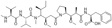
(2S)-2-[[(2R,3R)-3-methoxy-3-[(2S)-1-[(3R,4S,5S)-3-methoxy-5-methyl-4-[methyl-[(2S)-3-methyl-2-[[(2S)-3-methyl-2-(methylamino)butanoyl]amino]butanoyl]amino]heptanoyl]pyrrolidin-2-yl]-2-methylpropanoyl]amino]-3-phenylpropanoic acid
|
| 别名 |
MMAF; Monomethyl auristatin F; MMAF; 745017-94-1; Monomethyl Auristatin F; MMAF (GMP); (2S)-2-[[(2R,3R)-3-methoxy-3-[(2S)-1-[(3R,4S,5S)-3-methoxy-5-methyl-4-[methyl-[(2S)-3-methyl-2-[[(2S)-3-methyl-2-(methylamino)butanoyl]amino]butanoyl]amino]heptanoyl]pyrrolidin-2-yl]-2-methylpropanoyl]amino]-3-phenylpropanoic acid; MONOMETHYLAURISTATIN PHENYLALANINE; MFCD25976742; (S)-2-((2R,3R)-3-((S)-1-((3R,4S,5S)-4-((S)-N,3-Dimethyl-2-((S)-3-methyl-2-(methylamino)butanamido)butanamido)-3-methoxy-5-methylheptanoyl)pyrrolidin-2-yl)-3-methoxy-2-methylpropanamido)-3-phenylpropanoic acid; desmethyl-auristatin F;
|
| HS Tariff Code |
2934.99.9001
|
| 存储方式 |
Powder -20°C 3 years 4°C 2 years In solvent -80°C 6 months -20°C 1 month 注意: (1). 该产品在溶液状态不稳定,请现配现用。 (2). 请将本产品存放在密封且受保护的环境中,避免吸湿/受潮。 |
| 运输条件 |
Room temperature (This product is stable at ambient temperature for a few days during ordinary shipping and time spent in Customs)
|
| 溶解度 (体外实验) |
DMSO: 100~140 mg/mL (136.6~191.3 mM)
Ethanol: ~100 mg/mL (~136.6 mM) Water: ~100 mg/mL (~136.6 mM) |
|---|---|
| 溶解度 (体内实验) |
配方 1 中的溶解度: ≥ 3.5 mg/mL (4.78 mM) (饱和度未知) in 10% DMSO + 90% (20% SBE-β-CD in Saline) (这些助溶剂从左到右依次添加,逐一添加), 澄清溶液。
例如,若需制备1 mL的工作液,可将100 μL 35.0mg/mL澄清的DMSO储备液加入到900μL 20%SBE-β-CD生理盐水中,混匀。 *20% SBE-β-CD 生理盐水溶液的制备(4°C,1 周):将 2 g SBE-β-CD 溶解于 10 mL 生理盐水中,得到澄清溶液。 配方 2 中的溶解度: ≥ 3.5 mg/mL (4.78 mM) (饱和度未知) in 10% DMSO + 90% Corn Oil (这些助溶剂从左到右依次添加,逐一添加), 澄清溶液。 例如,若需制备1 mL的工作液,可将 100 μL 35.0 mg/mL 澄清 DMSO 储备液加入900 μL 玉米油中,混合均匀。 请根据您的实验动物和给药方式选择适当的溶解配方/方案: 1、请先配制澄清的储备液(如:用DMSO配置50 或 100 mg/mL母液(储备液)); 2、取适量母液,按从左到右的顺序依次添加助溶剂,澄清后再加入下一助溶剂。以 下列配方为例说明 (注意此配方只用于说明,并不一定代表此产品 的实际溶解配方): 10% DMSO → 40% PEG300 → 5% Tween-80 → 45% ddH2O (或 saline); 假设最终工作液的体积为 1 mL, 浓度为5 mg/mL: 取 100 μL 50 mg/mL 的澄清 DMSO 储备液加到 400 μL PEG300 中,混合均匀/澄清;向上述体系中加入50 μL Tween-80,混合均匀/澄清;然后继续加入450 μL ddH2O (或 saline)定容至 1 mL; 3、溶剂前显示的百分比是指该溶剂在最终溶液/工作液中的体积所占比例; 4、 如产品在配制过程中出现沉淀/析出,可通过加热(≤50℃)或超声的方式助溶; 5、为保证最佳实验结果,工作液请现配现用! 6、如不确定怎么将母液配置成体内动物实验的工作液,请查看说明书或联系我们; 7、 以上所有助溶剂都可在 Invivochem.cn网站购买。 |
| 制备储备液 | 1 mg | 5 mg | 10 mg | |
| 1 mM | 1.3662 mL | 6.8308 mL | 13.6616 mL | |
| 5 mM | 0.2732 mL | 1.3662 mL | 2.7323 mL | |
| 10 mM | 0.1366 mL | 0.6831 mL | 1.3662 mL |
1、根据实验需要选择合适的溶剂配制储备液 (母液):对于大多数产品,InvivoChem推荐用DMSO配置母液 (比如:5、10、20mM或者10、20、50 mg/mL浓度),个别水溶性高的产品可直接溶于水。产品在DMSO 、水或其他溶剂中的具体溶解度详见上”溶解度 (体外)”部分;
2、如果您找不到您想要的溶解度信息,或者很难将产品溶解在溶液中,请联系我们;
3、建议使用下列计算器进行相关计算(摩尔浓度计算器、稀释计算器、分子量计算器、重组计算器等);
4、母液配好之后,将其分装到常规用量,并储存在-20°C或-80°C,尽量减少反复冻融循环。
计算结果:
工作液浓度: mg/mL;
DMSO母液配制方法: mg 药物溶于 μL DMSO溶液(母液浓度 mg/mL)。如该浓度超过该批次药物DMSO溶解度,请首先与我们联系。
体内配方配制方法:取 μL DMSO母液,加入 μL PEG300,混匀澄清后加入μL Tween 80,混匀澄清后加入 μL ddH2O,混匀澄清。
(1) 请确保溶液澄清之后,再加入下一种溶剂 (助溶剂) 。可利用涡旋、超声或水浴加热等方法助溶;
(2) 一定要按顺序加入溶剂 (助溶剂) 。
|
|
|
InvivoChem的所有产品仅用于作科学研究,不面向患者销售
Copyright 2020 InvivoChem LLC | All Rights Reserved 粤ICP备20063088号-1


 COA
COA


 463611831
