| 规格 | 价格 | 库存 | 数量 |
|---|---|---|---|
| 10mg |
|
||
| 25mg |
|
||
| 50mg |
|
||
| 100mg |
|
||
| 250mg |
|
||
| 500mg |
|
||
| 1g |
|
||
| Other Sizes |
|

| 靶点 |
P2Y6 Receptor
The target of MRS 2578 is the purinergic receptor P2Y6, with an IC50 of 10.8 nM for human P2Y6 receptor and 9.1 nM for rat P2Y6 receptor [1] |
|---|---|
| 体外研究 (In Vitro) |
MRS2578 (1 μM) 完全阻止 1321N1 星形细胞瘤细胞在 TNFα 诱导的细胞凋亡过程中不受 UDP 保护[1]。在 HMEC-1 细胞中,MRS 2578 (10 μM) 完全消除 TNF-α 诱导的 NF-κB 报告基因活性。在 HMEC-1 细胞中,MRS 2578 (10 μM) 显着降低 TNF-α 诱导的促炎基因表达 [2]。
P2Y(6)核苷酸受体的生理作用可能涉及基于受体组织分布的心血管、免疫和消化功能,并且缺乏针对该受体的选择性拮抗剂。我们合成了一系列对称的芳基二异硫氰酸酯衍生物,并研究了它们抑制由五种重组P2Y受体亚型激活诱导的磷脂酶C(PLC)活性的能力。几种衍生物在抑制UDP对1321N1人星形胶质细胞中表达的人和大鼠P2Y(6)受体的作用方面比激活人P2Y(1)、P2Y(2)、P2X(4)和P2Y(11)受体更有效。1,2-二苯基乙烷(MRS2567)和1,4-二(苯基硫脲基)丁烷(MRS2578)的二异硫氰酸酯衍生物的抑制作用是浓度依赖性的,无法克服,IC(50)值分别为126+/-15 nM和37+/-16 nM(人)和101+/-27 nM和98+/-11 nM(大鼠)。1,4-苯二异硫氰酸酯衍生物(MRS2575)仅抑制人而不抑制大鼠P2Y(6)受体活性。MRS2567和MRS2578在10μM时不影响表达P2Y(2)和P2Y(4)受体的细胞的UTP(100nM)诱导的反应,也不影响P2Y(1)受体的2-甲硫基-ADP(30nM)诱导的响应或P2Y(11)受体的ATP(10μM)诱导反应。其他拮抗剂显示出混合选择性。选择性拮抗剂MRS2567、MRS2575和MRS2578(1microM)完全阻断了UDP对经历TNFα诱导凋亡的细胞的保护作用。因此,我们已经鉴定出P2Y(6)受体的强效、不可克服的拮抗剂,这些拮抗剂在PLC偶联的P2Y受体家族中具有选择性。[1] 选择性P2Y6受体拮抗剂MRS2578治疗以浓度依赖的方式抑制了机械牵张诱导的Rho活化,IC50值约为0.1μM(图5A和B)。[2] P2Y6受体拮抗剂MRS 2578在体外抑制介质诱导的血管内皮炎症[3] 在证明P2Y6受体转录物和蛋白质表达在炎症刺激下选择性增加后,我们接下来在体外研究了内皮P2Y6信号传导的功能后果。与体外模型一样,我们用含有p50/p65结合位点的NF-κB报告质粒转染HMEC-1细胞。用P2Y6激动剂尿苷二磷酸处理HMEC-1细胞仅导致NF-κB活性适度增加(1.64倍±0.45[倍;P<.05;补充图1)。然而,与之前关于P2Y6信号传导参与NF-κB活性的研究一致,26,27我们观察到在P2Y6拮抗剂MRS 2578存在的情况下,基础NF-κC活性受到严重抑制。NF-κB活性的降低与时间(图3A)和剂量(图3B)有关。因此,这些研究表明,P2Y6活化的下游靶点对于NF-κB活化是必要的,但还不够,并表明其在增强血管炎症方面具有间接作用[3]。 转染人或大鼠 P2Y6 受体的细胞实验中,MRS 2578 以浓度依赖性方式抑制 UDP 诱导的 P2Y6 受体激活,可阻断受体介导的细胞内 Ca²⁺ 浓度升高,且该抑制作用呈不可逆性 [1] - 选择性实验显示,MRS 2578 对其他嘌呤能受体(P2Y1、P2Y2、P2Y4、P2Y11、P2Y12、P2X1、P2X2、P2X4、P2X7)无明显抑制活性,即使浓度高达 10 μmol/L,对这些受体的抑制率仍低于 20% [1] - 人脐静脉内皮细胞(HUVECs)体外实验中,MRS 2578 预处理可显著抑制 UDP 诱导的炎症因子(IL-8、MCP-1、ICAM-1)mRNA 及蛋白表达,同时阻断 NF-κB 信号通路激活 [3] - 小鼠气道上皮细胞体外培养实验中,MRS 2578 可抑制 IL-4 诱导的 P2Y6 受体表达上调,减少趋化因子(CCL11、CCL24)的分泌,进而抑制嗜酸性粒细胞趋化 [4] - 大鼠心肌成纤维细胞实验中,MRS 2578 能阻断 P2Y6-Gα12/13 信号通路激活,抑制细胞增殖及胶原蛋白(Col1a1、Col3a1)的合成 [2] |
| 体内研究 (In Vivo) |
横主动脉缩窄 (TAC) 后,MRS2578(3 mg/kg;腹膜内注射;持续 3 天)可显着减少压力过载诱导的胶原沉积,而不影响心肌细胞肥大[4]。
抑制P2Y6受体可减轻体内压力超负荷诱导的心脏纤维化[2] 研究人员接下来研究了嘌呤能受体是否真的参与了体内压力超负荷诱导的心脏纤维化。TAC后使用MRS2578治疗显著抑制了压力超负荷诱导的胶原沉积,而不影响心肌细胞肥大(图6A-C)。MRS2578治疗显著抑制了压力超负荷引起的左心室功能障碍(图6D和E以及补充表3)。此外,MRS2578治疗抑制了压力超负荷引起的ANP、β-MHC、I型前胶原、骨膜炎蛋白和TGF-β2 mRNA表达的增加(图6F)。我们还发现,MRS2578抑制了压力超负荷诱导的Rho激活,TAC诱导骨膜炎蛋白、成熟TGF-βs和ACE蛋白表达的增加(图6G和H)。此外,我们发现苏拉明治疗还抑制了压力超负荷诱导的胶原沉积和左心室功能障碍(补充图7和补充表4)。这些结果表明,抑制P2Y6受体实际上可以减轻压力超负荷诱导的心脏纤维化和左心室功能障碍。 测量和主要结果:我们观察到气管内应用P2Y6R拮抗剂(MRS2578)和P2Y6R缺乏抑制了哮喘的主要特征,如支气管肺泡灌洗液嗜酸性粒细胞增多、气道重塑、Th2细胞因子产生和卵清蛋白-明矾模型中的支气管高反应性。在使用屋尘螨提取物诱导过敏性肺部炎症的模型中,MRS2578也能有效减少气道炎症。骨髓嵌合体实验揭示了P2Y6R在气道炎症中肺结构细胞表达的重要性。根据这一发现,我们发现实验性哮喘动物气道上皮细胞上P2Y6的表达强烈上调。关于潜在的机制,我们观察到MRS2578在体内抑制了肺上皮细胞释放IL-6和IL-8/KC,而肺内应用P2Y6R激动剂尿苷-5'-二磷酸会增加支气管肺泡中IL-6和KC的水平。此外,P2Y6受体的选择性激活在体外诱导小鼠和人肺上皮细胞释放IL-6和KC/IL-8。 结论:在急性和慢性过敏性气道炎症期间,气道上皮细胞上的P2Y6R表达上调,选择性阻断P2Y6R或P2Y6R缺乏对结构细胞的影响可减少实验性哮喘的主要特征。因此,阻断肺部P2Y6R可能是治疗过敏性气道炎症的靶点[4]。 小鼠压力超负荷心肌纤维化模型(主动脉缩窄术诱导)中,腹腔注射 MRS 2578(10 mg/kg,每日 1 次,持续 4 周)可显著降低心肌组织中胶原蛋白沉积,抑制成纤维细胞活化标志物(α-SMA)的表达,同时改善心脏舒张功能 [2] - 小鼠颈动脉结扎诱导的血管炎症模型中,MRS 2578 腹腔注射(10 mg/kg,每日 1 次,持续 14 天)可减少血管壁炎症细胞浸润(巨噬细胞、中性粒细胞),降低炎症因子(TNF-α、IL-6)及黏附分子(VCAM-1)的表达,抑制血管内膜增生 [3] - 小鼠过敏性气道炎症模型(卵清蛋白致敏激发)中,MRS 2578 腹腔注射(5 mg/kg,致敏后每 2 天 1 次,持续 14 天)可显著减轻气道嗜酸性粒细胞浸润,降低支气管肺泡灌洗液中 IL-4、IL-5、IL-13 等 Th2 细胞因子水平,改善气道高反应性及气道重塑(减少气道平滑肌增厚、黏液分泌)[4] |
| 酶活实验 |
P2Y6 受体活性抑制实验:将转染人或大鼠 P2Y6 受体的细胞接种于 96 孔板,培养至融合后加载 Ca²⁺ 荧光探针,加入不同浓度的 MRS 2578 预处理 30 分钟,再加入 UDP 刺激,通过荧光酶标仪实时检测细胞内 Ca²⁺ 荧光强度变化,计算 IC50 值 [1]
- 受体选择性实验:采用转染不同嘌呤能受体(P2Y1、P2Y2 等)的细胞,按上述 Ca²⁺ 检测方法,在 10 μmol/L 浓度下检测 MRS 2578 对各受体介导的 Ca²⁺ 响应的影响,评估其选择性 [1] |
| 细胞实验 |
核因子κB活性评价[3]
我们使用报告分析来评估核因子κB(NF-κB)活性。为了测量NF-κB的转录活性,将内皮细胞以2.5×10⁴个细胞/孔的密度铺在24孔板上,并让其粘附过夜。然后根据制造商的说明,使用GeneJuice转染试剂,用0.25μg NF-κB启动子报告子或对照pGL3载体转染单层4小时。将细胞暴露于P2Y6受体拮抗剂MRS2578或溶剂(二甲亚砜[DMSO])30分钟。随后,再加入10ng/mL TNF-α2小时。在孵育期结束时,在冰冷的磷酸盐缓冲盐水中洗涤细胞两次,并使用萤光素酶测定系统测量萤光素酶活性。为了使蛋白质浓度正常化,使用BCA蛋白质测定试剂盒测定蛋白质浓度。 MRS2578抑制炎性细胞因子mRNA HMEC-1细胞与10μM MRS2578预孵育30分钟。加入TNF-α(10ng/mL),在指定时间点后裂解细胞。使用补充表1中总结的引物集测定NF-κB诱导基因的mRNA水平。 原代正常人支气管上皮细胞的分离。[4] 正常人支气管上皮细胞取自移植肺的支气管或其支气管环。这项研究得到了弗莱堡当地伦理委员会的批准。支气管被纵向切开,用一次性手术刀进行机械解剖,随后在冰冷的汉克斯平衡盐溶液中清洗。将粘膜切成小块,在37°C的水浴中用Dispase II在80 ml PII溶液中消化90分钟,补充100μl DNase、青霉素和链霉素。使用100μm的细胞过滤器过滤粗溶液,在4°C下以1500 rpm离心5分钟,然后重新悬浮在补充了青霉素和链霉素的RPMI 1649培养基中15分钟,并放置在培养皿中15分钟。仔细收获非贴壁细胞并计数,然后使用Quantum 286作为上皮细胞培养基在6孔板中培养(1×10~6个细胞/孔)。细胞休息24小时。然后更换培养基,用指定浓度的UDP和MRS2578刺激细胞。24小时后,收集细胞培养上清液,通过ELISA进行细胞因子测量。 人和小鼠上皮细胞系A549、BEAS-2B和LA-4。[4] 人细胞系细胞(BEAS-2B)在RPMI 1640中培养,补充10%胎牛血清(FCS)、100 U/ml庆大霉素和1%谷氨酰胺。LA-4小鼠支气管上皮细胞在补充了15%FCS、100U/ml庆大霉素和1%谷氨酰胺的F12K营养混合物中培养。A549细胞在补充了5%FCS、100U/ml庆大霉素和1%谷氨酰胺的Eagle最低必需培养基中生长。对于每个实验,将1×106个细胞接种到24孔板中并静置24小时。然后更换培养基,用指定浓度的UDP和MRS2578刺激细胞。24小时后,收集细胞培养上清液,通过ELISA进行细胞因子测量。 内皮细胞炎症反应实验:HUVECs 接种后培养至 80% 融合,用不同浓度的 MRS 2578(1、10、100 nM)预处理 1 小时,再加入 UDP 刺激 6 小时(检测 mRNA)或 24 小时(检测蛋白);采用实时定量 PCR 检测 IL-8、MCP-1、ICAM-1 的 mRNA 表达,Western blot 检测 NF-κB p65 磷酸化水平 [3] - 心肌成纤维细胞增殖及胶原合成实验:分离大鼠心肌成纤维细胞,接种后用 MRS 2578(100 nM)预处理 30 分钟,加入 UDP 刺激 48 小时;通过 CCK-8 法检测细胞增殖活性,实时定量 PCR 检测 Col1a1、Col3a1 的 mRNA 表达,免疫荧光检测 α-SMA 蛋白表达 [2] - 气道上皮细胞趋化因子分泌实验:小鼠气道上皮细胞接种后,用 MRS 2578(10、100 nM)预处理 1 小时,加入 IL-4 刺激 24 小时;采用 ELISA 法检测细胞培养上清中 CCL11、CCL24 的浓度,实时定量 PCR 检测 P2Y6 受体 mRNA 表达 [4] |
| 动物实验 |
动物/疾病模型: 6周龄雄性C57BL/6J小鼠[4]
剂量: 3 mg/kg 给药途径: 腹腔注射;TAC术后每日一次,连续3天 实验结果: 显著抑制压力超负荷诱导的胶原沉积。 动物和TAC手术[2] 我们尝试构建表达p115-RGS的转基因C57BL/6J小鼠三次,最终仅获得一个品系用于本研究。我们构建了两个表达CA-Gα13的转基因小鼠品系(品系1和5)。本研究使用了品系5的杂合子。使用年龄匹配的雄性野生型C57BL/6J小鼠作为对照。对 8 至 10 周龄的雄性 p115-Tg 和野生型 C57BL/6J 小鼠进行 TAC 手术。术后 3 天,将装有生理盐水、MRS2578 或苏拉明的微型渗透泵(Alzet)腹腔植入 6 周龄雄性 C57BL/6J 小鼠体内。详细信息请参见 EMBO Journal Online(http://embojournal.org)的补充方法。 小鼠内毒素血症模型 [3] 使用 C57BL/6 小鼠或先前报道的 C57BL/6 背景下的 P2Y6−/− 小鼠19,或年龄、性别和体重匹配的同窝对照小鼠。小鼠麻醉后,经颈静脉注射 300 μg LPS(大肠杆菌 O26:B6)或载体。如文中所示,在应用LPS之前和之后1小时分别给予100 μL浓度为10 μM的拮抗剂MRS2578。所有动物实验均经当地动物伦理委员会批准,并按照相关指南进行。 卵清蛋白/明矾急性及慢性过敏性气道炎症模型。[4] 急性模型:雌性C57BL/6小鼠,以及C57BL/6背景下的P2Y6R−/−和P2Y6R+/+同窝小鼠(每组n = 5),进行假手术或卵清蛋白(OVA)致敏,并用III级OVA激发,具体方法如前所述。P2Y6R−/−小鼠的构建方法已在之前文献中描述(12);这些小鼠已回交至C57BL/6背景至少八代。简而言之,小鼠在第0天和第7天通过腹腔注射OVA/明矾或磷酸盐缓冲液(PBS)/明矾进行OVA或假致敏,并在第17天至第19天接受OVA气雾剂激发。每次过敏原激发前30分钟,用氯胺酮和赛拉嗪麻醉动物,并经气管内注射对照溶剂、受体拮抗剂MRS2578或激动剂UDP。实验重复三次。[4] 慢性模型:雌性C57BL/6小鼠(6-9周龄,每组n=8)在第0天和第7天通过腹腔注射进行假致敏或OVA致敏,随后每周三次接受OVA气雾剂激发,持续8周。在OVA气溶胶激发试验的最后两周,每周三次使用MRS2578进行治疗,每次治疗前30分钟给药。实验重复三次。[4] 在最后一次OVA暴露24小时后,在急性OVA模型和慢性OVA模型中,按照先前描述的方法进行气道高反应性测量、支气管肺泡灌洗液(BALF)的荧光激活细胞分选分析以及肺组织切除术(用于组织学和免疫组织化学分析)。在BALF和再刺激的纵隔淋巴结(MLN)中测量细胞因子水平。有关气道高反应性方法、细胞因子测量以及组织学和免疫组织化学的详细信息,请参见在线补充材料。 屋尘螨诱发的过敏性气道炎症。 [4] 雌性C57BL/6小鼠(6-9周龄,每组n=5)于第0天、第7天和第14天经气管内注射100 μg屋尘螨提取物(溶于80 μl PBS)。在MRS2578处理组中,于第7天和第14天将屋尘螨提取物与MRS2578混合注射。如前所述,于第17天评估动物的哮喘典型特征,例如气道高反应性、炎症和重塑,以及肠系膜淋巴结(MLN)再刺激细胞中的细胞因子水平。实验重复三次。详情请参见在线补充材料。 小鼠心脏纤维化实验:对8周龄C57BL/6小鼠进行横向主动脉缩窄术,建立压力负荷模型;假手术组仅行开胸手术,不进行主动脉缩窄。术后第一天起,模型组腹腔注射MRS 2578(10 mg/kg),对照组腹腔注射等体积生理盐水,每日一次,持续4周。实验结束时,检测心脏功能、心肌胶原含量及相关蛋白表达[2]。 - 小鼠血管炎症实验:对8周龄C57BL/6小鼠进行颈动脉结扎术。术后第一天起,腹腔注射MRS 2578(10 mg/kg)或生理盐水,每日一次,持续14天。处死小鼠后,分离结扎的颈动脉进行组织切片染色,以检测炎症细胞浸润和内膜增生,并采用实时定量PCR检测血管组织中炎症因子的表达[3] - 小鼠过敏性气道炎症实验:将6-8周龄的BALB/c小鼠腹腔注射卵清蛋白+铝佐剂致敏,14天后用卵清蛋白雾化激发,建立模型。致敏后第一天起,给药组每2天腹腔注射一次MRS 2578(5 mg/kg),持续14天。激发后,检测支气管肺泡灌洗液中的气道高反应性、细胞分类和细胞因子水平,并观察气道组织的病理变化[4] |
| 毒性/毒理 (Toxicokinetics/TK) |
体外实验表明,浓度为10 μmol/L的MRS 2578对P2Y6受体转染细胞、HUVECs、心脏成纤维细胞等无明显细胞毒性,细胞存活率高于90%[1][2][3]。体内实验表明,小鼠腹腔注射MRS 2578(最大剂量10 mg/kg,持续4周)后,未观察到明显的体重减轻、行为异常或肝肾功能指标(ALT、AST、BUN、Cr)升高[2][3][4]。
|
| 参考文献 |
|
| 其他信息 |
本研究表明,MRS2567 和 MRS2578 可阻断人源和鼠源 P2Y6 受体的激动剂效应,而 MRS2575 则选择性地阻断人源 P2Y6 受体的效应,但对鼠源 P2Y6 受体无影响。这是首个关于 P2Y6 受体选择性拮抗剂的报道,尽管其作用机制尚不完全清楚。此前的研究表明,DIDS 和 H2DIDS 在 10–100 μM 的浓度下可阻断 P2Y6 受体,但其效力明显低于 MRS2567、MRS2578 和 MRS2575。化合物 MRS2567 和 MRS2578 可在浓度低于 1 μM 时阻断 UDP 刺激的活性(对人 P2Y6 受体的 IC50 值分别为 126 ± 15 nM 和 37 ± 16 nM,对大鼠 P2Y6 受体的 IC50 值分别为 101 ± 27 nM 和 98 ± 11 nM)。有趣的是,MRS2575 是人 P2Y6 受体的选择性拮抗剂,其 IC50 值为 155 ± 49 nM,而对大鼠 P2Y6 受体无作用。由于该系列的其他化合物抑制了其他 P2Y 受体亚型,因此,此类二异硫氰酸酯类化合物可能有助于表征细胞外核苷酸的特定药理反应。例如,观察到以下混合选择性:MRS 2564 (P2Y6, P2Y11)、MRS 2576 (P2Y1, P2Y2, P2Y4, P2Y6) 和 MRS 2577 (P2Y4, P2Y6)。[1]
近期有报道称,造血细胞(如嗜酸性粒细胞和树突状细胞)上的 P2Y2R 和 P2X7R 信号通路参与了过敏性气道炎症的发生发展。此外,树突状细胞、嗜酸性粒细胞、肥大细胞、单核细胞和中性粒细胞上也存在 P2Y6R 的功能性表达,提示 P2Y6R 可能通过影响这些造血细胞的功能参与过敏性气道炎症的发病机制。因此,我们研究了在不同哮喘小鼠模型中,气管内应用 P2Y6R 特异性拮抗剂 MRS2578 的效果。事实上,MRS2578 可减轻急性及慢性 OVA-明矾模型以及屋尘螨提取物诱导的过敏性气道炎症模型中的多种哮喘特征。同样,P2Y6R 缺陷证实了 P2Y6R 在调节过敏性炎症中的重要性,因为用 OVA 致敏和激发后,P2Y6R 缺陷动物的支气管肺泡灌洗液 (BALF) 中嗜酸性粒细胞、淋巴细胞、中性粒细胞和巨噬细胞的数量均减少,纵隔淋巴结再刺激细胞产生的 IL-4、IL-5 和 IL-13 也减少。[4] MRS 2578 是一种强效且高选择性的 P2Y6 受体不可逆拮抗剂。其化学结构为二异硫氰酸酯衍生物,通过与P2Y6受体的特定位点结合,阻断UDP介导的信号转导[1] - 通过抑制P2Y6受体相关的炎症反应和成纤维细胞活化,MRS 2578在心脏纤维化、血管炎症和过敏性气道炎症等疾病模型中发挥保护作用,是研究P2Y6受体生理功能及相关疾病发病机制的重要工具药物[2][3][4] - 其作用机制与阻断P2Y6-Gα12/13-NF-κB信号通路密切相关,可抑制下游炎症因子的释放、细胞增殖和纤维化相关蛋白的合成[2][3] |
| 分子式 |
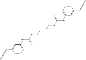
C20H20N6S4
|
|
|---|---|---|
| 分子量 |
472.67
|
|
| 精确质量 |
472.063
|
|
| 元素分析 |
C, 50.82; H, 4.27; N, 17.78; S, 27.13
|
|
| CAS号 |
711019-86-2
|
|
| 相关CAS号 |
|
|
| PubChem CID |
16078986
|
|
| 外观&性状 |
White to off-white solid powder
|
|
| 密度 |
1.3±0.1 g/cm3
|
|
| 沸点 |
652.7±65.0 °C at 760 mmHg
|
|
| 闪点 |
348.5±34.3 °C
|
|
| 蒸汽压 |
0.0±2.0 mmHg at 25°C
|
|
| 折射率 |
1.687
|
|
| LogP |
5.1
|
|
| tPSA |
201.2
|
|
| 氢键供体(HBD)数目 |
4
|
|
| 氢键受体(HBA)数目 |
6
|
|
| 可旋转键数目(RBC) |
9
|
|
| 重原子数目 |
30
|
|
| 分子复杂度/Complexity |
604
|
|
| 定义原子立体中心数目 |
0
|
|
| InChi Key |
QOHNRGHTJPFMSL-UHFFFAOYSA-N
|
|
| InChi Code |
InChI=1S/C20H20N6S4/c27-13-23-15-5-3-7-17(11-15)25-19(29)21-9-1-2-10-22-20(30)26-18-8-4-6-16(12-18)24-14-28/h3-8,11-12H,1-2,9-10H2,(H2,21,25,29)(H2,22,26,30)
|
|
| 化学名 |
1-(3-isothiocyanatophenyl)-3-[4-[(3-isothiocyanatophenyl)carbamothioylamino]butyl]thiourea
|
|
| 别名 |
|
|
| HS Tariff Code |
2934.99.9001
|
|
| 存储方式 |
Powder -20°C 3 years 4°C 2 years In solvent -80°C 6 months -20°C 1 month 注意: 请将本产品存放在密封且受保护的环境中,避免吸湿/受潮。 |
|
| 运输条件 |
Room temperature (This product is stable at ambient temperature for a few days during ordinary shipping and time spent in Customs)
|
| 溶解度 (体外实验) |
|
|||
|---|---|---|---|---|
| 溶解度 (体内实验) |
配方 1 中的溶解度: ≥ 2.5 mg/mL (5.29 mM) (饱和度未知) in 10% DMSO + 40% PEG300 + 5% Tween80 + 45% Saline (这些助溶剂从左到右依次添加,逐一添加), 澄清溶液。
例如,若需制备1 mL的工作液,可将100 μL 25.0 mg/mL澄清DMSO储备液加入到400 μL PEG300中,混匀;然后向上述溶液中加入50 μL Tween-80,混匀;加入450 μL生理盐水定容至1 mL。 *生理盐水的制备:将 0.9 g 氯化钠溶解在 100 mL ddH₂O中,得到澄清溶液。 配方 2 中的溶解度: 2.5 mg/mL (5.29 mM) in 10% DMSO + 90% (20% SBE-β-CD in Saline) (这些助溶剂从左到右依次添加,逐一添加), 悬浊液; 超声助溶。 例如,若需制备1 mL的工作液,可将 100 μL 25.0 mg/mL澄清DMSO储备液加入900 μL 20% SBE-β-CD生理盐水溶液中,混匀。 *20% SBE-β-CD 生理盐水溶液的制备(4°C,1 周):将 2 g SBE-β-CD 溶解于 10 mL 生理盐水中,得到澄清溶液。 View More
配方 3 中的溶解度: ≥ 2.08 mg/mL (4.40 mM) (饱和度未知) in 10% DMSO + 90% Corn Oil (这些助溶剂从左到右依次添加,逐一添加), 澄清溶液。 配方 4 中的溶解度: 30% propylene glycol, 5% Tween 80, 65% D5W: 30 mg/mL 1、请先配制澄清的储备液(如:用DMSO配置50 或 100 mg/mL母液(储备液)); 2、取适量母液,按从左到右的顺序依次添加助溶剂,澄清后再加入下一助溶剂。以 下列配方为例说明 (注意此配方只用于说明,并不一定代表此产品 的实际溶解配方): 10% DMSO → 40% PEG300 → 5% Tween-80 → 45% ddH2O (或 saline); 假设最终工作液的体积为 1 mL, 浓度为5 mg/mL: 取 100 μL 50 mg/mL 的澄清 DMSO 储备液加到 400 μL PEG300 中,混合均匀/澄清;向上述体系中加入50 μL Tween-80,混合均匀/澄清;然后继续加入450 μL ddH2O (或 saline)定容至 1 mL; 3、溶剂前显示的百分比是指该溶剂在最终溶液/工作液中的体积所占比例; 4、 如产品在配制过程中出现沉淀/析出,可通过加热(≤50℃)或超声的方式助溶; 5、为保证最佳实验结果,工作液请现配现用! 6、如不确定怎么将母液配置成体内动物实验的工作液,请查看说明书或联系我们; 7、 以上所有助溶剂都可在 Invivochem.cn网站购买。 |
| 制备储备液 | 1 mg | 5 mg | 10 mg | |
| 1 mM | 2.1156 mL | 10.5782 mL | 21.1564 mL | |
| 5 mM | 0.4231 mL | 2.1156 mL | 4.2313 mL | |
| 10 mM | 0.2116 mL | 1.0578 mL | 2.1156 mL |
1、根据实验需要选择合适的溶剂配制储备液 (母液):对于大多数产品,InvivoChem推荐用DMSO配置母液 (比如:5、10、20mM或者10、20、50 mg/mL浓度),个别水溶性高的产品可直接溶于水。产品在DMSO 、水或其他溶剂中的具体溶解度详见上”溶解度 (体外)”部分;
2、如果您找不到您想要的溶解度信息,或者很难将产品溶解在溶液中,请联系我们;
3、建议使用下列计算器进行相关计算(摩尔浓度计算器、稀释计算器、分子量计算器、重组计算器等);
4、母液配好之后,将其分装到常规用量,并储存在-20°C或-80°C,尽量减少反复冻融循环。
计算结果:
工作液浓度: mg/mL;
DMSO母液配制方法: mg 药物溶于 μL DMSO溶液(母液浓度 mg/mL)。如该浓度超过该批次药物DMSO溶解度,请首先与我们联系。
体内配方配制方法:取 μL DMSO母液,加入 μL PEG300,混匀澄清后加入μL Tween 80,混匀澄清后加入 μL ddH2O,混匀澄清。
(1) 请确保溶液澄清之后,再加入下一种溶剂 (助溶剂) 。可利用涡旋、超声或水浴加热等方法助溶;
(2) 一定要按顺序加入溶剂 (助溶剂) 。
|
|---|
|
|
|
|---|
|
|
 替卡格雷
替卡格雷
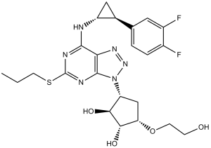
 AF-353 (Ro-4)
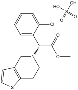 硫酸氢氯吡格雷
 普拉格雷
普拉格雷
InvivoChem的所有产品仅用于作科学研究,不面向患者销售
Copyright 2020 InvivoChem LLC | All Rights Reserved 粤ICP备20063088号-1


 COA
COA


 463611831
