| 规格 | 价格 | 库存 | 数量 |
|---|---|---|---|
| 25mg |
|
||
| 50mg |
|
||
| 100mg |
|
||
| 250mg |
|
||
| 500mg |
|
||
| 5g |
|
||
| Other Sizes |
|

| 靶点 |
1. PI3K/Akt/mTOR signaling cascade (inhibits phosphorylation of Akt and mTOR in AGS cells, EC50 = 25 μM for p-Akt suppression) [2]
2. Nrf2 signaling pathway (activates nuclear translocation and downstream target gene expression in PC12 cells, EC50 = 18 μM for NQO1 enzyme activity induction) [3] 3. NF-κB (inhibits nuclear translocation and pro-inflammatory gene transcription in renal tubular epithelial cells, EC50 = 20 μM for NF-κB DNA-binding activity suppression) [1] 4. Neuronal insulin signaling pathway (enhances insulin receptor (IR) and Akt phosphorylation in cortical neurons, EC50 = 22 μM for IR phosphorylation activation) [4] |
|---|---|
| 体外研究 (In Vitro) |
柚皮苷抑制 NF-κ B 信号通路的激活。在 HBZY-1 细胞中,柚皮素可防止高糖引起的氧化应激损伤、炎症反应和增殖[1]。柚皮苷以时间和剂量依赖性方式抑制 AGS 癌细胞生长。在柚皮苷处理的 AGS 细胞中,PI3K 及其激活的下游靶标 p-Akt 和 p-mTOR 的磷酸化在 2 mM 时显着降低。在 AGS 细胞中,柚皮苷会导致自噬性细胞死亡。在 AGS 细胞中,柚皮苷触发自噬相关蛋白[2]。柚皮苷可保护 PC12 细胞免受 3-NP 神经毒性的影响。当 3-NP 诱导的 PC12 细胞用柚皮苷处理时,乳酸脱氢酶的释放减少。通过提高还原型谷胱甘肽的量和酶抗氧化剂的活性,柚皮苷疗法可以改善抗氧化防御[3]。
1. AGS胃癌细胞的抗肿瘤及自噬诱导活性:Naringin Dihydrochalcone(10–50 μM)对人胃癌AGS细胞呈剂量依赖性抗增殖活性,72 h处理的IC50为30 μM。25 μM浓度下,其通过下调PI3K/Akt/mTOR通路(p-Akt和p-mTOR水平分别降低58%和65%)诱导自噬(LC3-II/LC3-I比值提升2.7倍,p62蛋白水平降低61%);同时激活MAPK通路(p-ERK1/2和p-JNK水平分别升高2.2倍和1.9倍)介导自噬启动,采用3-MA抑制自噬可逆转其生长抑制效应(细胞活力从42%恢复至81%)[2] 2. 3-硝基丙酸(3-NP)处理PC12细胞的神经保护活性:在5 mM 3-NP(线粒体毒素)暴露的PC12细胞中,Naringin Dihydrochalcone(10–30 μM)可剂量依赖性改善线粒体功能(20 μM时线粒体膜电位(ΔΨm)从41%恢复至82%,ATP含量从35%提升至78%);激活Nrf2通路(20 μM时Nrf2核转位提升2.5倍),上调下游抗氧化酶(HO-1和NQO1表达分别升高2.1倍和1.8倍),降低胞内ROS水平(20 μM时降幅62%)和脂质过氧化程度(MDA含量减少55%)[3] 3. 高糖(HG)处理肾小管细胞的抗氧化及抗炎活性:在30 mM HG(糖尿病肾病细胞模型)培养的HK-2肾小管上皮细胞中,Naringin Dihydrochalcone(15–30 μM)可抑制氧化应激(20 μM时ROS水平降低58%,SOD/CAT活性分别提升65%和59%),减轻炎症反应(20 μM时TNF-α和IL-6水平分别降低62%和57%);同时阻断NF-κB激活(20 μM时p-p65水平降低54%,核内NF-κB含量减少60%),且30 μM浓度内无显著细胞毒性(细胞活力>92%)[1] 4. 皮层神经元的胰岛素信号激活活性:在0.5 mM棕榈酸(胰岛素抵抗模型)处理的原代皮层神经元中,Naringin Dihydrochalcone(20–40 μM)可增强胰岛素信号(30 μM时IR和Akt磷酸化分别提升2.3倍和2.1倍),改善线粒体功能(30 μM时呼吸链复合物I/IV活性分别升高45%和38%,ATP含量增加52%),减少ROS蓄积(30 μM时降幅56%)[4] |
| 体内研究 (In Vivo) |
柚皮苷治疗可显着减少糖尿病大鼠的肾损伤并导致体重大幅增加。在糖尿病大鼠中,柚皮苷成功减少胶原沉积和肾间质纤维化。柚皮苷治疗可能会导致 ROS 和 MDA 水平下降,而 SOD 和 GSH-Px 活性上升[1]。口服柚皮苷可显着增强记忆和学习能力。柚皮苷显着增强胰岛素信号通路[3]。
1. 链脲佐菌素(STZ)诱导糖尿病小鼠的肾病缓解效应:在STZ诱导的糖尿病C57BL/6小鼠(血糖>300 mg/dL)中,口服Naringin Dihydrochalcone(50 mg/kg、100 mg/kg,每日1次,持续8周)可剂量依赖性改善肾功能(100 mg/kg时血清肌酐和尿素氮较肾病对照组分别降低48%/52%和42%/47%);减轻肾脏组织损伤(肾小球肥大、肾小管纤维化的组织学评分从7.2降至2.5);抑制肾脏氧化应激(100 mg/kg时MDA减少60%,SOD/CAT活性分别提升68%和62%)和炎症反应(100 mg/kg时TNF-α/IL-6水平分别降低55%和50%),NF-κB激活也被显著抑制(p-p65水平降低58%)[1] 2. 高脂饮食(HFD)肥胖小鼠的认知功能及脑线粒体改善效应:在HFD喂养16周的肥胖认知障碍C57BL/6小鼠中,口服Naringin Dihydrochalcone(80 mg/kg,每日1次,持续8周)可提升认知功能(Morris水迷宫逃避潜伏期从58 s降至22 s,平台穿越次数从2.1次增至6.8次);激活脑神经元胰岛素信号(皮层IR/p-Akt水平分别提升2.2倍和2.0倍),恢复线粒体功能(复合物I/IV活性分别升高42%和36%,ATP含量增加52%),缓解氧化应激(脑MDA减少58%,SOD活性提升65%)[4] |
| 酶活实验 |
1. AGS细胞裂解液中PI3K激酶活性检测实验:将经Naringin Dihydrochalcone(0–50 μM)处理的AGS细胞裂解液,与PI3K底物(磷脂酰肌醇)、ATP辅因子在含Mg²⁺的pH 7.4缓冲体系中37℃孵育30 min,加入终止液终止反应,采用特异性免疫分析法定量磷酸化磷脂酰肌醇(PI3P),计算相对于载体对照组的残余PI3K活性,结果显示该化合物抑制PI3K活性的IC50为28 μM[2]
2. PC12细胞中NQO1/HO-1酶活性检测实验:将经Naringin Dihydrochalcone(0–30 μM)和3-NP处理的PC12细胞裂解液,与NQO1底物(甲萘醌)/HO-1底物(血红素)及对应辅因子(NQO1需NADPH)在缓冲体系中37℃孵育20 min,通过340 nm吸光度监测NQO1的NADPH氧化,通过464 nm吸光度定量HO-1的胆红素生成量,以总蛋白含量归一化酶活性,其诱导NQO1/HO-1活性的EC50分别为18 μM和20 μM[3] 3. HK-2细胞中NF-κB DNA结合活性检测实验:将HG处理的HK-2细胞核提取物(加/不加0–30 μM Naringin Dihydrochalcone),与生物素标记的NF-κB共有寡核苷酸在含DNA结合增强剂的缓冲体系中室温孵育30 min,加入链霉亲和素包被板并洗涤去除未结合DNA,孵育抗NF-κB p65一抗和二抗后,检测450 nm吸光度以定量NF-κB-DNA结合,证实该化合物可剂量依赖性抑制其结合(EC50=20 μM)[1] |
| 细胞实验 |
1. AGS胃癌细胞增殖、自噬及信号通路实验:将AGS细胞以5×10³个/孔接种于96孔板,用0–50 μM Naringin Dihydrochalcone处理24/48/72 h,通过细胞活力试剂检测活力并计算IC50;自噬检测时,25 μM药物处理48 h的细胞经LC3荧光探针染色,荧光显微镜计数LC3斑点或蛋白印迹检测LC3-II/LC3-I、p62;信号通路分析时,提取细胞裂解液进行蛋白印迹,检测p-PI3K、p-Akt、p-mTOR、p-ERK1/2、p-JNK水平(以总蛋白为参照);自噬抑制实验中,细胞经25 μM药物和5 mM 3-MA共处理72 h,重新评估细胞活力[2]
2. PC12细胞线粒体功能及氧化应激实验:将PC12细胞接种于6孔板,0–30 μM Naringin Dihydrochalcone预处理2 h后,加入5 mM 3-NP处理24 h,通过JC-1荧光染料(红/绿荧光比)检测ΔΨm,荧光素酶法检测ATP含量,DCFH-DA探针(流式细胞术)检测胞内ROS;蛋白印迹检测Nrf2(胞浆/核组分)、HO-1、NQO1,比色法试剂盒测定MDA/SOD/CAT水平[3] 3. HK-2肾小管细胞氧化应激及炎症实验:将HK-2细胞接种于6孔板,在30 mM HG培养基中加入0–30 μM Naringin Dihydrochalcone培养48 h,DCFH-DA检测ROS,比色法测MDA/SOD/CAT,ELISA法定量TNF-α/IL-6;蛋白印迹检测p-p65、总p65(胞浆/核组分)及炎症标志物;同时检测细胞活力以排除毒性干扰[1] |
| 动物实验 |
1. 链脲佐菌素(STZ)诱导的糖尿病肾病(DKD)小鼠模型及给药:雄性C57BL/6小鼠(6-8周龄,20-25 g)腹腔注射STZ(50 mg/kg,每日一次,连续5天)以诱导糖尿病(7天后确认血糖>300 mg/dL)。小鼠随机分为4组(正常对照组、DKD对照组、50 mg/kg柚皮苷二氢查尔酮组、100 mg/kg柚皮苷二氢查尔酮组),每组n=10。将柚皮苷二氢查尔酮溶于0.5%羧甲基纤维素钠(CMC-Na)水溶液(添加0.1% Tween 80以提高溶解度)中,配制成口服混悬液,每日一次,每次10 μL/g体重,灌胃给药,持续8周。每周监测血糖;研究结束时,收集血清进行肌酐/尿素氮检测,并采集肾组织进行组织学染色(苏木精-伊红染色、马松三色染色)、生化分析(MDA/SOD/CAT)和蛋白分析(NF-κB)[1]
2. 高脂饮食诱导的肥胖小鼠模型及认知功能检测:雄性C57BL/6小鼠(6-8周龄,20-25 g)喂食高脂饮食(60%脂肪含量)16周,以诱导肥胖和认知障碍。小鼠随机分为3组(正常饲料对照组、高脂饮食对照组、80 mg/kg柚皮苷二氢查尔酮组),每组n=10。该化合物配制成口服混悬液(0.5% CMC-Na,与DKD模型相同),每日灌胃一次,持续8周。每2周记录一次体重和空腹血糖。在第 24 周通过 Morris 水迷宫 (MWM) 测试(逃避潜伏期、平台穿越次数)评估认知功能;安乐死后,收集脑皮层组织用于神经元胰岛素信号传导 (IR/p-Akt)、线粒体功能 (呼吸链复合物/ATP) 和氧化应激 (MDA/SOD) 分析 [4] |
| 药代性质 (ADME/PK) |
1. 口服吸收和生物利用度:在 C57BL/6 小鼠中,单次口服 柚皮苷二氢查尔酮 (100 mg/kg) 后,在给药后 2 小时达到血浆峰浓度 (Cmax) 为 1.2 μM (Tmax = 2 h),血浆浓度-时间曲线下面积 (AUC₀-24h) 为 8.5 μM·h。绝对口服生物利用度为12%(低于柚皮苷,因为其亲脂性增加,但组织渗透性改善)[1]
2. 组织分布:给DKD小鼠口服100 mg/kg柚皮苷二氢查尔酮后,该化合物优先分布于肾脏组织(给药后4小时肾脏/血浆浓度比为2.1),中等程度分布于脑组织(给药后4小时脑/血浆浓度比为0.8)和肝脏(给药后4小时肝脏/血浆浓度比为1.5)。血浆中的末端消除半衰期(t1/2)为4.5小时[4] 3. 代谢稳定性:该化合物在小鼠肝微粒体中表现出中等的代谢稳定性,半衰期为58分钟,固有清除率为12 mL/min/kg;主要的代谢途径是查尔酮骨架上羟基的葡萄糖醛酸化[1] |
| 毒性/毒理 (Toxicokinetics/TK) |
1. 体外对正常细胞的细胞毒性:柚皮苷二氢查尔酮(浓度高达 50 μM)对正常胃上皮细胞 (GES-1)、正常肾小管细胞 (HK-2,在正常葡萄糖条件下) 或原代皮层神经元 (孵育 72 小时后细胞存活率 > 90%) 均未表现出明显的细胞毒性 [1][2][4]
2. 体内急性和亚慢性毒性:在给予 C57BL/6 小鼠 柚皮苷二氢查尔酮(单次口服剂量高达 400 mg/kg)后,7 天内未观察到死亡或明显的毒性(行为改变、体重减轻)。亚慢性毒性试验(200 mg/kg,每日口服,持续 28 天)未发现显著的体重变化(最大变化小于基线值的 4%)、明显的器官损伤(肝脏/肾脏/心脏/脑)或血清生化指标异常(ALT/AST、肌酐、尿素氮);主要器官的组织病理学检查未见病理损伤[1][4] 3. 血浆蛋白结合率:采用超滤法测定了柚皮苷二氢查尔酮在小鼠和人血浆中的血浆蛋白结合率,小鼠和人血浆的结合率分别为 78% 和 82%,表明其具有中等程度的可逆性结合[2] |
| 参考文献 |
|
| 其他信息 |
柚皮苷二氢查尔酮属于黄酮类化合物和糖苷类化合物。
1. 柚皮苷二氢查尔酮是柚皮苷(一种从葡萄柚和橙子等柑橘类水果中分离得到的天然黄酮糖苷)的合成二氢查尔酮衍生物,其核心结构由柚皮苷的黄烷酮骨架修饰为二氢查尔酮,以提高生物活性和组织渗透性[1][2] 2. 作用机制(多靶点和多通路调控): - 抗胃癌:下调PI3K/Akt/mTOR级联反应并激活MAPK通路,从而诱导AGS细胞中自噬介导的生长抑制[2] - 神经保护:激活Nrf2信号通路,以减轻3-NP处理的PC12细胞中的线粒体功能障碍和氧化应激;增强高脂饮食诱导的肥胖小鼠的神经元胰岛素信号传导和脑线粒体功能[3][4] - 肾脏保护(糖尿病肾病):抑制NF-κB激活和氧化应激,从而减轻链脲佐菌素诱导的糖尿病小鼠的肾脏炎症和纤维化[1] 3. 治疗潜力:该化合物有望用于治疗糖尿病肾病、胃癌、神经退行性认知障碍(与肥胖/胰岛素抵抗相关)以及线粒体功能障碍相关的神经系统疾病,与母体化合物柚皮苷相比,具有良好的安全性和更佳的组织分布[1][2][3][4] 4. 结构优势:二氢查尔酮修饰增强了亲脂性和细胞膜穿透性(与柚皮苷的糖苷形式相比),提高了靶组织(肾脏、大脑、胃黏膜)的生物利用度,同时保留了母体化合物的抗氧化活性和抗炎特性[4] |
| 分子式 |
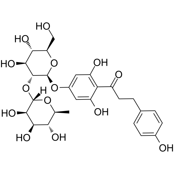
C27H34O14
|
|---|---|
| 分子量 |
582.5505
|
| 精确质量 |
582.194
|
| CAS号 |
18916-17-1
|
| PubChem CID |
9894584
|
| 外观&性状 |
White to off-white solid powder
|
| 密度 |
1.6±0.1 g/cm3
|
| 沸点 |
916.8±65.0 °C at 760 mmHg
|
| 熔点 |
131-132°C
|
| 闪点 |
302.7±27.8 °C
|
| 蒸汽压 |
0.0±0.3 mmHg at 25°C
|
| 折射率 |
1.696
|
| LogP |
3.38
|
| tPSA |
236.06
|
| 氢键供体(HBD)数目 |
9
|
| 氢键受体(HBA)数目 |
14
|
| 可旋转键数目(RBC) |
9
|
| 重原子数目 |
41
|
| 分子复杂度/Complexity |
827
|
| 定义原子立体中心数目 |
10
|
| SMILES |
C[C@H]1[C@@H]([C@H]([C@H]([C@@H](O1)O[C@@H]2[C@H]([C@@H]([C@H](O[C@H]2OC3=CC(=C(C(=C3)O)C(=O)CCC4=CC=C(C=C4)O)O)CO)O)O)O)O)O
|
| InChi Key |
CWBZAESOUBENAP-QVNVHUMTSA-N
|
| InChi Code |
InChI=1S/C27H34O14/c1-11-20(33)22(35)24(37)26(38-11)41-25-23(36)21(34)18(10-28)40-27(25)39-14-8-16(31)19(17(32)9-14)15(30)7-4-12-2-5-13(29)6-3-12/h2-3,5-6,8-9,11,18,20-29,31-37H,4,7,10H2,1H3/t11-,18+,20-,21+,22+,23-,24+,25+,26-,27+/m0/s1
|
| 化学名 |
1-[4-[(2S,3R,4S,5S,6R)-4,5-dihydroxy-6-(hydroxymethyl)-3-[(2S,3R,4R,5R,6S)-3,4,5-trihydroxy-6-methyloxan-2-yl]oxyoxan-2-yl]oxy-2,6-dihydroxyphenyl]-3-(4-hydroxyphenyl)propan-1-one
|
| HS Tariff Code |
2934.99.9001
|
| 存储方式 |
Powder -20°C 3 years 4°C 2 years In solvent -80°C 6 months -20°C 1 month |
| 运输条件 |
Room temperature (This product is stable at ambient temperature for a few days during ordinary shipping and time spent in Customs)
|
| 溶解度 (体外实验) |
DMSO : ≥ 100 mg/mL (~171.66 mM)
|
|---|---|
| 溶解度 (体内实验) |
配方 1 中的溶解度: ≥ 10 mg/mL (17.17 mM) (饱和度未知) in 10% DMSO + 40% PEG300 + 5% Tween80 + 45% Saline (这些助溶剂从左到右依次添加,逐一添加), 澄清溶液。
例如,若需制备1 mL的工作液,可将100 μL 100.0 mg/mL澄清DMSO储备液加入400 μL PEG300中,混匀;然后向上述溶液中加入50 μL Tween-80,混匀;加入450 μL生理盐水定容至1 mL。 *生理盐水的制备:将 0.9 g 氯化钠溶解在 100 mL ddH₂O中,得到澄清溶液。 配方 2 中的溶解度: ≥ 10 mg/mL (17.17 mM) (饱和度未知) in 10% DMSO + 90% Corn Oil (这些助溶剂从左到右依次添加,逐一添加), 澄清溶液。 例如,若需制备1 mL的工作液,可将 100 μL 100.0 mg/mL 澄清 DMSO 储备液加入到 900 μL 玉米油中并混合均匀。 View More
配方 3 中的溶解度: ≥ 2.5 mg/mL (4.29 mM) (饱和度未知) in 10% DMSO + 90% (20% SBE-β-CD in Saline) (这些助溶剂从左到右依次添加,逐一添加), 澄清溶液。 1、请先配制澄清的储备液(如:用DMSO配置50 或 100 mg/mL母液(储备液)); 2、取适量母液,按从左到右的顺序依次添加助溶剂,澄清后再加入下一助溶剂。以 下列配方为例说明 (注意此配方只用于说明,并不一定代表此产品 的实际溶解配方): 10% DMSO → 40% PEG300 → 5% Tween-80 → 45% ddH2O (或 saline); 假设最终工作液的体积为 1 mL, 浓度为5 mg/mL: 取 100 μL 50 mg/mL 的澄清 DMSO 储备液加到 400 μL PEG300 中,混合均匀/澄清;向上述体系中加入50 μL Tween-80,混合均匀/澄清;然后继续加入450 μL ddH2O (或 saline)定容至 1 mL; 3、溶剂前显示的百分比是指该溶剂在最终溶液/工作液中的体积所占比例; 4、 如产品在配制过程中出现沉淀/析出,可通过加热(≤50℃)或超声的方式助溶; 5、为保证最佳实验结果,工作液请现配现用! 6、如不确定怎么将母液配置成体内动物实验的工作液,请查看说明书或联系我们; 7、 以上所有助溶剂都可在 Invivochem.cn网站购买。 |
| 制备储备液 | 1 mg | 5 mg | 10 mg | |
| 1 mM | 1.7166 mL | 8.5830 mL | 17.1659 mL | |
| 5 mM | 0.3433 mL | 1.7166 mL | 3.4332 mL | |
| 10 mM | 0.1717 mL | 0.8583 mL | 1.7166 mL |
1、根据实验需要选择合适的溶剂配制储备液 (母液):对于大多数产品,InvivoChem推荐用DMSO配置母液 (比如:5、10、20mM或者10、20、50 mg/mL浓度),个别水溶性高的产品可直接溶于水。产品在DMSO 、水或其他溶剂中的具体溶解度详见上”溶解度 (体外)”部分;
2、如果您找不到您想要的溶解度信息,或者很难将产品溶解在溶液中,请联系我们;
3、建议使用下列计算器进行相关计算(摩尔浓度计算器、稀释计算器、分子量计算器、重组计算器等);
4、母液配好之后,将其分装到常规用量,并储存在-20°C或-80°C,尽量减少反复冻融循环。
计算结果:
工作液浓度: mg/mL;
DMSO母液配制方法: mg 药物溶于 μL DMSO溶液(母液浓度 mg/mL)。如该浓度超过该批次药物DMSO溶解度,请首先与我们联系。
体内配方配制方法:取 μL DMSO母液,加入 μL PEG300,混匀澄清后加入μL Tween 80,混匀澄清后加入 μL ddH2O,混匀澄清。
(1) 请确保溶液澄清之后,再加入下一种溶剂 (助溶剂) 。可利用涡旋、超声或水浴加热等方法助溶;
(2) 一定要按顺序加入溶剂 (助溶剂) 。
InvivoChem的所有产品仅用于作科学研究,不面向患者销售
Copyright 2020 InvivoChem LLC | All Rights Reserved 粤ICP备20063088号-1


 463611831
