| 规格 | 价格 | 库存 | 数量 |
|---|---|---|---|
| 250mg |
|
||
| 500mg |
|
||
| 1g |
|
||
| 2g |
|
||
| 5g |
|
||
| 10g |
|
||
| Other Sizes |
|

| 靶点 |
Neuraminidase; influenza A/H3N2, A/H1N2, A/H1N1, and B viruses
Sialidase (neuraminidase) inhibitor; inhibits viral sialidase activity of influenza virus. In canine mammary cancer cells (CMA07 and CMT-U27), it also inhibits endogenous mammalian sialidase activity, leading to increased sialylation. |
|---|---|
| 体外研究 (In Vitro) |
口服治疗后,磷酸奥司他韦(OP)是一种前药,很容易从胃肠道吸收。肝酯酶主要负责前药大量转化为羧酸奥司他韦 (OC) [1]。一种常用的抗流感唾液酸酶抑制剂是磷酸奥司他韦。根据单向方差分析测试,CMA07 和 CMT-U27 细胞系的代谢活性在 305 μM 磷酸奥司他韦处理后显着降低(分别为 p=0.005 和 p<0.0001)。另一方面,与对照细胞相比,当磷酸奥司他韦以 0.305 μM (p=0.9781)、3.05 μM (p=0.7436) 和 30.5 μM (p=0.9623) 处理时,没有观察到统计学上显着的变化。为了评估磷酸奥司他韦对 CMA07 和 CMT-U27 程序性细胞死亡的影响,采用 TUNEL 测试,考虑到 305 μM 磷酸奥司他韦给药会降低细胞代谢活性。与较低奥司他韦浓度或 PBS 相比,24 小时磷酸奥司他韦给药(特别是 305 μM)显着增加了 CMA07 (p=0.001) 和 CMT-U27 (p=0.0002) DNA 断裂[2]。
用奥司他韦磷酸盐(0.305 μM、3.05 μM、30.5 μM和305 μM)处理24小时,可抑制CMA07和CMT-U27犬乳腺癌细胞的唾液酸酶活性,表现为4-MuNana底物转化产生的荧光减弱。 在305 μM浓度下,奥司他韦磷酸盐显著降低两种细胞的代谢活性(MTS法),并增加DNA断裂(TUNEL法),表明具有细胞毒性和促凋亡作用。 在较低浓度(3.05 μM和30.5 μM)下,奥司他韦磷酸盐以剂量依赖的方式显著增强CMT-U27细胞的迁移(划痕实验)和侵袭能力(Matrigel侵袭实验)。 奥司他韦磷酸盐通过Western blot、二维电泳和荧光凝集素染色证实,能增加两种细胞系中α2,6-连接唾液酸化和SLe(x)的表达。 |
| 体内研究 (In Vivo) |
Ki-67抗原和caspase-3蛋白分别用于评估CMT-U27异种移植肿瘤细胞增殖和凋亡。奥司他韦治疗和未治疗小鼠之间的 Ki-67 和 caspase 3 (p=0.2) 表达几乎没有发现差异 [2]。磷酸奥司他韦治疗的小鼠在原发性肿瘤中显示出明显更多的炎症浸润(p=0.01)。
在接种CMT-U27细胞的裸鼠中,每日腹腔注射奥司他韦磷酸盐(100 mg/kg)可增加原发肿瘤中α2,6唾液酸化和SLe(x)的表达。 与对照组相比,治疗组小鼠肺转移灶数量有增加趋势(p=0.07),但未达到统计学显著性。 治疗组小鼠原发肿瘤中炎症浸润更明显,且出现更早的疾病进展迹象。 |
| 酶活实验 |
唾液酸酶活性测定[2]
与良性细胞相比,恶性细胞中唾液酰化水平较高。这可能取决于唾液酸转移酶和唾液酸酯酶的活性。为了评估磷酸奥司他韦对唾液酸酶活性的影响,我们在CMA07和CMT-U27犬乳腺肿瘤细胞中使用一种改良唾液酸(4-methyl-umbelliferyl-Nacetylneuraminic acid-4- munana)进行了体外实验。唾液酸酶的活性是通过获得唾液酸类似物4-MuNana在不同剂量的磷酸奥司他韦处理后转化为荧光化合物甲基-伞形酮(蓝色)来测定的。细胞在12 mm圆形玻璃中培养至融合后,用不同浓度奥司他韦磷酸盐(0.305 μM、3.05 μM、30.5 μM和305 μM奥司他韦磷酸盐溶解PBS)培养液孵育24 h,以药物载体(PBS)为对照。处理24小时后,将每个12mm圆形玻璃放在载玻片上,在2μ m 4-MuNana(2'-(4-甲基伞叶基)-α- d - n -乙酰神经氨酸)溶液中孵育。如前所述,在紫外光(激发波长为360 nm,发射波长为440 nm)下立即用荧光显微镜观察载玻片。用卡尔蔡司荧光显微镜分析载玻片并拍摄图像。 抗病毒活性[3] 将感染细胞(0.01 MOI)培养在含有增加利巴韦林或磷酸奥司他韦浓度的Opti-MEM (2 μg/mL TPCK-trypsin)中。在感染后约24 h (p.i.),去除等分,测定高斯荧光素酶活性。 使用荧光底物4-甲基伞形酮-N-乙酰神经氨酸(4-MuNana)评估唾液酸酶活性。细胞经奥司他韦磷酸盐处理24小时后,与2 μM 4-MuNana孵育。在紫外光下检测甲基伞形酮的荧光强度,荧光减弱表明奥司他韦磷酸盐抑制了唾液酸酶活性。 |
| 细胞实验 |
细胞形态分析[2]
CMA07和CMT-U27细胞以每孔1x10⁴个细胞的密度在6孔板中进行三次镀。以PBS为对照,研究了0.305 μM、3.05 μM和30.5 μM的磷酸奥司他韦浓度。在7天的时间里,使用对比倒置显微镜进行细胞融合和形态学分析。照片是在第0天和第7天在200倍放大镜下拍摄的。 细胞增殖试验[2] 以0.305 μM、3.05 μM、30.5 μM和305 μM磷酸奥司他韦和PBS为对照,分别在24孔板上培养CMA07和CMT-U27细胞。连续7天,每天在Neubauer氏室中以0.4%台锥蓝1:2的比例对细胞进行计数,细胞计数使用体积转换因子为1mm3,即1x10⁴。实验重复3次,记录生长曲线。 细胞生长试验[2] 在CMA07和CMT-U27细胞系中,使用市售的CellTiter 96水溶液试剂测定细胞生长,并根据制造商的说明进行。简单地说,细胞以每孔5x10³个细胞的密度在96孔板中一式三次镀。细胞贴壁后,分别以0.305 μM、3.05 μM、30.5 μM和305 μM的终浓度加入磷酸奥司他韦,以PBS为对照。加入MTS四氮唑试剂测定细胞代谢,在490nm处记录吸光度。在0、2、4、6、8、10、12、24和48小时进行测量。在没有细胞的培养孔中,在时间点0h进行额外的对照测量。实验进行了两次。 TUNEL分析[2] 将CMA07和CMT-U27细胞株培养于6孔板中,分别用不同浓度的磷酸奥司他韦(0.305 μM、3.05 μM、30.5 μM和305 μM, PBS为对照)处理。处理24小时后,收集培养基和tripsinized细胞,2000 rpm离心10分钟。细胞在PBS中洗涤,在冷甲醇中固定20分钟。固定后,将细胞重新悬浮于1ml PBS中进行细胞自旋处理。简单地说,使用聚赖氨酸包被载玻片将100 μL细胞悬液在cytospin3离心机中离心。然后根据制造商的说明,使用市售试剂盒(原位细胞死亡检测试剂盒,罗氏荧光素)标记DNA双链断裂,将载玻片用于原位细胞死亡检测。在488nm激发波长的荧光显微镜下观察载玻片,用ImageJ软件记录TUNEL阳性细胞与总细胞的比值,计算死亡细胞的百分比。本实验进行了两次。 愈合试验 在延时显微镜下,使用良性(CMA07)和高度转移(CMT-U27)犬乳腺肿瘤细胞系进行伤口愈合试验。简单地说,将20x104个细胞镀在24孔培养板上,达到高汇合后,用移液管尖端制作人工“伤口”。用磷酸奥司他韦浓度分别为0.305 μM、3.05 μM和30.5 μM的奥司他韦替换培养基,PBS作为对照。使用Axio Vision Release 4.8.2程序,每隔5分钟采集伤口图像,持续48小时。然后转换成视频。305 μM的奥司他韦磷酸盐由于其先前显示的细胞毒性而未进行细胞处理。本实验进行了两次。 荧光细胞化学[2] 细胞在玻璃罩中培养,培养基中分别添加0.305 μM、3.05 μM和30.5 μM的磷酸奥司他韦和PBS作为对照,培养24小时。然后用PBS洗涤细胞,用冷甲醇固定20分钟。固定后,用PBS重新水化细胞,并用10% BSA封闭20分钟。将植物凝集素SNA、MAL I和MAL II(生物素化的amurensis凝集素II, B-1265, Vector Laboratories)在5% BSA的PBS中稀释1:300,在载玻片上室温孵育1小时。然后用PBS洗涤三次,用链亲和素- fitc孵育1小时。PBS洗涤2次后,载玻片与DAPI在PBS中孵育10分钟,载玻片贴载于Vectashield贴载介质中进行荧光分析。在卡尔蔡司荧光显微镜下对载玻片进行分析并拍摄图像。 Western Blot分析[2] 将CMA07和CMT-U27细胞系细胞培养在6个孔板中汇合,在0.305 μM、3.05 μM和30.5 μM磷酸奥司他韦培养基中加入不同浓度的磷酸奥司他韦。孵育24小时后,用PBS洗涤细胞3次,用RIPA裂解缓冲液(50 mM Tris HCl, pH 8;150mm NaCl;NP-40 1%;0.5%去氧乙酸钠;0.1% SDS)含有完整的蛋白酶抑制剂混合物,1mM PMSF(苯基甲基磺酰氟)和1mM Na3VO4(正钒酸钠)。根据制造商的说明,使用Pierce BCA蛋白测定试剂盒的生物辛酸法测定蛋白质浓度。 细胞增殖实验:使用台盼蓝排除法连续7天每日计数。 代谢活性实验:采用MTS法在48小时内检测细胞代谢活性。 细胞凋亡实验:处理24小时后使用TUNEL法评估凋亡。 细胞迁移实验:通过划痕实验并在48小时内进行延时成像分析。 细胞侵袭实验:使用Matrigel包被的小室进行6小时侵袭实验。 糖基化分析:通过Western blot、二维电泳和荧光凝集素染色(SNA、MAL I、MAL II)分析唾液酸化结构的变化。 |
| 动物实验 |
实验小鼠分组及药物处理[2]
4-6周龄的雌性NIH(S)II-nu/nu裸鼠,使用25号针头将1×10⁶个活的CMT-U27犬乳腺癌细胞原位接种于乳腺脂肪垫。共接种8只小鼠。当结节体积达到约500mm³时,将小鼠(n=8)随机分为对照组(n=4)和治疗组(n=4)。两组小鼠每日腹腔注射100 μL PBS(对照组)或100 mg/kg的奥司他韦磷酸盐(购自药房,用PBS稀释)(治疗组),直至处死。使用游标卡尺测量肿瘤大小,并根据肿瘤的长×宽×高估算肿瘤体积(mm³)。为了观察转移情况,当所有小鼠的原发肿瘤平均体积达到约1000–1500 mm³时,均进行手术切除。小鼠通过腹腔注射100 μL含有50 mg/kg氯胺酮(IMALGENE 1000)和1 mg/kg盐酸美托咪定(Medetor)的混合液进行麻醉,然后切除肿瘤。每只小鼠使用2.5 mg/kg阿替美唑(Revertor)来拮抗麻醉作用。为预防疼痛,小鼠每8小时口服10 mg/kg盐酸曲马多(Tramal)溶液,持续24–48小时。动物在手术切除原发肿瘤后进行随访,观察是否存在侵袭和/或转移迹象。 小鼠感染[3] 将4至6周龄的雌性BALB/c小鼠在轻度异氟烷麻醉下,经鼻内接种30 μL PBS中含有的指定量病毒。每日监测小鼠体重。体重下降20%的小鼠被实施安乐死。在指定时间点,处死小鼠并取出肺脏进行进一步分析。采用TCID50法和荧光素酶法测定肺匀浆中的病毒载量。 抗病毒治疗方面,小鼠分别接受利巴韦林(80 mg/kg/天)或奥司他韦磷酸盐(20–50 mg/kg/天,溶于PBS)腹腔注射。治疗在感染前2小时开始,每日两次,直至实验结束。 雌性裸鼠接受原位接种CMT-U27细胞。当肿瘤体积达到约500 mm³时,将小鼠分为对照组(PBS)和治疗组(每日腹腔注射100 mg/kg磷酸奥司他韦)。当肿瘤体积达到约1000-1500 mm³时,进行手术切除,并监测小鼠的转移情况。对肺部进行组织学检查,以确定是否存在转移。 |
| 药代性质 (ADME/PK) |
吸收
口服磷酸奥司他韦后,可迅速从胃肠道吸收,并主要通过肝脏酯酶转化为活性代谢物奥司他韦羧酸盐。口服剂量的至少75%以活性代谢物的形式进入体循环。前药的暴露量相对于活性代谢物不足5%。前药和活性代谢物的血浆浓度均与剂量成正比,且不受与食物同服的影响。每日两次服用奥司他韦75mg胶囊后的药代动力学参数如下:奥司他韦和奥司他韦羧酸盐的Cmax分别为65ng/mL和348ng/mL,而奥司他韦和奥司他韦羧酸盐的AUC(0-12h)分别为112ng·h/mL和2719ng·h/mL。 消除途径 吸收后,超过90%的奥司他韦通过转化为奥司他韦羧酸盐而消除,随后完全通过肾脏排泄。临床研究发现,口服放射性标记剂量中仅有不到20%经粪便排出。 分布容积 奥司他韦羧酸盐在人体内的稳态平均分布容积约为23至26升,该容积大致相当于细胞外液。由于神经氨酸酶活性存在于细胞外,奥司他韦羧酸盐可分布至流感病毒传播的所有部位。 清除率 该药物的肾清除率(18.8升/小时)超过肾小球滤过率(7.5升/小时),表明除肾小球滤过外,还存在肾小管分泌。 蛋白结合率:磷酸奥司他韦:中等(42%)。奥司他韦羧酸盐:极低 < 3%。 奥司他韦羧酸盐:24 例受试者静脉给药后,分布容积为 23 至 26 升。 口服磷酸奥司他韦易被吸收,随后主要通过肝酯酶大量转化为活性形式——奥司他韦羧酸盐。口服剂量中至少 75% 以奥司他韦羧酸盐的形式进入体循环。口服剂量中,以磷酸奥司他韦形式进入体循环的比例不足 5%。 清除:肾脏:奥司他韦羧酸盐主要通过肾脏排泄清除(> 99%)。肾清除率 (18.8 升/小时) 超过肾小球滤过率 (7.5 升/小时),表明存在肾小管分泌。粪便:口服放射性标记剂量后,粪便排泄量<20%。 有关奥司他韦(共8项)的更多吸收、分布和排泄(完整)数据,请访问HSDB记录页面。 代谢/代谢物 奥司他韦主要在肝脏中通过酯酶转化为活性代谢物奥司他韦羧酸盐。奥司他韦羧酸盐不再进一步代谢。奥司他韦和奥司他韦羧酸盐均不是细胞色素P450同工酶的底物或抑制剂。体内尚未发现这两种化合物的II期结合物。 奥司他韦主要在肝脏中通过酯酶转化为奥司他韦羧酸盐。奥司他韦及其羧酸盐均不是细胞色素P450同工酶的底物或抑制剂。 生物转化:肝脏代谢;奥司他韦乙酯前药经广泛水解转化为活性星状形式——奥司他韦羧酸盐。 生物半衰期 口服给药后,大多数受试者血浆中奥司他韦的浓度在1至3小时内下降,而大多数受试者口服给药后血浆中奥司他韦羧酸盐的浓度在6至10小时内下降。 消除:奥司他韦的消除时间为1至3小时,奥司他韦羧酸盐的消除时间为6至10小时。 |
| 毒性/毒理 (Toxicokinetics/TK) |
肝毒性
在奥司他韦的临床试验中,2%的受试者出现血清转氨酶升高,但所有患者均无症状且症状短暂,也未见出现伴有黄疸的临床明显肝损伤的报告。奥司他韦组的ALT升高率通常与安慰剂组或对照药物组相似。自1999年获批以来,奥司他韦已在流感季节性爆发期间广泛使用。曾有少数接受奥司他韦治疗的患者出现轻度肝损伤的个案报道,但这些损伤与奥司他韦之间的关系尚未得到充分证实。目前尚无因使用奥司他韦而导致急性肝衰竭或慢性肝病的报告。此外,部分流感患者在急性期会出现血清酶升高,甚至轻度黄疸,且与是否接受任何治疗无关。 可能性评分:D(可能是临床上明显的肝损伤的罕见原因)。 妊娠和哺乳期影响 ◉哺乳期用药概述 有限的数据表明,奥司他韦及其活性代谢物很少分泌到乳汁中。母亲每日服用150毫克,乳汁中的药物浓度较低,预计不会对母乳喂养的婴儿造成任何不良影响。 2 周龄以上的婴儿可以直接服用奥司他韦,剂量远高于母乳中的剂量。 ◉ 对母乳喂养婴儿的影响 截至修订日期,未找到相关的已发表信息。 ◉ 对哺乳和母乳的影响 截至修订日期,未找到相关的已发表信息。 药物和哺乳数据库 (LactMed) 11.1.5 相互作用 与丙磺舒合用会导致活性代谢物浓度增加约两倍,这是由于肾脏中活性阴离子管分泌减少所致。 Thomson/Micromedex. 医疗保健专业人员药物信息。第 1 卷,科罗拉多州格林伍德村,2006 年,第 11 页。 2305 危险物质数据库 (HSDB) 体外研究表明,奥司他韦及其羧酸盐均不是 P450 混合功能氧化酶或葡萄糖醛酸转移酶的良好底物。西咪替丁是一种非特异性细胞色素 P450 同工酶抑制剂,可竞争性抑制碱性或阳离子药物在肾小管的分泌,但它对奥司他韦及其羧酸盐的血浆浓度没有影响。 《医师案头参考手册》第 60 版,Thomson PDR,Montvale,NJ,2006 年,第 2305 页。 2811 危险物质数据库 (HSDB) 与阿莫西林合用不会改变两种化合物的血浆浓度,表明对阴离子分泌途径的竞争较弱。 蛋白结合 活性奥司他韦羧酸盐代谢物与人血浆蛋白的结合率约为3%,可忽略不计;而奥司他韦与人血浆蛋白的结合率为42%,不足以引起显著的基于置换的药物相互作用。 在MDCK细胞中进行的体外抗病毒试验中,所测试浓度的磷酸奥司他韦(最高浓度达到用于IC50测定的最高浓度)未显示明显的细胞毒性迹象,这可从试验中未报告细胞活力受损的情况推断得出。 在体内小鼠研究中,每日20或50 mg/kg剂量的磷酸奥司他韦未引起明显的不良反应或直接归因于其毒性。文中有所报道。该研究重点关注其对流感病毒感染的保护作用。 |
| 参考文献 |
|
| 其他信息 |
磷酸奥司他韦是一种磷酸盐,含有奥司他韦分子。
磷酸奥司他韦是奥司他韦的磷酸盐,奥司他韦是一种合成的乙酯衍生物前药,具有抗病毒活性。奥司他韦通过阻断流感病毒表面的神经氨酸酶,干扰宿主细胞释放完整的病毒颗粒。 奥司他韦是一种乙酰胺基环己烯,是唾液酸的结构同系物,可抑制神经氨酸酶。 另见:奥司他韦酸(含活性部分);奥司他韦(含活性部分)。奥司他韦羧酸盐(含活性部分)。 药物适应症 治疗和预防流感 磷酸奥司他韦是一种抗流感前药,经羧酸酯酶转化为活性奥司他韦羧酸盐。 本研究表明,它可能抑制癌细胞中的哺乳动物唾液酸酶,从而改变唾液酸化,并可能增加肿瘤的侵袭性。 该研究使用犬乳腺肿瘤细胞作为人类乳腺癌研究的模型。 |
| 分子式 |
C16H31N2O8P
|
|---|---|
| 分子量 |
410.3997
|
| 精确质量 |
410.181
|
| 元素分析 |
C, 46.83; H, 7.61; N, 6.83; O, 31.19; P, 7.55
|
| CAS号 |
204255-11-8
|
| 相关CAS号 |
Oseltamivir;196618-13-0;Oseltamivir acid;187227-45-8;Oseltamivir-d5 phosphate;Oseltamivir-d3 phosphate
|
| PubChem CID |
78000
|
| 外观&性状 |
White to off-white solid powder
|
| 密度 |
1.08g/cm3
|
| 沸点 |
473.3ºC at 760 mmHg
|
| 熔点 |
196-198°C
|
| 闪点 |
240ºC
|
| LogP |
1.448
|
| tPSA |
178.22
|
| 氢键供体(HBD)数目 |
5
|
| 氢键受体(HBA)数目 |
9
|
| 可旋转键数目(RBC) |
8
|
| 重原子数目 |
27
|
| 分子复杂度/Complexity |
468
|
| 定义原子立体中心数目 |
3
|
| SMILES |
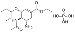
CCC(CC)O[C@@H]1C=C(C[C@@H]([C@H]1NC(=O)C)N)C(=O)OCC.OP(=O)(O)O
|
| InChi Key |
PGZUMBJQJWIWGJ-ONAKXNSWSA-N
|
| InChi Code |
InChI=1S/C16H28N2O4.H3O4P/c1-5-12(6-2)22-14-9-11(16(20)21-7-3)8-13(17)15(14)18-10(4)19;1-5(2,3)4/h9,12-15H,5-8,17H2,1-4H3,(H,18,19);(H3,1,2,3,4)/t13-,14+,15+;/m0./s1
|
| 化学名 |
ethyl (3R,4R,5S)-4-acetamido-5-amino-3-pentan-3-yloxycyclohexene-1-carboxylate;phosphoric acid
|
| 别名 |
GS-4071, GS-4104, GS4071, GS4104, GS 4071, Oseltamivir phosphate; 204255-11-8; Tamiflu; Oseltamivir (phosphate); Oseltamir Phosphate; Ro 64-0796/002; Oseltamivir (as phosphate); 4A3O49NGEZ; GS 4104, Tamiflu
|
| HS Tariff Code |
2934.99.9001
|
| 存储方式 |
Powder -20°C 3 years 4°C 2 years In solvent -80°C 6 months -20°C 1 month 注意: 请将本产品存放在密封且受保护的环境中,避免吸湿/受潮。 |
| 运输条件 |
Room temperature (This product is stable at ambient temperature for a few days during ordinary shipping and time spent in Customs)
|
| 溶解度 (体外实验) |
H2O : ~100 mg/mL (~243.66 mM)
DMSO : ~100 mg/mL (~243.66 mM) |
|---|---|
| 溶解度 (体内实验) |
配方 1 中的溶解度: ≥ 2.5 mg/mL (6.09 mM) (饱和度未知) in 10% DMSO + 40% PEG300 + 5% Tween80 + 45% Saline (这些助溶剂从左到右依次添加,逐一添加), 澄清溶液。
例如,若需制备1 mL的工作液,可将100 μL 25.0 mg/mL澄清DMSO储备液加入到400 μL PEG300中,混匀;然后向上述溶液中加入50 μL Tween-80,混匀;加入450 μL生理盐水定容至1 mL。 *生理盐水的制备:将 0.9 g 氯化钠溶解在 100 mL ddH₂O中,得到澄清溶液。 配方 2 中的溶解度: ≥ 2.5 mg/mL (6.09 mM) (饱和度未知) in 10% DMSO + 90% (20% SBE-β-CD in Saline) (这些助溶剂从左到右依次添加,逐一添加), 澄清溶液。 例如,若需制备1 mL的工作液,可将 100 μL 25.0 mg/mL澄清DMSO储备液加入900 μL 20% SBE-β-CD生理盐水溶液中,混匀。 *20% SBE-β-CD 生理盐水溶液的制备(4°C,1 周):将 2 g SBE-β-CD 溶解于 10 mL 生理盐水中,得到澄清溶液。 请根据您的实验动物和给药方式选择适当的溶解配方/方案: 1、请先配制澄清的储备液(如:用DMSO配置50 或 100 mg/mL母液(储备液)); 2、取适量母液,按从左到右的顺序依次添加助溶剂,澄清后再加入下一助溶剂。以 下列配方为例说明 (注意此配方只用于说明,并不一定代表此产品 的实际溶解配方): 10% DMSO → 40% PEG300 → 5% Tween-80 → 45% ddH2O (或 saline); 假设最终工作液的体积为 1 mL, 浓度为5 mg/mL: 取 100 μL 50 mg/mL 的澄清 DMSO 储备液加到 400 μL PEG300 中,混合均匀/澄清;向上述体系中加入50 μL Tween-80,混合均匀/澄清;然后继续加入450 μL ddH2O (或 saline)定容至 1 mL; 3、溶剂前显示的百分比是指该溶剂在最终溶液/工作液中的体积所占比例; 4、 如产品在配制过程中出现沉淀/析出,可通过加热(≤50℃)或超声的方式助溶; 5、为保证最佳实验结果,工作液请现配现用! 6、如不确定怎么将母液配置成体内动物实验的工作液,请查看说明书或联系我们; 7、 以上所有助溶剂都可在 Invivochem.cn网站购买。 |
| 制备储备液 | 1 mg | 5 mg | 10 mg | |
| 1 mM | 2.4366 mL | 12.1832 mL | 24.3665 mL | |
| 5 mM | 0.4873 mL | 2.4366 mL | 4.8733 mL | |
| 10 mM | 0.2437 mL | 1.2183 mL | 2.4366 mL |
1、根据实验需要选择合适的溶剂配制储备液 (母液):对于大多数产品,InvivoChem推荐用DMSO配置母液 (比如:5、10、20mM或者10、20、50 mg/mL浓度),个别水溶性高的产品可直接溶于水。产品在DMSO 、水或其他溶剂中的具体溶解度详见上”溶解度 (体外)”部分;
2、如果您找不到您想要的溶解度信息,或者很难将产品溶解在溶液中,请联系我们;
3、建议使用下列计算器进行相关计算(摩尔浓度计算器、稀释计算器、分子量计算器、重组计算器等);
4、母液配好之后,将其分装到常规用量,并储存在-20°C或-80°C,尽量减少反复冻融循环。
计算结果:
工作液浓度: mg/mL;
DMSO母液配制方法: mg 药物溶于 μL DMSO溶液(母液浓度 mg/mL)。如该浓度超过该批次药物DMSO溶解度,请首先与我们联系。
体内配方配制方法:取 μL DMSO母液,加入 μL PEG300,混匀澄清后加入μL Tween 80,混匀澄清后加入 μL ddH2O,混匀澄清。
(1) 请确保溶液澄清之后,再加入下一种溶剂 (助溶剂) 。可利用涡旋、超声或水浴加热等方法助溶;
(2) 一定要按顺序加入溶剂 (助溶剂) 。
 TTHP 512
 VNT-101
 Antiviral agent 70
 VF-57a
InvivoChem的所有产品仅用于作科学研究,不面向患者销售
Copyright 2020 InvivoChem LLC | All Rights Reserved 粤ICP备20063088号-1


 COA
COA
 463611831
