| 规格 | 价格 | 库存 | 数量 |
|---|---|---|---|
| 10 mM * 1 mL in DMSO |
|
||
| 1mg |
|
||
| 2mg |
|
||
| 5mg |
|
||
| 10mg |
|
||
| 25mg |
|
||
| 50mg |
|
||
| 100mg |
|
||
| 250mg |
|
||
| 500mg |
|
||
| Other Sizes |
|

| 靶点 |
EGFR (IC50 = 0.7 nM)
PD168393 (PD-168393) selectively inhibits epidermal growth factor receptor (EGFR) tyrosine kinase (IC₅₀ = 2 nM for recombinant EGFR; Ki = 0.7 nM for EGFR ATP-binding site) [1] PD168393 (PD-168393) shows no significant inhibitory activity against insulin receptor, platelet-derived growth factor receptor (PDGFR), or c-Src (IC₅₀ > 1000 nM) [2] |
|---|---|
| 体外研究 (In Vitro) |
PD 168393 对接至 EGFR TK 的 ATP 结合袋中。 PD168393 在连续暴露期间完全抑制 A431 细胞中的 EGF 依赖性受体自身磷酸化,甚至在不含化合物的培养基中 8 小时后也能持续抑制。 PD168393 抑制 MDA-MB-453 细胞中调蛋白诱导的酪氨酸磷酸化,IC50 为 5.7 nM。 PD168393 对胰岛素、PDGF 和基本 FGFR TK 以及 PKC 无活性。 PD168393 抑制 HS-27 人成纤维细胞中 EGF 介导的酪氨酸磷酸化,IC50 为 1-6 nM,但对 FGF 或 PDGF 介导的酪氨酸磷酸化几乎没有影响。 PD168393 在 3T3-Her2 细胞中显示出对 Her2 诱导的酪氨酸磷酸化的快速有效抑制,IC50 约为 100 nM。 D168393 还抑制 3T3-Her2 细胞中 PLCγ1/Stat1/Dok1/δ-catenin 的磷酸化(Fyb 除外)。激酶测定:PD168393 是一种有效的、细胞渗透性的、不可逆的 EGFR 抑制剂,IC50 为 0.70 nM,不可逆地烷基化 Cys-773,对胰岛素、PDGFR、FGFR 和 PKC 无活性。目标:EGFR IC 50:0.7 nM (1) PD 168393 抑制 A431 人表皮样癌细胞中的 EGFr 自身磷酸化,其效力比 PD 174265 高 9 倍以上。(2) PD 168393 减少 TNF-α 的产生和 ERK1/磷酸化LPS 在心肌细胞中诱导 2 和 p38。 (3) PD168393 在低至 0.03 umol/L 的浓度下即可完全抑制 AKT 和 ERK 磷酸化。 (4) PD168393可以诱导ErbB2阳性肺癌和乳腺癌细胞系凋亡并抑制细胞生长。 (5) 通过抑制磷酸化 p44/42 ERK 来确定,PD168393 会破坏 HaCaT 细胞中的 MEK1/p44/42 ERK 信号传导。细胞测定:(1) PD 168393 抑制 A431 人表皮样癌细胞中的 EGFr 自磷酸化,其效力比 PD 174265 高 9 倍。(2) PD 168393 减少 TNF-α 的产生以及由 TNF-α 诱导的 ERK1/2 和 p38 磷酸化心肌细胞中的脂多糖。 (3) PD168393 在低至 0.03 umol/L 的浓度下即可完全抑制 AKT 和 ERK 磷酸化。 (4) PD168393可以诱导ErbB2阳性肺癌和乳腺癌细胞系凋亡并抑制细胞生长。
PD168393(PD-168393)剂量依赖性抑制EGFR过表达的肿瘤细胞系增殖,包括A431表皮样癌细胞(IC₅₀=0.04μM)和MDA-MB-468乳腺癌细胞(IC₅₀=0.06μM)。浓度≥0.1μM时,可阻断这些细胞中表皮生长因子(EGF)诱导的EGFR磷酸化及下游ERK1/2信号通路[1] PD168393(PD-168393)在0.2μM浓度下,可分别抑制A431细胞的迁移和侵袭约70%和65%,其机制与下调基质金属蛋白酶-9(MMP-9)表达有关[2] 在原代皮质神经元中,PD168393(PD-168393)(1μM)通过阻断EGFR磷酸化及下游PI3K/Akt信号,抑制EGFR介导的神经突生长[3] |
| 体内研究 (In Vivo) |
PD 168393 在裸鼠 A431 人表皮样癌异种移植物中产生 115% 的肿瘤生长抑制,同时 EGFR 磷酸酪氨酸含量降低 50%。 PD 168393 还显示出低血浆浓度。
在本研究中,研究人员评估了EGFR抑制剂PD168393(PD)对小鼠挫伤性SCI模型髓鞘形成的影响。研究发现,经PD168393处理的小鼠脊髓损伤后,髓鞘碱性蛋白(MBP)的表达显著升高。PD168393处理后,病变中的胶质前体细胞和少突胶质细胞(OLs)密度增加,细胞凋亡减弱。此外,PD168393治疗降低了脊髓受损区域OX42+小胶质细胞和胶质纤维酸性蛋白+星形胶质细胞的数量。因此,他们得出结论,SCI后EGFR抑制的治疗作用包括促进受损脊髓的髓鞘再生,增加少突胶质前体细胞和OLs,以及抑制星形胶质细胞和小胶质细胞/巨噬细胞的活化[3]。 PD168393(PD-168393)以15mg/kg/天的剂量腹腔注射给药21天,可抑制裸鼠A431异种移植瘤的生长。与对照组相比,肿瘤体积减少约68%,瘤内EGFR磷酸化水平显著下调[1] 在7,12-二甲基苯并(a)蒽(DMBA)和12-O-十四酰佛波醇-13-乙酸酯(TPA)诱导的小鼠皮肤癌发生模型中,PD168393(PD-168393)(10mg/kg/天,腹腔注射,持续14周)使皮肤乳头状瘤数量减少约55%[2] |
| 酶活实验 |
PD168393 是一种有效的、细胞渗透性的、不可逆的 EGFR 抑制剂,可不可逆地烷基化 Cys-773。它对 PDGFR、FGFR、PKC 和胰岛素无活性。其 IC50 为 0.70 nM。目标:EGFR IC 50 = 0.7 nM (1) PD 168393 在抑制 A431 人表皮样癌细胞中 EGFr 自磷酸化方面的效力比 PD 174265 高 9 倍以上。 (2) 在脂多糖 (LPS) 刺激的心肌细胞中,PD 168393 减少 TNF-α 的产生以及 ERK1/2 和 p38 的磷酸化。 (3) 在低至0.03 umol/L的浓度下,PD168393完全抑制AKT和ERK的磷酸化。 (4) 在ErbB2阳性肺癌和乳腺癌细胞系中,PD168393可能导致细胞凋亡并抑制细胞生长。 (5)磷酸化p44/42 ERK的抑制表明PD168393干扰HaCaT细胞中的MEK1/p44/42 ERK信号传导。
将重组人EGFR激酶结构域与ATP及特异性多肽底物在系列稀释的PD168393(PD-168393)存在下孵育,反应在37°C下进行60分钟,采用放射免疫法检测磷酸化底物。通过与溶媒对照组的放射性对比计算抑制率,从量效曲线中得出IC₅₀值[1] 采用相同方案检测PD168393(PD-168393)对重组胰岛素受体、PDGFR和c-Src激酶的抑制活性以评估选择性,反应条件保持一致,通过确定IC₅₀值证实其对EGFR的选择性靶向[2] |
| 细胞实验 |
(1) 在 A431 人表皮样癌细胞中,PD 168393 抑制 EGFr 自磷酸化的效力比 PD 174265 高 9 倍以上。 (2) 在脂多糖 (LPS) 刺激的心肌细胞中,PD 168393 减少 TNF-α 的产生以及 ERK1/2 和 p38 的磷酸化。 (3) 在低至0.03 umol/L的浓度下,PD168393完全抑制AKT和ERK的磷酸化。 (4) 在ErbB2阳性肺癌和乳腺癌细胞系中,PD168393可能导致细胞凋亡并抑制细胞生长。
将A431和MDA-MB-468细胞以5×10³个细胞/孔接种到96孔板中,用PD168393(PD-168393)(0.01-1μM)处理72小时,采用四唑盐法检测细胞活性并计算IC₅₀值。蛋白质印迹分析中,用0.1-0.5μM药物处理细胞并加入EGF刺激,裂解后与抗磷酸化EGFR、ERK1/2和GAPDH的抗体孵育[1] 用PD168393(PD-168393)(0.1-0.5μM)处理A431细胞24小时,采用Boyden小室进行迁移和侵袭实验,通过逆转录-聚合酶链反应(RT-PCR)定量MMP-9 mRNA的表达[2] 分离原代皮质神经元并接种到24孔板中,用PD168393(PD-168393)(0.5-2μM)预处理1小时后,用EGF刺激细胞。48小时后在显微镜下观察神经突生长,通过蛋白质印迹法检测磷酸化EGFR和Akt[3] |
| 动物实验 |
无胸腺裸鼠(携带A431人表皮样癌细胞)
58 mg/kg 腹腔注射 体内疗效。[1] 为了说明不可逆性的优势,表2直接比较了PD168393(不可逆)和PD174265(可逆)在活细胞中对靶点调控的作用。PD168393抑制A431人表皮样癌细胞中EGFR自磷酸化的效力比PD174265高9倍以上。在MDA-MB-453人乳腺癌细胞中,针对heregulin介导的酪氨酸磷酸化的抑制作用差异更为显著,PD168393的效力比PD174265高30倍以上。图 6a 非常直观地展示了不可逆抑制的治疗优势。该图直接比较了 PD168393 和 PD174265 对裸鼠体内异种移植的 A431 人表皮样癌的体内活性。PD168393 在每日一次腹腔注射给药的情况下,对肿瘤生长的抑制作用远优于 PD174265。PD168393 的肿瘤生长抑制率达到 115%,在本实验中,该值定义为治疗组肿瘤达到三次体积倍增所需时间的中位数减去对照组肿瘤达到三次体积倍增所需时间的中位数,并以治疗持续时间(15 天)的百分比表示。相比之下,PD174265 的肿瘤生长抑制率仅为 13%。这两种化合物的抗肿瘤活性与其抑制EGFR磷酸酪氨酸含量的能力相关。注射后4小时,两种化合物均使磷酸化水平降低了约80%(图6b)。然而,注射可逆化合物PD 174265的小鼠在8小时后,磷酸化水平恢复至对照组的75%,并在24小时后恢复至100%。相比之下,注射PD168393的小鼠在24小时后EGFR的磷酸酪氨酸含量仍降低了50%。尽管在所有检测时间点,PD168393的血浆浓度均低于PD 174265(数据未显示),但其治疗优势依然存在。[1] 携带A431异种移植瘤(100-150 mm³)的裸鼠被随机分为对照组和治疗组。将PD-168393 (PD-168393)溶于DMSO,并用生理盐水稀释(最终DMSO浓度≤5%),然后以15 mg/kg/天的剂量腹腔注射,连续21天。每3天测量一次肿瘤体积,并处死小鼠收集肿瘤组织,用于EGFR磷酸化Western blot分析[1]。雌性CD-1小鼠用DMBA(100 μg/只)诱导,并用TPA(2 μg/只)每周两次促进,持续14周。从第4周开始,小鼠接受PD-168393 (PD-168393)(10 mg/kg/天,腹腔注射)治疗,持续10周。每周计数皮肤乳头状瘤的数量,并收集皮肤组织进行组织病理学分析[2]。 |
| 药代性质 (ADME/PK) |
PD-168393 (PD-168393) 在小鼠单次口服 15 mg/kg 剂量后的生物利用度约为 28%。血浆半衰期约为 3.5 小时,给药后 1 小时达到最大血浆浓度 (Cmax) 为 1.8 μg/mL [1]。在大鼠中,腹腔注射 10 mg/kg 的 PD-168393 (PD-168393) 后,24 小时 AUC₀ 为 12.6 μg·h/mL。该药物主要分布于肝脏和肿瘤组织,肿瘤/血浆浓度比约为 2.1 [2]。
|
| 毒性/毒理 (Toxicokinetics/TK) |
小鼠以15 mg/kg/天(腹腔注射)的剂量接受PD168393(PD-168393)治疗21天后,体重略有下降(约6%),但未出现明显的肝肾毒性。血清ALT、AST和肌酐水平均在正常范围内[1]。在长期毒性研究(14周,10 mg/kg/天,腹腔注射)中,大鼠未出现血液学异常或胃肠道副作用。通过平衡透析法测定,PD168393(PD-168393)在人血浆中的血浆蛋白结合率约为90%[2]。
|
| 参考文献 | |
| 其他信息 |
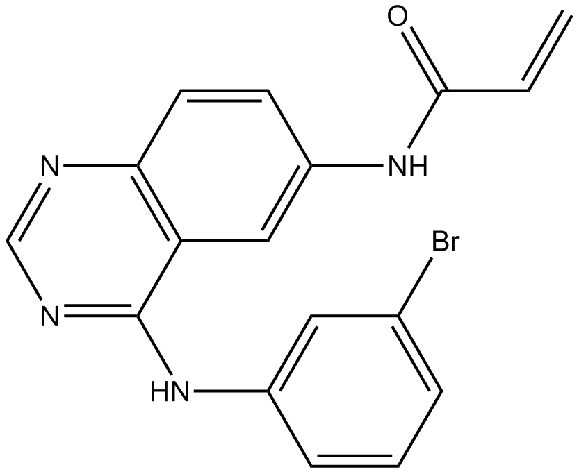
PD168393 是一种喹唑啉类化合物,其 4 位和 6 位分别带有溴苯胺基和丙烯酰胺基取代基。它是一种表皮生长因子受体拮抗剂。PD168393 属于喹唑啉类、丙烯酰胺类、取代苯胺类、溴苯类和仲酰胺类化合物。PD168393 是一种表皮生长因子受体抑制剂。EGFR 抑制剂 PD-168393 是一种具有抗肿瘤活性的喹唑啉酮类化合物。PD-168393 是一种细胞渗透性、不可逆且选择性的配体依赖性表皮生长因子 (EGF) 受体 (EGFR) 抑制剂。该化合物以1:1的化学计量比与EGFR的催化结构域结合,并通过烷基化ATP结合口袋内的半胱氨酸残基(Cys-773)来灭活EGFR酪氨酸激酶活性,从而抑制表达EGFR的肿瘤细胞增殖。
本发明公开了一类高亲和力抑制剂,该类抑制剂通过特异性共价修饰ATP结合口袋中的半胱氨酸残基,选择性地靶向并不可逆地灭活表皮生长因子受体酪氨酸激酶。一系列采用质谱、分子建模、定点诱变和活细胞14C标记研究的实验明确证明,这些化合物以1:1的化学计量比选择性地与表皮生长因子受体的催化结构域结合,并烷基化Cys-773。这些化合物在溶液中基本不发生反应,但当它们与ATP结合口袋结合时,会受到该特定氨基酸的快速亲核攻击。这些抑制剂中丙烯酰胺基团相对于Cys-773的分子取向和位置,完全支持了基于ATP结合位点同源建模分子模型的分子对接实验结果。此外,还有证据表明这些化合物以类似的方式与erbB2相互作用,但对本研究中测试的其他受体酪氨酸激酶或细胞内酪氨酸激酶没有活性。最后,6-丙烯酰胺基-4-苯胺基喹唑啉与一种效力相当但可逆的类似物之间的直接比较表明,不可逆抑制剂PD168393在人表皮样癌异种移植模型中具有远优于可逆类似物的体内抗肿瘤活性,且在治疗剂量下未观察到明显的毒性。该化合物的活性谱是具有巨大治疗潜力的新一代酪氨酸激酶抑制剂的典型代表,有望在增生性疾病的治疗中发挥重要作用。[1] Her2/neu (Her2) 是一种酪氨酸激酶,属于 EGF 受体 (EGFR)/ErbB 家族,在 20-30% 的人类乳腺癌中过表达。我们利用基于质谱的定量蛋白质组学方法,进一步表征了 Her2 信号转导通路。我们构建了稳定转染的过表达 Her2 或空载体的细胞系,并表征了 EGFR 和 Her2 选择性酪氨酸激酶抑制剂 PD168393 对这些细胞的影响。我们采用 SILAC(细胞培养中氨基酸稳定同位素标记)方法,同时监测三种条件下的蛋白质,并对 462 种蛋白质进行了定量分析。在这些蛋白质中,198种在Her2过表达细胞中酪氨酸磷酸化水平显著升高,81种则显著降低。用PD168393处理Her2过表达细胞后,大多数Her2触发的磷酸化事件可迅速逆转。鉴定出的磷酸化蛋白包括许多已知的Her2信号分子,以及一些此前未与Her2关联的已知EGFR信号蛋白,例如Stat1、Dok1和δ-catenin。重要的是,还鉴定出几种此前未被表征的Her2信号蛋白,包括Axl酪氨酸激酶、衔接蛋白Fyb和钙结合蛋白Pdcd-6/Alg-2。我们还鉴定出Her2蛋白中的一个磷酸化位点Y877,该位点位于激酶结构域的激活环中,不同于已知的C端尾部自磷酸化位点,可能对Her2信号通路的调控具有重要意义。我们利用网络建模,结合磷酸化蛋白质组学结果和文献整理的蛋白质-蛋白质相互作用数据,揭示了一些先前未鉴定的Her2信号蛋白的功能。[2] 预防脱髓鞘和促进裸露轴突的髓鞘再生是脊髓损伤(SCI)的一种有前景的治疗策略。据报道,表皮生长因子受体(EGFR)抑制剂有利于SCI后的神经功能恢复和轴突再生。然而,其在脊髓损伤后轴突脱髓鞘和髓鞘再生中的作用尚不清楚。在本研究中,我们评估了EGFR抑制剂PD168393(PD)对小鼠脊髓挫伤模型中髓鞘形成的影响。我们发现,PD治疗小鼠脊髓损伤部位髓鞘碱性蛋白(MBP)的表达显著升高。PD168393治疗后,神经胶质前体细胞和少突胶质细胞(OLs)的密度增加,损伤部位的细胞凋亡减少。此外,PD168393治疗还减少了脊髓损伤区域OX42阳性小胶质细胞和胶质纤维酸性蛋白(GFAP)阳性星形胶质细胞的数量。因此,我们得出结论,脊髓损伤后EGFR抑制剂的治疗作用包括促进受损脊髓的髓鞘再生、增加少突胶质细胞前体细胞和少突胶质细胞的数量,以及抑制星形胶质细胞和小胶质细胞/巨噬细胞的活化。[3] PD168393 (PD-168393)是一种可逆的小分子抑制剂,它与EGFR酪氨酸激酶的ATP结合位点结合,阻断EGF介导的细胞增殖、迁移和存活信号通路。[1] 除了抗肿瘤活性外,PD168393 (PD-168393)在研究EGFR介导的神经突生长方面也展现出潜力,并可能有助于深入了解EGFR在神经发育和神经退行性疾病中的作用。[3] 该药物被广泛用作临床前研究的工具化合物。该药物用于研究EGFR信号通路,但由于药代动力学特性欠佳,尚未进入临床试验阶段[2] |
| 分子式 |
C17H13BRN4O
|
|
|---|---|---|
| 分子量 |
369.22
|
|
| 精确质量 |
368.027
|
|
| 元素分析 |
C, 55.30; H, 3.55; Br, 21.64; N, 15.17; O, 4.33
|
|
| CAS号 |
194423-15-9
|
|
| 相关CAS号 |
|
|
| PubChem CID |
4708
|
|
| 外观&性状 |
Light yellow to khaki solid powder
|
|
| 密度 |
1.6±0.1 g/cm3
|
|
| 沸点 |
571.1±50.0 °C at 760 mmHg
|
|
| 熔点 |
279℃
|
|
| 闪点 |
299.2±30.1 °C
|
|
| 蒸汽压 |
0.0±1.6 mmHg at 25°C
|
|
| 折射率 |
1.744
|
|
| LogP |
3.72
|
|
| tPSA |
70.4
|
|
| 氢键供体(HBD)数目 |
2
|
|
| 氢键受体(HBA)数目 |
4
|
|
| 可旋转键数目(RBC) |
4
|
|
| 重原子数目 |
23
|
|
| 分子复杂度/Complexity |
433
|
|
| 定义原子立体中心数目 |
0
|
|
| SMILES |
BrC1=C([H])C([H])=C([H])C(=C1[H])N([H])C1C2C([H])=C(C([H])=C([H])C=2N=C([H])N=1)N([H])C(C([H])=C([H])[H])=O
|
|
| InChi Key |
HTUBKQUPEREOGA-UHFFFAOYSA-N
|
|
| InChi Code |
InChI=1S/C17H13BrN4O/c1-2-16(23)21-13-6-7-15-14(9-13)17(20-10-19-15)22-12-5-3-4-11(18)8-12/h2-10H,1H2,(H,21,23)(H,19,20,22)
|
|
| 化学名 |
N-[4-(3-bromoanilino)quinazolin-6-yl]prop-2-enamide
|
|
| 别名 |
PD 168393; PD-168393; 4-[(3-Bromophenyl)amino]-6-acrylamidoquinazoline; pd 168393; N-(4-((3-bromophenyl)amino)quinazolin-6-yl)acrylamide; n-{4-[(3-bromophenyl)amino]quinazolin-6-yl}prop-2-enamide; N-[4-(3-bromoanilino)quinazolin-6-yl]prop-2-enamide; PD168393
|
|
| HS Tariff Code |
2934.99.9001
|
|
| 存储方式 |
Powder -20°C 3 years 4°C 2 years In solvent -80°C 6 months -20°C 1 month |
|
| 运输条件 |
Room temperature (This product is stable at ambient temperature for a few days during ordinary shipping and time spent in Customs)
|
| 溶解度 (体外实验) |
|
|||
|---|---|---|---|---|
| 溶解度 (体内实验) |
配方 1 中的溶解度: ≥ 2.5 mg/mL (6.77 mM) (饱和度未知) in 10% DMSO + 40% PEG300 + 5% Tween80 + 45% Saline (这些助溶剂从左到右依次添加,逐一添加), 澄清溶液。
例如,若需制备1 mL的工作液,可将100 μL 25.0 mg/mL澄清DMSO储备液加入到400 μL PEG300中,混匀;然后向上述溶液中加入50 μL Tween-80,混匀;加入450 μL生理盐水定容至1 mL。 *生理盐水的制备:将 0.9 g 氯化钠溶解在 100 mL ddH₂O中,得到澄清溶液。 配方 2 中的溶解度: 30% PEG400+0.5% Tween80+5% propylene glycol: 30mg/mL 请根据您的实验动物和给药方式选择适当的溶解配方/方案: 1、请先配制澄清的储备液(如:用DMSO配置50 或 100 mg/mL母液(储备液)); 2、取适量母液,按从左到右的顺序依次添加助溶剂,澄清后再加入下一助溶剂。以 下列配方为例说明 (注意此配方只用于说明,并不一定代表此产品 的实际溶解配方): 10% DMSO → 40% PEG300 → 5% Tween-80 → 45% ddH2O (或 saline); 假设最终工作液的体积为 1 mL, 浓度为5 mg/mL: 取 100 μL 50 mg/mL 的澄清 DMSO 储备液加到 400 μL PEG300 中,混合均匀/澄清;向上述体系中加入50 μL Tween-80,混合均匀/澄清;然后继续加入450 μL ddH2O (或 saline)定容至 1 mL; 3、溶剂前显示的百分比是指该溶剂在最终溶液/工作液中的体积所占比例; 4、 如产品在配制过程中出现沉淀/析出,可通过加热(≤50℃)或超声的方式助溶; 5、为保证最佳实验结果,工作液请现配现用! 6、如不确定怎么将母液配置成体内动物实验的工作液,请查看说明书或联系我们; 7、 以上所有助溶剂都可在 Invivochem.cn网站购买。 |
| 制备储备液 | 1 mg | 5 mg | 10 mg | |
| 1 mM | 2.7084 mL | 13.5421 mL | 27.0841 mL | |
| 5 mM | 0.5417 mL | 2.7084 mL | 5.4168 mL | |
| 10 mM | 0.2708 mL | 1.3542 mL | 2.7084 mL |
1、根据实验需要选择合适的溶剂配制储备液 (母液):对于大多数产品,InvivoChem推荐用DMSO配置母液 (比如:5、10、20mM或者10、20、50 mg/mL浓度),个别水溶性高的产品可直接溶于水。产品在DMSO 、水或其他溶剂中的具体溶解度详见上”溶解度 (体外)”部分;
2、如果您找不到您想要的溶解度信息,或者很难将产品溶解在溶液中,请联系我们;
3、建议使用下列计算器进行相关计算(摩尔浓度计算器、稀释计算器、分子量计算器、重组计算器等);
4、母液配好之后,将其分装到常规用量,并储存在-20°C或-80°C,尽量减少反复冻融循环。
计算结果:
工作液浓度: mg/mL;
DMSO母液配制方法: mg 药物溶于 μL DMSO溶液(母液浓度 mg/mL)。如该浓度超过该批次药物DMSO溶解度,请首先与我们联系。
体内配方配制方法:取 μL DMSO母液,加入 μL PEG300,混匀澄清后加入μL Tween 80,混匀澄清后加入 μL ddH2O,混匀澄清。
(1) 请确保溶液澄清之后,再加入下一种溶剂 (助溶剂) 。可利用涡旋、超声或水浴加热等方法助溶;
(2) 一定要按顺序加入溶剂 (助溶剂) 。
The irreversible inhibitor PD168393 overcomes lapatinib resistance caused by the ErbB2 T798I mutation.PLoS One.2014 Sep 19;9(9):e106349. |
Antitumor effects of lapatinib and PD168393 in the ErbB2 positive lung cancer cell line, Calu3 and the ErbB2 positive breast cancer cell line, SkBr3 after 72 hour treatment.PLoS One.2014 Sep 19;9(9):e106349. |
Clonogenic survival assay shows that PHLDA1 overexpression could significantly enhance lapatinib sensitivity in breast cancer cells.PLoS One.2014 Sep 19;9(9):e106349. |
InvivoChem的所有产品仅用于作科学研究,不面向患者销售
Copyright 2020 InvivoChem LLC | All Rights Reserved 粤ICP备20063088号-1


 COA
COA


 463611831
