| 规格 | 价格 | 库存 | 数量 |
|---|---|---|---|
| 1mg |
|
||
| 5mg |
|
||
| 10mg |
|
||
| 25mg |
|
||
| 50mg |
|
||
| 100mg |
|
||
| 250mg |
|
||
| 500mg |
|
||
| 1g |
|
||
| Other Sizes |
|

| 靶点 |
Aryl hydrocarbon receptor/AhR (IC50 = 127 nM)
StemRegenin 1 (SR1) is a selective antagonist of the aryl hydrocarbon receptor (AhR), with an IC50 of 10 nM for human AhR in an AhR-responsive luciferase reporter assay. It does not bind to other nuclear receptors (e.g., PPARγ, RARα) at concentrations up to 1 μM [1] |
|---|---|
| 体外研究 (In Vitro) |
StemRegenin 1 (SR1)对芳烃受体具有拮抗作用。(AhR)。在EC50小于120 nM的情况下,stemregenin 1在5 ~ 7天后可增加CD34+细胞的数量。综上所述,StemRegenin 1诱导CD34+细胞生长是通过直接结合AhR抑制介导的(IC50=40 nM)。StemRegenin 1也能减少光亲和配体(PAL)的结合。StemRegenin 1 (SR1)是一种芳烃受体拮抗剂,能强烈促进人CD34+细胞的体外生长。用stemgenin 1治疗可降低VentX表达水平,加速CD34+细胞增殖[2]。
人脐带血(UCB)来源CD34+造血干细胞(HSCs)扩增实验:分离CD34+细胞,在添加细胞因子(SCF、TPO、Flt3-L、IL-6)的StemSpan培养基中培养。SR1(10 nM)处理7天,CD34+细胞数量较溶媒对照组(DMSO)增加8.2倍;CD34+CD38-细胞(原始HSCs标志物)比例从5.2%升至18.3%(流式细胞术)。集落形成单位(CFU)实验显示,粒-巨噬细胞集落(CFU-GM)增加6.5倍,红细胞集落(CFU-E)增加5.8倍,混合谱系集落(CFU-Mix)增加4.3倍[1] - 长期培养起始细胞(LTC-IC)实验:SR1(10 nM)处理14天的UCB CD34+细胞,与小鼠骨髓基质细胞共培养5周。LTC-ICs(反映长期HSC功能)数量较对照组增加7.1倍,表明SR1可维持HSCs的自我更新能力[1] - AhR下游基因抑制实验:SR1(10 nM)处理UCB CD34+细胞后,AhR靶基因(CYP1A1、CYP1B1)的mRNA表达分别降低75%和68%(qPCR分析),证实其通过拮抗AhR发挥作用[1] |
| 体内研究 (In Vivo) |
为了计算StemRegenin 1 (SR1)扩增细胞的SRC频率,进行第二次移植实验。最终培养物的一部分,相当于30至3000个初始CB CD34+细胞,在对照条件下或StemRegenin 1 (SR1)扩增21天,移植到NSG小鼠中(图3A)。泊松统计显示,未培养细胞的SRC频率为1/703,细胞因子扩增后的SRC频率为1/597,StemRegenin 1 (SR1)扩增后的SRC频率为1/40(图3B)。这表示用于启动培养的2.5 × 105 CB CD34+细胞中SRC的总含量为356,对照培养中为403,在StemRegenin 1 (SR1)培养后为6283(图3C和表S5)。用StemRegenin 1 (SR1)培养后NSG植入量的增加在7个独立实验中是可重复的(表S6)。根据培养后注射的细胞数量计算SRC数量显示,对照培养物的SRC频率比新鲜细胞减少了1000倍,但SRC总含量没有减少,这可能反映了这些培养物中的大多数细胞是失去移植NSG小鼠能力的祖细胞。纳入StemRegenin 1 (SR1)降低了这种影响,导致SRC频率相对于对照培养物增加8倍。这一结果,加上细胞总数增加2倍,可能导致SRC含量增加17倍。[1]
SR1 促进 CB CD34+ 细胞的扩增,有助于早期和持续植入 NOD.Cg-Prkdcscid Il2rgtm1Wjl/SzJ (NSG) 小鼠。 NOD/SCID小鼠HSC移植实验:将SR1(10 nM)处理7天的UCB CD34+细胞(1×10⁵细胞/只)静脉注射到亚致死剂量照射(2.5 Gy)的NOD/SCID小鼠体内。移植后4周,小鼠骨髓中人CD45+细胞比例为28.5%,显著高于溶媒处理组的8.2%(流式细胞术);骨髓中人CD34+细胞增加9.3倍,骨髓细胞CFU实验显示人源CFUs增加6.7倍,表明SR1扩增的HSCs具有更强的植入能力和多谱系重建能力[1] |
| 酶活实验 |
AhR荧光素酶测定。[1]
将胰酶化的HepG2细胞(2 × 10⁶)用Fugene6在Opti-MEM中瞬时转染20 μg pGodLuc6.1。将转染的细胞洗涤后,用20 mL添加10个胎牛血清的MEM重悬,以50 μL/孔的浓度,接种于384孔板中。转染24 h后,用化合物处理细胞1 h,再加入TCDD (3 nM)。TCDD诱导24小时后,使用Bright-Glo™荧光素酶测定系统(Promega)检测荧光素酶活性。 AhR配体结合试验。[1] 实验按照文献描述方法进行。 AhR响应性荧光素酶报告基因实验:HEK293细胞以5×10⁴细胞/孔接种于24孔板,含10% FBS的DMEM培养24小时。用转染试剂共转染0.5 μg人AhR表达质粒、0.5 μg AhR响应性荧光素酶报告质粒(含二噁英响应元件DRE)和0.1 μg β-半乳糖苷酶质粒(内参)。24小时后,更换为含SR1(0.1-1000 nM)和10 nM 2,3,7,8-四氯二苯并-p-二噁英(TCDD,AhR激动剂)的无血清DMEM,继续孵育24小时。用报告基因裂解缓冲液裂解细胞,luminometer检测荧光素酶活性,β-半乳糖苷酶活性校正转染效率,非线性回归计算IC50[1] |
| 细胞实验 |
人CD34+细胞用1 μm SR1处理1周或模拟处理(对照)。然后收集处理后的细胞,分析VentX mRNA的表达水平。F、GFP-或GFP- ventx转染的CD34+细胞(2 × 10⁴)分选,用1 μm SR1处理1周或模拟处理。然后计算细胞总数并绘制。[2]
人UCB CD34+细胞分离与扩增实验:收集UCB样本,密度梯度离心分离单个核细胞(MNCs),免疫磁珠分选(CD34+微珠)纯化CD34+细胞。纯化的CD34+细胞(1×10⁴细胞/mL)在添加重组人SCF(100 ng/mL)、TPO(50 ng/mL)、Flt3-L(50 ng/mL)和IL-6(20 ng/mL)的StemSpan SFEM培养基中,于37°C、5% CO₂条件下培养。SR1终浓度为0.5、1、5、10或50 nM(溶媒对照:0.1% DMSO)。培养7或14天后,收集细胞,流式细胞术(抗CD34-PE和抗CD38-FITC抗体)分析CD34+和CD34+CD38-细胞比例,血细胞计数板计数细胞以计算扩增倍数[1] - CFU实验:将培养后的CD34+细胞(1×10³细胞/孔)接种于含细胞因子(SCF、GM-CSF、EPO、IL-3)的甲基纤维素培养基24孔板,37°C、5% CO₂培养14天。倒置显微镜下根据形态学标准计数集落(CFU-GM、CFU-E、CFU-Mix)[1] - LTC-IC实验:小鼠骨髓基质细胞(MS-5细胞系)接种于6孔板,培养至融合。将SR1处理或溶媒处理的CD34+细胞(5×10³细胞/孔)加入基质层,在MyeloCult培养基中37°C、5% CO₂共培养,每周更换一半培养基,持续5周。收集细胞并接种于甲基纤维素培养基计数CFUs,LTC-IC定义为共培养5周后仍能形成CFU的细胞[1] |
| 动物实验 |
CD34+细胞在NSG小鼠体内的移植。[1]
将250,000个脐带血来源的CD34+细胞在对照条件下(DMSO,0.01%)或StemRegenin 1 (SR1) (0.75 μM)处理下培养3周。此时,对照组细胞扩增了1080倍,而StemRegenin 1 (SR1)处理组细胞扩增了2024倍(相对于起始细胞数)。移植30至30,000个未培养的CD34+脐带血来源细胞,或相当于30至10,000个起始细胞的最终培养细胞的一部分。将细胞经眼眶后静脉途径注射到经亚致死剂量照射(图2A实验为300 rad,图3A-B实验为200 rad)的6至10周龄NSG小鼠体内。照射后24小时内进行移植。通过流式细胞术分析经眼眶后静脉窦或骨髓采集的血液样本,并使用抗人CD45和抗小鼠CD45抗体监测移植情况。移植后13至16周处死小鼠;收集骨髓(来自双侧股骨和胫骨)、脾脏和胸腺进行分析。对于二次移植,将每只受体小鼠50%的骨髓移植到另一只经亚致死剂量照射的NSG小鼠体内。移植后15周,收集二次移植小鼠的骨髓并进行流式细胞术分析。 [1] NOD/SCID小鼠造血干细胞移植方案:雌性NOD/SCID小鼠(6-8周龄,18-22 g)饲养于SPF级条件下(22±2°C,12小时光照/黑暗循环,自由摄食饮水)。移植前24小时,小鼠接受单次2.5 Gy亚致死剂量γ射线照射。脐带血CD34+细胞用SR1(10 nM)或载体处理7天,然后重悬于PBS中,浓度为1×10⁵个细胞/100 μL。每只小鼠经尾静脉注射100 μL细胞悬液。移植后4周,小鼠用二氧化碳处死,并从股骨和胫骨中冲洗出骨髓。骨髓细胞用抗人CD45-APC抗体染色,并通过流式细胞术分析以确定人细胞植入情况。对于CFU分析,将骨髓细胞接种于甲基纤维素培养基中以计数人CFU[1] |
| 毒性/毒理 (Toxicokinetics/TK) |
体外毒性:用 SR1 (0.5-50 nM) 处理 UCB CD34+ 细胞 14 天,未观察到明显的细胞毒性——所有浓度下细胞活力均保持在 90% 以上(台盼蓝染色)[1]
- 体内毒性:在移植了 SR1 扩增的 HSC 的 NOD/SCID 小鼠中,移植后 4 周未观察到异常行为、体重减轻(<5% 的基线值)或主要器官(肝脏、肾脏、脾脏)的组织病理学变化[1] |
| 参考文献 |
|
| 其他信息 |
StemRegenin 1 是一种芳烃受体抑制剂。
StemRegenin 1 (SR1)是一种小分子芳烃受体拮抗剂,专为体外扩增人造血干细胞 (HSC) 而开发。其作用机制是抑制通常限制 HSC 自我更新的 AhR 信号通路,从而促进 HSC 增殖,同时保持其多系分化和移植能力 [1] - 使用 SR1 进行 HSC 体外扩增解决了造血干细胞移植 (HSCT) 的一个关键限制:脐带血或外周血移植中 HSC 数量不足。 SR1扩增的造血干细胞在免疫缺陷小鼠中显示出增强的植入能力,支持其在造血干细胞移植治疗血液系统疾病(例如白血病、淋巴瘤)中的潜在临床应用[1] - 文献[2]主要关注VentX转录因子在造血干细胞扩增中的作用,并未提及干细胞再生素1 (SR1)或其生物学功能[2] |
| 分子式 |
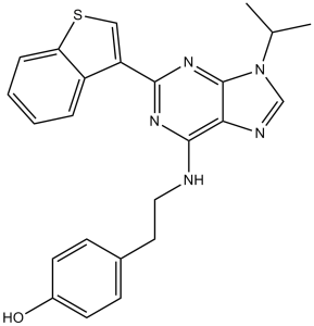
C24H23N5OS
|
|
|---|---|---|
| 分子量 |
429.54
|
|
| 精确质量 |
429.162
|
|
| 元素分析 |
C, 67.11; H, 5.40; N, 16.30; O, 3.72; S, 7.46
|
|
| CAS号 |
1227633-49-9
|
|
| 相关CAS号 |
|
|
| PubChem CID |
46199207
|
|
| 外观&性状 |
White to off-white solid powder
|
|
| 密度 |
1.4±0.1 g/cm3
|
|
| 沸点 |
622.9±65.0 °C at 760 mmHg
|
|
| 闪点 |
330.5±34.3 °C
|
|
| 蒸汽压 |
0.0±1.9 mmHg at 25°C
|
|
| 折射率 |
1.721
|
|
| LogP |
5.1
|
|
| tPSA |
104.1
|
|
| 氢键供体(HBD)数目 |
2
|
|
| 氢键受体(HBA)数目 |
6
|
|
| 可旋转键数目(RBC) |
6
|
|
| 重原子数目 |
31
|
|
| 分子复杂度/Complexity |
586
|
|
| 定义原子立体中心数目 |
0
|
|
| InChi Key |
BGFHMYJZJZLMHW-UHFFFAOYSA-N
|
|
| InChi Code |
InChI=1S/C24H23N5OS/c1-15(2)29-14-26-21-23(25-12-11-16-7-9-17(30)10-8-16)27-22(28-24(21)29)19-13-31-20-6-4-3-5-18(19)20/h3-10,13-15,30H,11-12H2,1-2H3,(H,25,27,28)
|
|
| 化学名 |
4-[2-[[2-(1-benzothiophen-3-yl)-9-propan-2-ylpurin-6-yl]amino]ethyl]phenol
|
|
| 别名 |
|
|
| HS Tariff Code |
2934.99.9001
|
|
| 存储方式 |
Powder -20°C 3 years 4°C 2 years In solvent -80°C 6 months -20°C 1 month |
|
| 运输条件 |
Room temperature (This product is stable at ambient temperature for a few days during ordinary shipping and time spent in Customs)
|
| 溶解度 (体外实验) |
|
|||
|---|---|---|---|---|
| 溶解度 (体内实验) |
配方 1 中的溶解度: ≥ 2.5 mg/mL (5.82 mM) (饱和度未知) in 10% DMSO + 40% PEG300 + 5% Tween80 + 45% Saline (这些助溶剂从左到右依次添加,逐一添加), 澄清溶液。
例如,若需制备1 mL的工作液,可将100 μL 25.0 mg/mL澄清DMSO储备液加入到400 μL PEG300中,混匀;然后向上述溶液中加入50 μL Tween-80,混匀;加入450 μL生理盐水定容至1 mL。 *生理盐水的制备:将 0.9 g 氯化钠溶解在 100 mL ddH₂O中,得到澄清溶液。 配方 2 中的溶解度: 2.5 mg/mL (5.82 mM) in 10% DMSO + 90% (20% SBE-β-CD in Saline) (这些助溶剂从左到右依次添加,逐一添加), 悬浊液; 超声助溶。 例如,若需制备1 mL的工作液,可将 100 μL 25.0 mg/mL澄清DMSO储备液加入900 μL 20% SBE-β-CD生理盐水溶液中,混匀。 *20% SBE-β-CD 生理盐水溶液的制备(4°C,1 周):将 2 g SBE-β-CD 溶解于 10 mL 生理盐水中,得到澄清溶液。 View More
配方 3 中的溶解度: ≥ 2.5 mg/mL (5.82 mM) (饱和度未知) in 10% DMSO + 90% Corn Oil (这些助溶剂从左到右依次添加,逐一添加), 澄清溶液。 1、请先配制澄清的储备液(如:用DMSO配置50 或 100 mg/mL母液(储备液)); 2、取适量母液,按从左到右的顺序依次添加助溶剂,澄清后再加入下一助溶剂。以 下列配方为例说明 (注意此配方只用于说明,并不一定代表此产品 的实际溶解配方): 10% DMSO → 40% PEG300 → 5% Tween-80 → 45% ddH2O (或 saline); 假设最终工作液的体积为 1 mL, 浓度为5 mg/mL: 取 100 μL 50 mg/mL 的澄清 DMSO 储备液加到 400 μL PEG300 中,混合均匀/澄清;向上述体系中加入50 μL Tween-80,混合均匀/澄清;然后继续加入450 μL ddH2O (或 saline)定容至 1 mL; 3、溶剂前显示的百分比是指该溶剂在最终溶液/工作液中的体积所占比例; 4、 如产品在配制过程中出现沉淀/析出,可通过加热(≤50℃)或超声的方式助溶; 5、为保证最佳实验结果,工作液请现配现用! 6、如不确定怎么将母液配置成体内动物实验的工作液,请查看说明书或联系我们; 7、 以上所有助溶剂都可在 Invivochem.cn网站购买。 |
| 制备储备液 | 1 mg | 5 mg | 10 mg | |
| 1 mM | 2.3281 mL | 11.6404 mL | 23.2807 mL | |
| 5 mM | 0.4656 mL | 2.3281 mL | 4.6561 mL | |
| 10 mM | 0.2328 mL | 1.1640 mL | 2.3281 mL |
1、根据实验需要选择合适的溶剂配制储备液 (母液):对于大多数产品,InvivoChem推荐用DMSO配置母液 (比如:5、10、20mM或者10、20、50 mg/mL浓度),个别水溶性高的产品可直接溶于水。产品在DMSO 、水或其他溶剂中的具体溶解度详见上”溶解度 (体外)”部分;
2、如果您找不到您想要的溶解度信息,或者很难将产品溶解在溶液中,请联系我们;
3、建议使用下列计算器进行相关计算(摩尔浓度计算器、稀释计算器、分子量计算器、重组计算器等);
4、母液配好之后,将其分装到常规用量,并储存在-20°C或-80°C,尽量减少反复冻融循环。
计算结果:
工作液浓度: mg/mL;
DMSO母液配制方法: mg 药物溶于 μL DMSO溶液(母液浓度 mg/mL)。如该浓度超过该批次药物DMSO溶解度,请首先与我们联系。
体内配方配制方法:取 μL DMSO母液,加入 μL PEG300,混匀澄清后加入μL Tween 80,混匀澄清后加入 μL ddH2O,混匀澄清。
(1) 请确保溶液澄清之后,再加入下一种溶剂 (助溶剂) 。可利用涡旋、超声或水浴加热等方法助溶;
(2) 一定要按顺序加入溶剂 (助溶剂) 。
| NCT Number | Recruitment | interventions | Conditions | Sponsor/Collaborators | Start Date | Phases |
| NCT02765997 | Withdrawn | Drug: Larotrectinib Sulfate Procedure: Bone Scan |
Biological: Unmanipulated UCB Biological: SR-1 UCB |
Masonic Cancer Center, University of Minnesota |
April 2017 | Phase 2 |
|
|---|
|
|
InvivoChem的所有产品仅用于作科学研究,不面向患者销售
Copyright 2020 InvivoChem LLC | All Rights Reserved 粤ICP备20063088号-1


 COA
COA
")
")
")
 463611831
