| 规格 | 价格 | 库存 | 数量 |
|---|---|---|---|
| 10 mM * 1 mL in DMSO |
|
||
| 1mg |
|
||
| 2mg |
|
||
| 5mg |
|
||
| 10mg |
|
||
| 25mg |
|
||
| 50mg |
|
||
| 100mg |
|
||
| 250mg |
|
||
| Other Sizes |
|

| 靶点 |
Store-operated calcium entry channel Orai
|
|---|---|
| 体外研究 (In Vitro) |
CRAC 通道孔由 Orai 形成,可被 Synta66 抑制。在米勒神经胶质细胞中,Synta66 (10 μM) 降低了峰值 SOCE。在 Trpc1−/− Müller 细胞中,synta66 (10 μM) 会阻止 orai 通道介导残余 SOC 电流 [1]。由注射 CaCl2 引起的 Ca2+ 进入信号几乎被 Synta66 (10 μM) 完全阻断,而血小板中储存的 Ca2+ 的动员仅略微降低 10% 至 30%。 Synta66 (10 μM) 可抑制血浆和全血血栓中的人血小板活化。在小鼠中,Synta66 (10 μM) 还可以预防血栓形成和血小板反应 [2]。 Synta66 (10 μM) 抑制人肥大细胞系 LAD2 的表达。 Synta66 (10 μM) 对人肺肥大细胞 (HLMC) 中 FcεRI 刺激的前列腺素 D2 和细胞因子释放具有不同的影响,并强烈抑制 FcεRI 刺激的组胺和 TNFα 产生 [3]。
|
| 酶活实验 |
SOCE阻断剂抑制血浆和全血血栓形成中的人血小板活化[2]
在血浆或全血系统中,亲脂性抑制剂通常需要以比非血浆缓冲系统高10倍至50倍的浓度添加,以影响血小板功能。28 SOCE抑制剂似乎也是如此。当添加到富含血小板的血浆中时,需要100μmol/L的Synta66、2APB或GSK-7975A的浓度来抑制康夫新诱导的Ca2+升高和41%至49%的PS暴露(数据未显示)。为了验证这些抑制剂是否影响血小板促凝活性,测量了富含血小板血浆中Synta66、2APB或GSK-7975A(100μmol/L)对凝血酶生成的影响。在用1pmol/L组织因子触发后,Synta66、2APB和GSK-7975A的凝血酶生成峰值分别降低到对照组的29±2%、58±2%和28±2% |
| 细胞实验 |
游离[Ca2+]ER降低后,CRAC通道以STIM依赖的方式被激活(Prakriya和Lewis,2015)。为了评估这些Ca2+选择性通道对Müller神经胶质SOCE的贡献,我们在假定的选择性抑制剂Synta66和GSK7975A存在的情况下耗尽了ER储存。Synta66(10μm)将峰值SOCE从511.0±78.5衰减到349.9±40.1nm(N=2;p<0.01),而拮抗剂对基础[Ca2+]i没有显著影响(未处理的对照组为221.5±29.2nm,Synta66处理的细胞为251.7±31.3nm;图5)。同样,野生型细胞中的SOCE反应被GSK-7975A(10μm;图5C-E)部分拮抗。2-APB/SKF 96365/Gd3+消除了Orai抑制细胞中残留的SOCE(图5A,B)。[1]
核心体温激活STIM1,使STIM1与Orai1分离(Xiao等人,2011),并可能刺激Müller细胞中表达的TRPV4热通道(Ryskamp等人,2014)。因此,ICRAC在RT时的激活效果并不理想(Somasundaram等人,1996)。为了确定胶质细胞SOCE是否受温度调节,我们比较了对照组和Synta66处理细胞的过冲反应幅度。从室温到32°C的温度升高导致SOCE适度增加,但没有统计学意义(图5C)。32°C时的SOCE振幅为0.723±0.165,在Synta66存在的情况下降至0.371±0.058(n=6/9个细胞)。尽管减少了约49%,但由于反应的可变性很大,结果没有达到显著性。这些数据表明,Orai与TRPC[Ca2+]SOCE反应的相对分数可能会在核心体温下持续存在。[1] |
| 动物实验 |
野生型或嵌合型Orai1−/−小鼠按指示注射载体溶液或2APB(3 mg/kg)。注射后60分钟采集血样,并按上述方法测量胶原诱导的血栓形成。为诱导小鼠局灶性脑缺血,采用管腔内丝线短暂阻断大脑中动脉(MCA)60分钟(短暂性MCA阻断模型),具体方法见其他文献20。MCA供血区再灌注后立即注射载体溶液或2APB(3 mg/kg)。动物于短暂性MCA阻断后第1天处死,脑组织切片用2% 2,3,5-氯化三苯基四氮唑染色,以量化缺血脑体积(校正水肿)[2]。
|
| 参考文献 |
|
| 其他信息 |
内质网 (ER) 是星形胶质细胞 Ca²⁺ 信号传导的核心。我们旨在阐明小鼠 Müller 细胞中内质网钙库补充的储存操纵性钙内流的分子机制。通过在无钙生理盐水中阻断钙螯合转运蛋白诱导钙库耗竭,可协同激活经典瞬时受体电位 1 (TRPC1) 通道和 Orai 通道。我们通过电生理特性、药理学阻断剂以及 Trpc1 基因敲除来鉴定储存操纵性 TRPC1 通道。我们通过离子通透性、电压依赖性以及对选择性 Orai 拮抗剂 Synta66 和 GSK7975A 的敏感性来鉴定钙释放激活电流 (ICRAC)。耗竭诱导的钙离子内流始于穆勒氏细胞的终足和顶端突起,触发Ca²⁺波向细胞体离心传播。终足区域的电镜分析显示,高密度的内质网池遮蔽了视网膜神经节细胞(RGC)的胞体和轴突,以及原浆星形胶质细胞、血管内皮细胞和终足玻璃体表面的内质网-线粒体连接。小鼠视网膜表达编码Stim和所有已知Orai基因的转录本;穆勒氏胶质细胞主要表达基质相互作用分子1(STIM1),而STIM2主要局限于外丛状层和RGC层。TRPC1的缺失促进了眼压升高诱导的穆勒氏胶质增生,提示TRPC通道可能在机械应激过程中发挥神经保护作用。通过表征Müller细胞中储存操纵型信号通路的特性,这些研究扩展了目前对这些细胞在视网膜生理和病理学中功能作用的认识,同时也为中枢神经系统星形胶质细胞中钙信号机制的复杂性提供了进一步的证据。[1]
目的:血小板Orai1通道介导储存操纵型Ca(2+)内流(SOCE),这是促凝活性和动脉血栓形成所必需的。药理学阻断这些通道可能提供一种新的抗血栓治疗方法。因此,本研究旨在确定针对血小板Orai1的SOCE阻滞剂的血栓保护作用。方法和结果:筛选候选抑制剂对洗涤人血小板中SOCE的影响。测试的拮抗剂包括已知化合物SKF96365、2-氨基乙基二苯基硼酸酯和MRS1845,以及新型化合物Synta66和GSK-7975A。SOCE抑制效力顺序为:Synta66、2-氨基乙基二苯基硼酸酯、GSK-7975A、SKF96365、MRS1845。前三种化合物的特异性已通过Orai1缺陷小鼠的血小板进行验证。在血浆和全血中评估了这些化合物对促凝活性和高剪切力血栓形成的抑制作用。在血浆存在的情况下,所有三种化合物均能抑制血小板反应并抑制血流条件下的血栓形成。使用小鼠卒中模型,通过短暂性大脑中动脉闭塞在体内诱导动脉血栓形成。术后给予2-氨基乙基二苯基硼酸酯可显著缩小脑梗死面积。结论:血浆可溶性SOCE阻滞剂,如2-氨基乙基二苯基硼酸酯,可抑制血小板依赖性凝血和血栓形成。血小板Orai1通道是预防血栓事件导致脑梗死的新靶点。[2] 肥大细胞通过FcεRI受体的异常激活会导致炎症介质的释放和过敏性疾病症状。钙离子内流是肥大细胞信号传导的关键调节因子,是预先形成的介质胞吐以及类花生酸、细胞因子和趋化因子合成所必需的。啮齿动物和人类肥大细胞的研究已证实Orai钙通道是FcεRI启动的介质释放的关键因素。然而,迄今为止,TRPC钙通道在FcεRI介导的人类肥大细胞信号传导中的作用尚未见报道。本文证实了Orai 1、2和3以及TRPC1和6在原代人肺肥大细胞和LAD2人肥大细胞系中的表达,但我们仅发现Orai通道而非TRPC通道对FcεRI介导的钙离子内流有功能性贡献。利用Orai选择性拮抗剂(Synta66)进行的钙成像实验表明,Orai参与了人肥大细胞中FcεRI介导的信号传导。然而,使用TRPC3/6选择性拮抗剂和激动剂(分别为GSK-3503A和GSK-2934A)并未发现TRPC6参与人肥大细胞中FcεRI介导的钙信号传导的证据。同样,在人肥大细胞中,STIM1 调控的 TRPC1 失活(通过将 STIM1-KK684-685EE - TRPC1 门控突变体转染细胞进行测试)未能改变 LAD2 人肥大细胞中 FcεRI 介导的钙信号传导。介质释放实验证实,FcεRI 介导的钙离子通过 Orai 内流是组胺和 TNFα 释放所必需的,但在细胞因子和类花生酸的生成中发挥不同的作用。[3] |
| 分子式 |
C20H17N2O3F
|
|---|---|
| 分子量 |
352.359
|
| 精确质量 |
352.122
|
| CAS号 |
835904-51-3
|
| PubChem CID |
11337104
|
| 外观&性状 |
Typically exists as White to gray solids at room temperature
|
| 密度 |
1.3±0.1 g/cm3
|
| 沸点 |
422.4±45.0 °C at 760 mmHg
|
| 闪点 |
209.3±28.7 °C
|
| 蒸汽压 |
0.0±1.0 mmHg at 25°C
|
| 折射率 |
1.611
|
| LogP |
2.52
|
| tPSA |
60.4Ų
|
| 氢键供体(HBD)数目 |
1
|
| 氢键受体(HBA)数目 |
5
|
| 可旋转键数目(RBC) |
5
|
| 重原子数目 |
26
|
| 分子复杂度/Complexity |
456
|
| 定义原子立体中心数目 |
0
|
| SMILES |
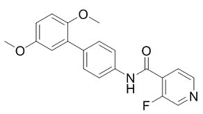
O=C(C1C(F)=CN=CC=1)NC1C=CC(C2C(OC)=CC=C(OC)C=2)=CC=1
|
| InChi Key |
GFEIWXNLDKUWIK-UHFFFAOYSA-N
|
| InChi Code |
InChI=1S/C20H17FN2O3/c1-25-15-7-8-19(26-2)17(11-15)13-3-5-14(6-4-13)23-20(24)16-9-10-22-12-18(16)21/h3-12H,1-2H3,(H,23,24)
|
| 化学名 |
N-[4-(2,5-dimethoxyphenyl)phenyl]-3-fluoropyridine-4-carboxamide
|
| 别名 |
Synta-66; CHEMBL3403742; SCHEMBL1829334; CHEBI:231608; GLXC-03244; GSK1349571A;
|
| HS Tariff Code |
2934.99.9001
|
| 存储方式 |
Powder -20°C 3 years 4°C 2 years In solvent -80°C 6 months -20°C 1 month |
| 运输条件 |
Room temperature (This product is stable at ambient temperature for a few days during ordinary shipping and time spent in Customs)
|
| 溶解度 (体外实验) |
DMSO : ~77.5 mg/mL (~219.95 mM)
|
|---|---|
| 溶解度 (体内实验) |
配方 1 中的溶解度: 2.58 mg/mL (7.32 mM) in 10% DMSO + 40% PEG300 + 5% Tween80 + 45% Saline (这些助溶剂从左到右依次添加,逐一添加), 悬浮液; 超声和加热处理
例如,若需制备1 mL的工作液,可将100 μL 25.8 mg/mL澄清DMSO储备液加入400 μL PEG300中,混匀;然后向上述溶液中加入50 μL Tween-80,混匀;加入450 μL生理盐水定容至1 mL。 *生理盐水的制备:将 0.9 g 氯化钠溶解在 100 mL ddH₂O中,得到澄清溶液。 配方 2 中的溶解度: ≥ 2.58 mg/mL (7.32 mM) (饱和度未知) in 10% DMSO + 90% (20% SBE-β-CD in Saline) (这些助溶剂从左到右依次添加,逐一添加), 悬浮液。 例如,若需制备1 mL的工作液,可将 100 μL 25.8mg/mL澄清的DMSO储备液加入到900μL 20%SBE-β-CD生理盐水中,混匀。 *20% SBE-β-CD 生理盐水溶液的制备(4°C,1 周):将 2 g SBE-β-CD 溶解于 10 mL 生理盐水中,得到澄清溶液。 View More
配方 3 中的溶解度: 2.58 mg/mL (7.32 mM) in 10% DMSO + 90% Corn Oil (这些助溶剂从左到右依次添加,逐一添加), 澄清溶液。 1、请先配制澄清的储备液(如:用DMSO配置50 或 100 mg/mL母液(储备液)); 2、取适量母液,按从左到右的顺序依次添加助溶剂,澄清后再加入下一助溶剂。以 下列配方为例说明 (注意此配方只用于说明,并不一定代表此产品 的实际溶解配方): 10% DMSO → 40% PEG300 → 5% Tween-80 → 45% ddH2O (或 saline); 假设最终工作液的体积为 1 mL, 浓度为5 mg/mL: 取 100 μL 50 mg/mL 的澄清 DMSO 储备液加到 400 μL PEG300 中,混合均匀/澄清;向上述体系中加入50 μL Tween-80,混合均匀/澄清;然后继续加入450 μL ddH2O (或 saline)定容至 1 mL; 3、溶剂前显示的百分比是指该溶剂在最终溶液/工作液中的体积所占比例; 4、 如产品在配制过程中出现沉淀/析出,可通过加热(≤50℃)或超声的方式助溶; 5、为保证最佳实验结果,工作液请现配现用! 6、如不确定怎么将母液配置成体内动物实验的工作液,请查看说明书或联系我们; 7、 以上所有助溶剂都可在 Invivochem.cn网站购买。 |
| 制备储备液 | 1 mg | 5 mg | 10 mg | |
| 1 mM | 2.8380 mL | 14.1900 mL | 28.3801 mL | |
| 5 mM | 0.5676 mL | 2.8380 mL | 5.6760 mL | |
| 10 mM | 0.2838 mL | 1.4190 mL | 2.8380 mL |
1、根据实验需要选择合适的溶剂配制储备液 (母液):对于大多数产品,InvivoChem推荐用DMSO配置母液 (比如:5、10、20mM或者10、20、50 mg/mL浓度),个别水溶性高的产品可直接溶于水。产品在DMSO 、水或其他溶剂中的具体溶解度详见上”溶解度 (体外)”部分;
2、如果您找不到您想要的溶解度信息,或者很难将产品溶解在溶液中,请联系我们;
3、建议使用下列计算器进行相关计算(摩尔浓度计算器、稀释计算器、分子量计算器、重组计算器等);
4、母液配好之后,将其分装到常规用量,并储存在-20°C或-80°C,尽量减少反复冻融循环。
计算结果:
工作液浓度: mg/mL;
DMSO母液配制方法: mg 药物溶于 μL DMSO溶液(母液浓度 mg/mL)。如该浓度超过该批次药物DMSO溶解度,请首先与我们联系。
体内配方配制方法:取 μL DMSO母液,加入 μL PEG300,混匀澄清后加入μL Tween 80,混匀澄清后加入 μL ddH2O,混匀澄清。
(1) 请确保溶液澄清之后,再加入下一种溶剂 (助溶剂) 。可利用涡旋、超声或水浴加热等方法助溶;
(2) 一定要按顺序加入溶剂 (助溶剂) 。
 SKF-96365
 CRAC channel inhibitor-1
 Zegocractin (CM-4620)
 GSK-5498A
InvivoChem的所有产品仅用于作科学研究,不面向患者销售
Copyright 2020 InvivoChem LLC | All Rights Reserved 粤ICP备20063088号-1


 COA
COA
 463611831
