| 规格 | 价格 | 库存 | 数量 |
|---|---|---|---|
| 10 mM * 1 mL in DMSO |
|
||
| 1mg |
|
||
| 5mg |
|
||
| 25mg |
|
||
| 50mg |
|
||
| 100mg |
|
||
| 250mg |
|
||
| 500mg |
|
||
| Other Sizes |
|
| 靶点 |
VEGFR2 (IC50 = 6.5 nM); VEGFR3 (IC50 = 15 nM); EphB2 (IC50 = 24 nM); VEGFR1 (IC50 = 30 nM); PDGFRα (IC50 = 40 nM)
Vascular Endothelial Growth Factor Receptor 1 (VEGFR1), VEGFR2, and VEGFR3, tyrosine kinases involved in angiogenesis. For Tivozanib (AV951; KRN-951), literature [1] reported: VEGFR1 (IC50 = 0.21 nM), VEGFR2 (IC50 = 0.16 nM), VEGFR3 (IC50 = 0.24 nM) via HTRF kinase assay. It showed weak inhibition of PDGFRβ (IC50 = 4.1 nM) and no activity against EGFR or c-Kit (IC50 > 1 μM) [1] - Consistent with [1], [2] confirmed VEGFR2 (Ki = 0.08 nM) via equilibrium binding assay; VEGFR1 (Ki = 0.12 nM), VEGFR3 (Ki = 0.15 nM) [2] |
|---|---|
| 体外研究 (In Vitro) |
AV-951是一种新型的尿素和喹啉衍生物。AV-951可阻止内皮细胞增殖和vegf依赖性的有丝分裂原活化蛋白激酶的激活。[1]
KRN951是一种针对vegfr的新型酪氨酸激酶抑制剂,具有抗肿瘤血管生成和抗生长活性。KRN951在体外亚纳摩尔IC50值(IC50 = 0.16 nmol/L)下有效抑制内皮细胞中vegf诱导的VEGFR-2磷酸化。它还能抑制配体诱导的血小板衍生生长因子受体β (pdgfr - β)和c-Kit的磷酸化(IC50分别为1.72和1.63 nmol/L)。KRN951阻断了vegf依赖性而非非依赖性的丝裂原活化蛋白激酶的激活和内皮细胞的增殖。此外,还能抑制vegf介导的人脐静脉内皮细胞的迁移。[1] VEGFR依赖内皮细胞活性:在HUVECs(VEGFR2依赖)中,Tivozanib(0.001 μM–1 μM)抑制VEGF诱导的增殖,MTT法(72小时)IC50=0.02 μM;0.1 μM处理24小时可抑制管腔形成80%。Western blot显示HUVECs经0.05 μM处理1小时后p-VEGFR2减少90% [1] - 肾细胞癌(RCC)细胞:在786-O(透明细胞RCC,VEGF过表达)和ACHN(RCC)细胞中,Tivozanib(0.01 μM–10 μM)抑制增殖,CCK-8法(72小时)IC50分别为786-O 0.15 μM、ACHN 0.2 μM。0.5 μM处理786-O细胞24小时后,ELISA检测显示VEGF分泌减少65% [2] - 血管生成相关信号:在HUVECs中,Tivozanib(0.01 μM–0.5 μM)剂量依赖性减少p-ERK和p-AKT(VEGFR2下游分子):0.1 μM处理2小时后,两者均减少75%(Western blot) [1] |
| 体内研究 (In Vivo) |
体内研究表明,特别是口服剂量为1mg /kg的AV-951,还可以降低肿瘤异种移植物的微血管密度并抑制VEGFR2磷酸化水平。在胸腺发育不全的大鼠中,AV-951几乎完全抑制异种肿瘤的生长(TGI>85%)。[1]另一项使用大鼠腹膜播散性肿瘤模型的研究表明,AV-951可以延长荷瘤大鼠MST后的生存期,最长可达53.5天。当应用于各种人类肿瘤异种移植物,如肺癌、乳腺癌、结肠癌、卵巢癌、胰腺癌和前列腺癌时,AV-951表现出抗肿瘤活性。[2]
给胸大鼠po后,KRN951降低了异种肿瘤移植物内的微血管密度,并减弱了肿瘤内皮中VEGFR-2的磷酸化水平。它也显示抗肿瘤活性,对多种人类肿瘤异种移植物,包括肺癌,乳腺癌,结肠癌,卵巢癌,胰腺癌和前列腺癌。此外,动态对比增强磁共振成像(DCE-MRI)分析显示,肿瘤血管高通透性的显著降低与KRN951的抗肿瘤活性密切相关。这些发现表明,KRN951是一种高效的抗血管生成和抗肿瘤药物,DCE-MRI将有助于在临床环境中检测KRN951的早期反应。KRN951目前处于I期临床开发阶段,用于治疗晚期癌症患者。[1] RCC异种移植模型:6周龄雄性裸鼠接种786-O细胞,随机分为3组(每组n=8):溶媒组(0.5%甲基纤维素+0.1%吐温80)、Tivozanib 0.5 mg/kg组、1 mg/kg组。药物口服每日一次,连续28天。肿瘤体积减少率:0.5 mg/kg组60%、1 mg/kg组85%;肿瘤重量减少率:0.5 mg/kg组55%、1 mg/kg组78%。免疫组化显示1 mg/kg组微血管密度(CD31染色)减少70% [1] - 肝转移模型:7周龄雌性裸鼠建立ACHN肝转移模型后,用Tivozanib 1 mg/kg(口服每日一次)处理35天。转移结节数量较溶媒组减少65%,血清VEGF从450 pg/mL降至180 pg/mL [2] |
| 酶活实验 |
AV-951 针对各种重组受体和非受体酪氨酸激酶(例如 VEGFR1、VEGFR2、VEGFR3、c-Kit、PDGFRβ、Flt-3 和 FGFR1)的 IC50 值通过使用 1 进行四次无细胞激酶测定来确定。 μM ATP。
激酶选择性。[1] 在1 μmol/L ATP条件下进行四次无细胞激酶试验,测定Tivozanib (AV951; KRN-951)的IC50值;Tivozanib (AV951; KRN-951)抗多种重组受体和非受体酪氨酸激酶。重组酶来自ProQinase GmbH。 [1] 以细胞为基础的试验确定Tivozanib (AV951; KRN-951);如前所述,Tivozanib (AV951; KRN-951)抑制受体酪氨酸激酶的配体依赖性磷酸化。简单地说,将细胞在含有0.5%胎牛血清(FBS)的适当基本培养基中饥饿过夜。加入Tivozanib (AV951; KRN-951)或0.1% DMSO后,细胞孵育1小时,然后用同源配体在37℃下刺激。除VEGFR-3(10分钟)、c-Met(10分钟)和c-Kit(15分钟)外,受体磷酸化诱导时间为5分钟。除VEGF-C(一种大鼠重组蛋白)外,实验中使用的所有配体均为人重组蛋白。细胞裂解后,用适当的抗体对受体进行免疫沉淀,并用磷酸酪氨酸进行免疫印迹。印迹的定量和IC50值的计算如前所述进行。 [1] 丝裂原活化蛋白激酶活化。[1] 按照前面的描述对其进行了评估。简单地说,HUVECs在含有0.5% FBS的基本培养基(EBM-2)中饥饿16小时。Tivozanib (AV951; KRN-951)作用1小时,用50 ng/mL VEGF、25 ng/mL碱性成纤维细胞生长因子或20 ng/mL EGF刺激HUVECs。细胞裂解液进行SDS-PAGE,然后用磷酸化的p44/42丝裂原活化蛋白激酶(MAPK)抗体对磷酸化的MAPKs进行免疫印迹。[1] 通透性试验和VEGFR‐2磷酸化检测。[2] 恶性腹水对内皮细胞通透性和VEGFR‐2磷酸化的影响,以及Tivozanib (AV951;KRN-951)对这些影响进行了评价。将第25天从经载体处理的腹膜播散性肿瘤模型中采集的腹水样本进行汇总,并将所得上清用于这些实验。我们检查碘化丙啶摄取作为通透性的措施在体外测定。western blotting检测VEGFR‐2磷酸化水平。为了进行渗透性试验,将HUVEC培养在90%的融合度下,在含有0.5%胎牛血清的基本培养基(EBM‐2)中进行血清饥饿过夜。然后用磷酸盐缓冲盐水、恶性腹水和10 nM浓度的Tivozanib (AV951; KRN-951);培养板中加入Tivozanib (AV951; KRN-951)。单独培养基或50 ng/mL无腹水的VEGF作为内部对照。孵育7 h后,收获细胞,碘化丙啶(1µg/mL)处理,进行FACS分析。通过测定HUVEC对碘化丙啶的吸收来评估其通透性。对于western blotting,除了用腹水刺激时间为10分钟外,采用相同的方法处理HUVEC。细胞裂解后,用抗VEGFR - 2抗体免疫沉淀VEGFR蛋白,然后用抗磷酸酪氨酸抗体免疫印迹,如前所述。[2] VEGFR1/2/3 HTRF激酶实验(文献[1]):将重组人VEGFR1(791–1338位氨基酸)、VEGFR2(786–1356位氨基酸)或VEGFR3(803–1363位氨基酸)与生物素化肽底物(Ac-EAIYAAPFAKKK-NH2,20 μM)、Eu标记抗磷酸酪氨酸抗体及ATP(10 μM)共同孵育于激酶缓冲液(25 mM Tris-HCl pH 7.5、10 mM MgCl₂、1 mM DTT)中。加入系列稀释的Tivozanib(0.001 nM–10 nM),30°C孵育60分钟。检测时间分辨荧光(激发光340 nm,发射光620 nm),计算IC50 [1] - VEGFR结合实验(文献[2]):重组VEGFR1/2/3与Tivozanib(0.001 nM–10 nM)在结合缓冲液(25 mM Tris-HCl pH 7.5、150 mM NaCl)中37°C孵育24小时。平衡透析分离游离/结合药物,HPLC定量游离药物浓度,推导Ki值 [2] |
| 细胞实验 |
使用基于人脐静脉内皮细胞 (HUVEC) 和正常人真皮成纤维细胞的测定来评估 Tivozanib (AV951/KRN-951) 抑制酪氨酸激酶受体配体依赖性磷酸化的能力。在含有 0.5% 胎牛血清 (FBS) 的适当基础培养基中,细胞在第二天处于饥饿状态。将细胞与 Tivozanib (AV951/KRN-951) 或 0.1% DMSO 一起孵育一小时后,在 37 °C 下用同源配体刺激。除了 VEGFR3、c-Met 和 c-Kit 分别诱导 10 分钟和 15 分钟外,受体磷酸化持续 5 分钟。 VEGF-C 是一种大鼠重组蛋白,是检测中使用的唯一非人重组蛋白的配体。细胞裂解后,用适当的抗体进行免疫沉淀后,对受体进行磷酸酪氨酸免疫印迹。印迹定量和IC50值计算均已完成。
内皮细胞增殖。[1] 将HUVECs接种于含5% FBS的M-199中,以4000个细胞/200 μL/孔的密度接种于胶原包被的96孔板中。24小时后,加入Tivozanib (AV951/KRN-951),然后加入20 ng/mL VEGF或10 ng/mL bFGF,培养72小时。加入胸腺嘧啶(1 μCi/mL) [3H],继续培养12小时。然后收集细胞,用液体闪烁计数器测量其放射性。[1] 趋化性分析。[1] 采用96孔微室板评估HUVEC迁移。细胞在含有0.1%牛血清白蛋白(BSA)的EBM-2中饥饿5小时。然后,收集细胞,在含有0.1% BSA的EBM-2中重悬,并置于上腔。将含有10 ng/mL VEGF、0.1% FBS和0.1% BSA的培养基置于底室,开始细胞迁移。当有指示时,在上、下腔均加入Tivozanib (AV951/KRN-951)。孵育22 h后,用4 μg/mL钙黄素AM染色HBSS细胞。在激发/发射波长为485/530 nm的荧光板阅读器中,通过腔室底部直接测量通过荧光阻断膜孔迁移的细胞的荧光。[1] 细胞毒性检测。[1] 这些试验按前面所述进行。简单地说,将细胞接种于96孔板中,并在含有10%胎牛血清的培养基中培养。在开始培养后约24小时加入Tivozanib (AV951/KRN-951),细胞孵育72小时。采用WST-1试剂检测细胞活力[1]。 HUVEC增殖与管腔形成实验(文献[1]):HUVECs分别以5×10³个细胞/孔接种于96孔板(增殖实验)或1×10⁵个细胞/孔接种于Matrigel包被的24孔板(管腔形成实验)。加入Tivozanib(0.001 μM–1 μM)+VEGF(50 ng/mL),37°C、5% CO₂孵育。增殖实验72小时后MTT法检测计算IC50;管腔形成实验24小时后成像并定量总管长 [1] - RCC细胞实验(文献[2]):786-O/ACHN细胞以5×10³个细胞/孔接种于96孔板,用Tivozanib(0.01 μM–10 μM)处理72小时。CCK-8法检测活力;0.5 μM药物处理24小时后ELISA分析VEGF分泌。786-O细胞经0.1 μM处理2小时后,Western blot检测p-VEGFR2/p-ERK [2] |
| 动物实验 |
小鼠:将癌细胞皮下注射到无胸腺大鼠的右侧腹部。手术切除体积达 1500 mm³ 的肿瘤,并将较小的肿瘤组织块(20-30 mg)皮下植入接受辐射的大鼠的右侧腹部。从随机分组的第 0 天开始,口服给予 KRN951(0.2 或 1 mg/kg)或载体。使用游标卡尺测量并计算肿瘤体积,每周两次。
肿瘤异种移植模型。使用无胸腺大鼠 (RH-rnu/rnu)。在用 γ 射线源(7 Gy,Co60)进行全身照射 24 小时后,将癌细胞皮下接种到大鼠的右侧腹部。肿瘤形成后,将体积约为 1500 mm³ 的肿瘤进行手术切除,并将较小的肿瘤碎片(20-30 mg)皮下植入经照射大鼠的右侧腹部。在随机分组当天(第 0 天),开始口服给予Tivozanib (AV951/KRN-951)(0.2 或 1 mg/kg)或载体。每周两次使用游标卡尺测量肿瘤体积,并按公式(长度 × 宽度²)× 0.5 计算。相对肿瘤体积 (RTV) 的计算公式为:第 x 天的 RTV = 第 x 天的肿瘤体积 / 第 0 天的肿瘤体积。肿瘤生长抑制率 (TGI%) 的计算方法如前所述。RTV 的统计分析采用非配对 t 检验。DCE-MRI。将新鲜的 Calu-6 肿瘤碎片皮下植入无胸腺大鼠 (RH-rnu/rnu) 体内。当肿瘤体积达到 274 至 287 mm³ 时(第 -1 天),将大鼠随机分组。随机分组后第二天(第 0 天)开始每日一次口服给予Tivozanib (AV951/KRN-951)或赋形剂,持续 2 周(第 0-13 天)。MRI 实验在配备柔性接收线圈(圆极化)的全身磁体上进行,磁场强度为 1.5 T。动态对比增强磁共振成像 (DCE-MRI) 分别于第 -1 天(治疗开始前)、第 2 天、第 13 天和第 21 天进行。在第 2 天和第 13 天,大鼠在口服给予Tivozanib (AV951/KRN-951) 4 小时后进行成像。在将动物放入磁体之前,通过尾静脉插管注射造影剂。实验过程中,大鼠经肌注氯胺酮和赛拉嗪混合液(体积比2:1,剂量分别为70 mg/kg和15 mg/kg)麻醉。麻醉后的大鼠仰卧放置于谐振腔内。通过预扫描成像序列确定大鼠的精确位置。[1] 肿瘤血管直径的测量。在MRI研究期间,另取三组携带Calu-6肿瘤的大鼠(RH-rnu/rnu,每组三只)用于使用荧光染料H33342(24)测量肿瘤血管直径。大鼠接受Tivozanib (AV951/KRN-951)(0.2 或 1 mg/kg)或载体治疗 14 天(从第 0 天到第 13 天),并在第 13 天静脉注射 H33342(20 mg/kg)1 分钟后处死。取出肿瘤,并从每个肿瘤的五个层面制备 10 μm 厚的冰冻切片,各层面之间至少间隔 200 μm。使用尼康落射荧光显微镜在紫外光照射下观察肿瘤切片,以识别周围环绕着荧光 H33342 标记细胞晕圈的血管。使用 Win ROOF 软件测量晕圈内的管腔直径作为血管直径。统计分析采用 Mann-Whitney 检验。[1] 肿瘤血管平滑肌肌动蛋白阳性周细胞覆盖的组织学分析。通过皮下注射细胞,在无胸腺大鼠体内建立了Calu-6肿瘤异种移植模型。当肿瘤体积平均达到273至275 mm³时,将大鼠随机分组,然后分别口服给予Tivozanib (AV951/KRN-951)或载体,持续2周。在用抗CD31抗体对内皮细胞进行染色后,使用Cy3标记的单克隆抗α-平滑肌肌动蛋白抗体对肿瘤周细胞进行免疫荧光染色。使用LSM 510系统以100倍放大倍率采集组织图像。随机选取每个切片的六个视野(每个视野0.8489 mm²)进行分析,排除周围结缔组织和中心坏死组织。为了避免操作者偏倚,在对组织切片进行盲法编码后,使用 Win ROOF 软件对 CD31 阳性细胞及其周围 α-平滑肌肌动蛋白阳性区域内的细胞数量进行定量分析。[1] 替沃扎尼 (AV951/KRN-951) 的药代动力学分析。无胸腺大鼠(F344/N JcL-rnu,每组 4 只雌性)口服替沃扎尼 (AV951/KRN-951),并在给药后 72 小时内按预定时间间隔从尾静脉采集血样。向每个血清样本中加入适量的内标物 KRN633。用乙腈对血清样本进行脱蛋白处理,并用高效液相色谱-串联质谱法分析上清液。采用非房室模型分析计算药代动力学参数。按照先前描述的方法[1]模拟了重复口服0.2 mg/kg剂量后Tivozanib (AV951/KRN-951)的稳态血清浓度。 Tivozanib (AV951/KRN-951)悬浮于溶剂(0.5%甲基纤维素的蒸馏水溶液)中,并储存于4°C。每周配制新鲜溶液。[2] 实验设计:接种RCN-9细胞的大鼠随机分为三组,每日口服Tivozanib (AV951/KRN-951)(1或3 mg/kg)或0.5%甲基纤维素溶剂对照。这些治疗分别在肿瘤移植后第4天或第14天开始,并分别持续10天或11天。治疗结束后,处死大鼠并评估肿瘤进展情况。同时收集腹水并测量其体积。在显微镜下观察肠系膜中每个被脂肪组织包围的透明窗口。然后计数肠系膜窗口内有血管的百分比以及肠系膜窗口上肿瘤结节的数量(无论有无血管)。[2] 在随后的生存研究中,接种了RCN-9细胞的大鼠被随机分配到载体处理组或1 mg/kg替沃扎尼(AV951/KRN-951)处理组(每组n = 10)。分别从肿瘤接种当天或移植后14天开始进行治疗。使用Kaplan-Meier法绘制结果图,并使用log-rank检验分析生存差异。P值<0.05被认为具有统计学意义。 [2] 肿瘤血管成像。接种RCN-9细胞的大鼠,分别接受或不接受Tivozanib (AV951/KRN-951)治疗,麻醉后静脉注射异硫氰酸荧光素标记的葡聚糖(分子量200,000)。处死动物后,用4%多聚甲醛固定肠系膜,并将其置于载玻片上。然后,在显微镜下拍摄每个肠系膜窗口相关的血管。客观记录血管连接点和路径的数量(作为血管分叉特征)、血管的面积和长度(作为血管生成密度)以及血管的迂曲度,并使用血管生成图像分析仪(Kurabo,大阪,日本)进行定量评估。这些实验每组使用四只大鼠,并分析每只动物的 12-15 个不同视野。[2] 786-O RCC 异种移植方案(文献[1]):将 5×10⁶ 个 786-O 细胞皮下植入 6 周龄雄性裸鼠体内。当肿瘤体积达到约 100 mm³ 时,将 Tivozanib 溶解于 0.5% 甲基纤维素 + 0.1% Tween 80 溶液中,每日口服一次(0.5 mg/kg 或 1 mg/kg),持续 28 天。每 3 天测量一次肿瘤体积(长×宽²/2);小鼠于第28天处死,肿瘤组织进行CD31免疫组化染色[1] - ACHN肝转移方案(文献[2]):将2×10⁶个ACHN细胞经尾静脉注射至7周龄雌性裸鼠体内,诱导肝转移。7天后,每日一次口服Tivozanib(1 mg/kg,溶于0.5%羟丙基甲基纤维素溶液),持续35天。取出肝脏组织计数转移结节;采用ELISA法检测血清VEGF水平[2] |
| 药代性质 (ADME/PK) |
吸收、分布和排泄
替沃扎尼的中位达峰时间 (Tmax) 为 10 小时,但范围可从 3 小时到 24 小时不等。一项针对 8 名健康受试者的药代动力学研究显示,放射性标记的替沃扎尼的 Cmax 和 AUC 分别为 12.1 ± 5.67 ng/mL 和 1084 ± 417.0 ng·h/mL。替沃扎尼的稳态浓度是在正常剂量的 6-7 倍浓度下达到的。 替沃扎尼主要经粪便排泄。健康志愿者口服1.34 mg放射性标记的替沃扎尼后,79%的给药剂量存在于粪便中(其中26%为原药),12%仅以代谢物的形式存在于尿液中。 替沃扎尼的表观分布容积(V/F)为123 L。 替沃扎尼的表观清除率(CL/F)约为0.75 L/h。 代谢/代谢物 替沃扎尼主要通过CYP3A4代谢。健康志愿者口服1.34 mg放射性标记的替沃扎尼后,血清中检测到的放射性药物中90%为未代谢的替沃扎尼。 生物半衰期 根据处方信息,替沃扎尼的半衰期约为111小时。临床研究信息显示其半衰期为4-5天。 大鼠药代动力学(文献[1]):雄性Sprague-Dawley大鼠(8周龄)口服替沃扎尼1 mg/kg:口服生物利用度=62%,Cmax=3.5 μM,Tmax=1.2 h,末端t₁/₂=7.8 h。静脉注射 0.2 mg/kg:CL = 8.3 mL/min/kg,Vss = 1.1 L/kg [1] - 人血浆蛋白结合率:99%(平衡透析,[1][2]) - 代谢(文献[2]):在人肝微粒体中,替沃扎尼主要通过 CYP3A4 (70%) 和 CYP2D6 (20%) 代谢;尿液中原形药物排泄量 < 6% [2] |
| 毒性/毒理 (Toxicokinetics/TK) |
肝毒性
在已发表的替沃扎尼预注册临床试验中,血清ALT或AST升高发生率在10%至29%之间,其中1%至4%的治疗患者ALT或AST升高超过正常值上限(ULN)的5倍。一些临床试验报告了临床上明显的肝损伤病例,包括肝功能衰竭导致的死亡,但所有病例均归因于肝转移或其他基础肝脏疾病。自获批并广泛应用于临床以来,尚未有因替沃扎尼引起临床上明显的肝损伤或肝功能衰竭的报道,但其临床应用仍然有限。 可能性评分:E(未经证实但怀疑是临床上明显的肝损伤的原因)。 蛋白结合 体外实验表明,替沃扎尼主要与白蛋白结合,结合率≥99%。 体外细胞毒性:在正常人肾近端小管细胞(RPTEC)和包皮成纤维细胞中,替沃扎尼(浓度高达10 μM,作用72小时)的细胞活力>80%,表明其非特异性毒性较低[1][2]。 体内急性毒性:大鼠口服替沃扎尼1 mg/kg(28天)后出现轻度高血压(10%的动物,收缩压升高<20 mmHg)。且无肝肾损伤(ALT/AST/肌酐正常)[1] - 无严重毒性:接受替沃扎尼1 mg/kg(口服,35天)治疗的小鼠未出现体重减轻、嗜睡或器官组织病理学改变[2] |
| 参考文献 | |
| 其他信息 |
药效学
替沃扎尼通过抑制生长因子受体治疗肾细胞癌。在小鼠和大鼠中,替沃扎尼可抑制肿瘤血管生成、肿瘤生长和血管通透性。临床试验表明,替沃扎尼常引起高血压;开始治疗前必须控制高血压。一项替沃扎尼心脏安全性研究报告了心脏QT间期延长,但这些反应在临床上并不严重。在临床研究中,血清可溶性VEGFR2 (sVEGFR2) 水平随时间推移而降低,且随着替沃扎尼暴露量的增加,这种效应增强,因此sVEGFR2可作为VEGFR抑制的药效学标志物。 替沃扎尼(AV951;KRN-951)是一种强效、选择性的口服VEGFR1/2/3抑制剂,用于治疗血管生成依赖性癌症(例如肾细胞癌、转移性实体瘤)[1][2] - 其作用机制包括与VEGFR1/2/3的ATP结合口袋结合,抑制酪氨酸激酶活化和下游ERK/AKT信号通路,从而抑制血管生成和肿瘤生长[1][2] - 由于其对VEGFR的高选择性,替沃扎尼在肾细胞癌异种移植模型和转移模型中显示出强大的抗肿瘤活性,且脱靶毒性极低[1][2] |
| 分子式 |
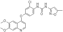
C22H19CLN4O5
|
|---|---|
| 分子量 |
454.86
|
| 精确质量 |
454.104
|
| 元素分析 |
C, 58.09; H, 4.21; Cl, 7.79; N, 12.32; O, 17.59
|
| CAS号 |
475108-18-0
|
| 相关CAS号 |
Tivozanib hydrochloride hydrate;682745-41-1; Tivozanib;475108-18-0; 682745-40-0 (hydrate)
|
| PubChem CID |
9911830
|
| 外观&性状 |
Light brown to brown solid powder
|
| 密度 |
1.4±0.1 g/cm3
|
| 沸点 |
550.4±50.0 °C at 760 mmHg
|
| 熔点 |
220-233
|
| 闪点 |
286.7±30.1 °C
|
| 蒸汽压 |
0.0±1.5 mmHg at 25°C
|
| 折射率 |
1.680
|
| LogP |
4.31
|
| tPSA |
107.74
|
| 氢键供体(HBD)数目 |
2
|
| 氢键受体(HBA)数目 |
7
|
| 可旋转键数目(RBC) |
6
|
| 重原子数目 |
32
|
| 分子复杂度/Complexity |
631
|
| 定义原子立体中心数目 |
0
|
| SMILES |
ClC1C([H])=C(C([H])=C([H])C=1N([H])C(N([H])C1C([H])=C(C([H])([H])[H])ON=1)=O)OC1C([H])=C([H])N=C2C([H])=C(C(=C([H])C2=1)OC([H])([H])[H])OC([H])([H])[H]
|
| InChi Key |
SPMVMDHWKHCIDT-UHFFFAOYSA-N
|
| InChi Code |
InChI=1S/C22H19ClN4O5/c1-12-8-21(27-32-12)26-22(28)25-16-5-4-13(9-15(16)23)31-18-6-7-24-17-11-20(30-3)19(29-2)10-14(17)18/h4-11H,1-3H3,(H2,25,26,27,28)
|
| 化学名 |
1-[2-chloro-4-(6,7-dimethoxyquinolin-4-yl)oxyphenyl]-3-(5-methyl-1,2-oxazol-3-yl)urea
|
| 别名 |
Tivozanib; KRN-951, AV-951; AV951; AV 951; KRN951; KRN 951
|
| HS Tariff Code |
2934.99.9001
|
| 存储方式 |
Powder -20°C 3 years 4°C 2 years In solvent -80°C 6 months -20°C 1 month |
| 运输条件 |
Room temperature (This product is stable at ambient temperature for a few days during ordinary shipping and time spent in Customs)
|
| 溶解度 (体外实验) |
|
|||
|---|---|---|---|---|
| 溶解度 (体内实验) |
配方 1 中的溶解度: ≥ 2.5 mg/mL (5.50 mM) (饱和度未知) in 10% DMSO + 40% PEG300 + 5% Tween80 + 45% Saline (这些助溶剂从左到右依次添加,逐一添加), 澄清溶液。
例如,若需制备1 mL的工作液,可将100 μL 25.0 mg/mL澄清DMSO储备液加入到400 μL PEG300中,混匀;然后向上述溶液中加入50 μL Tween-80,混匀;加入450 μL生理盐水定容至1 mL。 *生理盐水的制备:将 0.9 g 氯化钠溶解在 100 mL ddH₂O中,得到澄清溶液。 配方 2 中的溶解度: ≥ 2.5 mg/mL (5.50 mM) (饱和度未知) in 10% DMSO + 90% (20% SBE-β-CD in Saline) (这些助溶剂从左到右依次添加,逐一添加), 澄清溶液。 例如,若需制备1 mL的工作液,可将 100 μL 25.0 mg/mL澄清DMSO储备液加入900 μL 20% SBE-β-CD生理盐水溶液中,混匀。 *20% SBE-β-CD 生理盐水溶液的制备(4°C,1 周):将 2 g SBE-β-CD 溶解于 10 mL 生理盐水中,得到澄清溶液。 View More
配方 3 中的溶解度: 0.5% methylcellulose: 30mg/mL 1、请先配制澄清的储备液(如:用DMSO配置50 或 100 mg/mL母液(储备液)); 2、取适量母液,按从左到右的顺序依次添加助溶剂,澄清后再加入下一助溶剂。以 下列配方为例说明 (注意此配方只用于说明,并不一定代表此产品 的实际溶解配方): 10% DMSO → 40% PEG300 → 5% Tween-80 → 45% ddH2O (或 saline); 假设最终工作液的体积为 1 mL, 浓度为5 mg/mL: 取 100 μL 50 mg/mL 的澄清 DMSO 储备液加到 400 μL PEG300 中,混合均匀/澄清;向上述体系中加入50 μL Tween-80,混合均匀/澄清;然后继续加入450 μL ddH2O (或 saline)定容至 1 mL; 3、溶剂前显示的百分比是指该溶剂在最终溶液/工作液中的体积所占比例; 4、 如产品在配制过程中出现沉淀/析出,可通过加热(≤50℃)或超声的方式助溶; 5、为保证最佳实验结果,工作液请现配现用! 6、如不确定怎么将母液配置成体内动物实验的工作液,请查看说明书或联系我们; 7、 以上所有助溶剂都可在 Invivochem.cn网站购买。 |
| 制备储备液 | 1 mg | 5 mg | 10 mg | |
| 1 mM | 2.1985 mL | 10.9924 mL | 21.9848 mL | |
| 5 mM | 0.4397 mL | 2.1985 mL | 4.3970 mL | |
| 10 mM | 0.2198 mL | 1.0992 mL | 2.1985 mL |
1、根据实验需要选择合适的溶剂配制储备液 (母液):对于大多数产品,InvivoChem推荐用DMSO配置母液 (比如:5、10、20mM或者10、20、50 mg/mL浓度),个别水溶性高的产品可直接溶于水。产品在DMSO 、水或其他溶剂中的具体溶解度详见上”溶解度 (体外)”部分;
2、如果您找不到您想要的溶解度信息,或者很难将产品溶解在溶液中,请联系我们;
3、建议使用下列计算器进行相关计算(摩尔浓度计算器、稀释计算器、分子量计算器、重组计算器等);
4、母液配好之后,将其分装到常规用量,并储存在-20°C或-80°C,尽量减少反复冻融循环。
计算结果:
工作液浓度: mg/mL;
DMSO母液配制方法: mg 药物溶于 μL DMSO溶液(母液浓度 mg/mL)。如该浓度超过该批次药物DMSO溶解度,请首先与我们联系。
体内配方配制方法:取 μL DMSO母液,加入 μL PEG300,混匀澄清后加入μL Tween 80,混匀澄清后加入 μL ddH2O,混匀澄清。
(1) 请确保溶液澄清之后,再加入下一种溶剂 (助溶剂) 。可利用涡旋、超声或水浴加热等方法助溶;
(2) 一定要按顺序加入溶剂 (助溶剂) 。
A Phase 1b/2a, Open-Label, Multi-Center Study of AV-951 in Combination with Paclitaxel in Subjects with Advanced or Metastatic Breast Cancer
CTID: null
Phase: Phase 2 Status: Completed
Date:
Effects of KRN951 on VEGFR-2 phosphorylation levels on tumor endothelium and tumor microvessel density.Cancer Res.2006 Sep 15;66(18):9134-42. |
DCE-MRI analysis of tumor vascular permeability. Athymic rats bearing Calu-6 tumors were randomized at day −1 and then treated with 0.2 mg/kg KRN951 (○), 1 mg/kg KRN951 (▴), or vehicle (•) once daily for 14 days (days 0-13).Cancer Res.2006 Sep 15;66(18):9134-42. |
Effects of KRN951 on tumor vessel diameter and pericyte coverage.Cancer Res.2006 Sep 15;66(18):9134-42. |
 VEGFR2-IN-7
 SYHA1813
 VEGFR-2-IN-38
 BHEP
BHEP
InvivoChem的所有产品仅用于作科学研究,不面向患者销售
Copyright 2020 InvivoChem LLC | All Rights Reserved 粤ICP备20063088号-1


 COA
COA
")
")
")
 463611831
