| 规格 | 价格 | 库存 | 数量 |
|---|---|---|---|
| 10 mM * 1 mL in DMSO |
|
||
| 1mg |
|
||
| 5mg |
|
||
| 10mg |
|
||
| 25mg |
|
||
| 50mg |
|
||
| 100mg |
|
||
| 250mg |
|
||
| 500mg |
|
||
| 1g |
|
||
| Other Sizes |
|

| 靶点 |
The primary target of U-104 is the tumor-associated carbonic anhydrase IX (CA IX), a zinc-dependent enzyme highly expressed in hypoxic tumor cells. For recombinant human CA IX, the IC50 in the carbonic anhydrase activity assay was 1.2 nM [1]
; It exhibited high selectivity for CA IX over other CA isoforms: the IC50 for human recombinant CA II (a ubiquitous isoform) was >1000 nM (≈833-fold lower affinity than CA IX), and no significant inhibition of CA I/IV was observed (IC50 > 1000 nM) [1] |
|---|---|
| 体外研究 (In Vitro) |
U-104 (SLC-0111) 是一种强效外泌体抑制剂[3]。对于 CA I (Ki=5080 nM) 和 CA II (Ki=9640 nM),U-104 表现出适度的抑制作用[1]。在缺氧条件下,U-104 (50 μM) 抑制 4T1 细胞中癌症干细胞群的间充质表型。该效果持续72小时。在转移性 MDA-MB-231 LM2 -4Luc+ 细胞中,U-104 (<50 μM) 显着且剂量依赖性地减少迁移,导致细胞发育为类似于亲代 MDA-MB-231 细胞的紧凑集落[2] 。
1. 抑制缺氧乳腺癌细胞中CA IX活性:在缺氧条件(1% O₂,48小时)培养的MDA-MB-231(三阴性乳腺癌,TNBC)细胞中,U-104以剂量依赖性方式抑制CA IX活性。10 nM时,CA IX活性较缺氧溶媒对照组降低58%;20 nM(≈2×IC50)时,抑制率达82%。该抑制作用可逆转肿瘤细胞微环境酸化(20 nM U-104处理后,细胞外pH从6.5升至7.2)[1] 。 2. 对缺氧乳腺癌细胞的抗增殖活性:U-104选择性抑制缺氧乳腺癌细胞增殖。72小时MTS实验显示: - 缺氧MDA-MB-231细胞:IC50=15 nM [1] - 缺氧MCF-7(雌激素受体阳性乳腺癌)细胞:IC50=18 nM [1] - 常氧(21% O₂)MDA-MB-231/MCF-7细胞:IC50>200 nM(无显著抗增殖作用)[1] 。 3. 耗竭乳腺癌干细胞(BCSC):在MDA-MB-231细胞中,U-104降低缺氧条件下BCSC(CD44⁺/CD24⁻表型)比例。10 nM时,BCSC比例从缺氧对照组的35%降至18%;20 nM时进一步降至8%(流式细胞术分析)[2] 。U-104(20 nM)还可抑制BCSC自我更新:超低吸附板中形成的肿瘤球数量较对照组减少70%,干性相关蛋白(Sox2、Oct4、Nanog)表达下调55-65%(Western blot)[2] 。 4. 抑制肿瘤细胞迁移与侵袭:U-104(20 nM)使缺氧MDA-MB-231细胞迁移能力降低60%(划痕实验),侵袭能力降低55%(Matrigel Transwell实验)[1] 。Western blot显示,U-104(20 nM)下调血管生成与转移关键介质VEGF(65%)和MMP-9(70%)[1] 。 |
| 体内研究 (In Vivo) |
在原位植入 MDA-MB-231 LM2-4Luc+ 细胞的小鼠中,U-104(19、38 mg/kg;每天;持续 27 天)抑制原发性肿瘤的形成。 4T1 实验性转移小鼠模型表现出 U-104(19 毫克/千克;每天;持续 27 天;5 天)对转移形成的抑制作用[1]。当 MDA-MB-231 LM2-4Luc+ 细胞原位植入 NOD/SCID 小鼠时,U-104(38 mg/kg;腹腔注射;11-27 天)可显着抑制癌症干细胞的数量并减缓原代细胞的形成。肿瘤[2]。 ?原位植入 4T1 细胞的 Balb/c 小鼠在给予 U-104(50 mg/kg;口服强饲;连续 4 天,暂停 1 天;10 至 30 天)时,肿瘤形成明显延迟[2 ]。
1. 抑制异种移植模型中乳腺癌生长:6-8周龄雌性裸鼠皮下注射5×10⁶个缺氧预处理的MDA-MB-231细胞(0.1 mL PBS与基质胶1:1混合)。待肿瘤体积达~100 mm³时,小鼠随机分为3组: - 溶媒对照组:0.5%甲基纤维素PBS溶液(口服,每日1次,连续21天); - U-104 25 mg/kg组:口服,每日1次,连续21天; - U-104 50 mg/kg组:口服,每日1次,连续21天。 25 mg/kg组肿瘤生长抑制率(TGI)为65%,50 mg/kg组TGI达80% [1] 。50 mg/kg组肿瘤重量仅为对照组的20%,免疫组化(IHC)染色显示肿瘤组织中CA IX阳性细胞减少75% [1] 。 2. 抑制乳腺癌转移:在MDA-MB-231肺转移模型(尾静脉注射2×10⁶个细胞)中,U-104(25 mg/kg,口服,每日1次,连续28天)处理组小鼠肺转移结节数较对照组减少70%(对照组平均17个/鼠,处理组平均5个/鼠)[1] 。 3. 体内耗竭BCSC:在携带MDA-MB-231异种移植瘤的裸鼠中,口服U-104(25 mg/kg/天,连续14天)可使肿瘤中CD44⁺/CD24⁻ BCSC比例从对照组的32%降至9%(肿瘤单细胞悬液流式分析)[2] 。这种耗竭与停药后肿瘤复发潜伏期延长2.5倍(从20天延长至50天)相关 [2] 。 |
| 酶活实验 |
1. 重组人CA IX活性测定(pH-stat法):实验在37°C下通过pH电极进行。反应缓冲液含25 mM Tris-HCl(pH 7.5)、10 mM NaCl及10 nM重组人CA IX蛋白。U-104在反应缓冲液中系列稀释(0.1-50 nM),与CA IX预孵育10分钟。加入37°C CO₂饱和水启动反应(CO₂水合为H₂CO₃,导致溶液pH下降),记录2分钟内的pH下降速率。CA IX活性以相对于溶媒对照组的pH变化速率百分比计算,IC50通过四参数逻辑模型拟合抑制曲线得到 [1]
。 2. CA II选择性实验:实验流程与CA IX测定一致,但以10 nM重组人CA II为酶源,且U-104测试浓度最高达1000 nM。结果显示1000 nM时CA II活性抑制率<10%,证实U-104对CA IX的选择性 [1] 。 |
| 细胞实验 |
1. 缺氧细胞培养与抗增殖(MTS)实验:MDA-MB-231/MCF-7细胞以5×10³个细胞/孔接种于96孔板,常氧(21% O₂,5% CO₂)孵育过夜。随后转移至低氧培养箱(1% O₂,5% CO₂,94% N₂)培养48小时以诱导CA IX表达,加入U-104(0.5-200 nM),继续缺氧培养72小时。每孔加入20 μL MTS试剂,孵育2小时后读取490 nm处吸光度,定义抑制增殖50%的U-104浓度为IC50 [1]
。 2. 乳腺癌干细胞(BCSC)检测(流式细胞术):MDA-MB-231细胞缺氧培养48小时后,用U-104(10-30 nM)处理72小时。胰酶消化收集细胞,冷PBS洗涤,加入荧光标记抗体(CD44-PE、CD24-FITC)4°C避光染色30分钟,流式细胞术分析,计算CD44⁺/CD24⁻细胞比例 [2] 。 3. 肿瘤球形成实验:缺氧MDA-MB-231细胞经U-104(10-30 nM)处理72小时后,以1×10³个细胞/孔接种于超低吸附6孔板,培养基为干细胞培养基(DMEM/F12添加B27、EGF、bFGF)。培养10天后,计数直径>50 μm的肿瘤球,计算相对于对照组的球形成率 [2] 。 4. Western blot分析:缺氧MDA-MB-231/MCF-7细胞经U-104(10-30 nM)处理24小时后,用含蛋白酶抑制剂的RIPA缓冲液裂解,4°C、12,000×g离心15分钟。取30 μg蛋白进行10% SDS-PAGE电泳,转移至PVDF膜,5%脱脂牛奶TBST溶液室温封闭1小时。膜与一抗(抗CA IX、抗HIF-1α、抗VEGF、抗Sox2)4°C孵育过夜,再与HRP标记二抗孵育1小时,ECL显影,ImageJ定量条带强度 [1, 2] 。 |
| 动物实验 |
溶于 55.6% PEG 400、11.1% 乙醇和 33% 水中;5 mg/kg;灌胃给药。
将 4T1 细胞原位移植到 Balb/c 小鼠体内。 1. 裸鼠乳腺癌皮下异种移植模型:雌性裸鼠(6-8 周龄,每组 n=6)用异氟烷麻醉。将 5×10⁶ 个 MDA-MB-231 细胞(在缺氧条件下预培养 48 小时)悬浮于 0.1 mL PBS 和 Matrigel 的 1:1 混合液中,然后皮下注射到每只小鼠的右侧腹部。当肿瘤平均体积达到约 100 mm³(细胞注射后 7 天)时,将小鼠随机分为三组: - 载体对照组:灌胃给予 0.5% 甲基纤维素 PBS 溶液,每日一次,连续 21 天; - U-104 25 mg/kg 组:将 U-104 悬浮于 0.5% 甲基纤维素溶液中,浓度为 5 mg/mL,每日一次,连续 21 天; - U-104 50 mg/kg 组:将 U-104 悬浮于 0.5% 甲基纤维素溶液中,浓度为 10 mg/mL,每日一次,连续 21 天。 每 2 天使用数字游标卡尺测量肿瘤体积(体积 = 长 × 宽² / 2),并每周记录体重。治疗结束后,处死小鼠,切除肿瘤并称重,收集肿瘤组织进行免疫组化染色(抗CA IX)[1]。 2. 裸鼠乳腺癌肺转移模型:雌性裸鼠(6-8周龄,每组n=6)经尾静脉注射2×10⁶个缺氧预处理的MDA-MB-231细胞(0.2 mL PBS)。细胞注射后1天,小鼠接受U-104(25 mg/kg,口服,每日一次)或载体处理,持续28天。治疗结束后,处死小鼠,取出肺脏并用4%多聚甲醛固定,在体视显微镜下计数肺表面的转移结节[1]。 3. 体内BCSC耗竭实验:将5×10⁶个CD44⁺/CD24⁻ MDA-MB-231细胞(经流式细胞术纯化)皮下注射到6-8周龄雌性裸鼠(每组n=5)体内。当肿瘤体积达到约100 mm³时,小鼠接受U-104(25 mg/kg,口服,每日一次)或载体处理,持续14天。切除肿瘤,将其解离成单细胞,并通过流式细胞术分析CD44⁺/CD24⁻细胞的比例。为了进行复发潜伏期分析,在停药后监测剩余小鼠的肿瘤复发情况[2] 。 |
| 毒性/毒理 (Toxicokinetics/TK) |
1. 小鼠急性毒性:雌性 CD-1 小鼠(6-8 周龄,每剂量 n=6)口服给予 U-104,剂量分别为 50、100、200 mg/kg。在 50 和 100 mg/kg 剂量下,未观察到死亡或明显的毒性(体重减轻 <4%,血清 ALT、AST 和肌酐水平正常)。在 200 mg/kg 剂量下,6 只小鼠中有 1 只在 7 天内死亡,存活的小鼠出现短暂的体重下降(6%),但肝肾功能指标未见显著变化 [1]
。 2. 裸鼠慢性毒性:在为期 21 天的异种移植研究中,U-104(25-50 mg/kg,口服,每日一次)未引起显著的体重下降(较基线变化 <5%)或血清生化指标(ALT、AST、肌酐)异常 [1] 。在为期 14 天的 BCSC 耗竭研究中,未观察到对小鼠行为或器官形态(肝脏、肾脏、脾脏)的不良影响 [2] 。 |
| 参考文献 |
|
| 其他信息 |
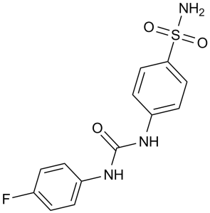
CAIX抑制剂SLC-0111是一种磺酰胺类碳酸酐酶抑制剂,具有潜在的抗肿瘤活性。给药后,CAIX抑制剂SLC-0111抑制肿瘤相关碳酸酐酶IX(CAIX),CAIX是一种缺氧诱导的跨膜糖蛋白,可催化二氧化碳和水可逆快速转化为碳酸、质子和碳酸氢根离子。这可防止肿瘤细胞外微环境酸化和细胞质碱化,从而增加表达CAIX的缺氧肿瘤细胞的死亡。CAIX在多种肿瘤中过表达,并在细胞内和细胞外pH调节、癌细胞进展、存活、迁移和侵袭中发挥关键作用。它还与化疗和放疗耐药性有关。
1. 化学类别和设计背景:U-104 是一种合成的磺酰胺衍生的选择性碳酸酐酶 IX (CA IX) 抑制剂。它的设计目的是靶向 CA IX 的独特活性位点(在缺氧肿瘤中过表达),避免抑制普遍存在的 CA 同工酶(例如 CA II),从而降低脱靶毒性 [1] 。 2. 抗肿瘤作用机制:U-104 通过三个关键途径发挥作用:(1) 抑制 CA IX 活性以逆转肿瘤微环境酸化,从而抑制肿瘤细胞增殖和迁移;(2) 下调 HIF-1α/VEGF 信号通路以抑制血管生成和转移; (3) 清除 CD44⁺/CD24⁻ 乳腺癌干细胞以降低肿瘤复发风险 [1, 2] 。 3. 治疗潜力:U-104 在临床前研究中显示出治疗缺氧性乳腺癌,特别是三阴性乳腺癌 (TNBC) 的潜力。TNBC 是一种 CA IX 高表达、转移率高且预后不良的亚型。U-104 能够抑制原发肿瘤生长和转移,并清除乳腺癌干细胞 (BCSC),从而满足 TNBC 治疗中尚未满足的需求 [1, 2] 。 |
| 分子式 |
C13H12FN3O3S
|
|
|---|---|---|
| 分子量 |
309.32
|
|
| 精确质量 |
309.058
|
|
| CAS号 |
178606-66-1
|
|
| 相关CAS号 |
|
|
| PubChem CID |
310360
|
|
| 外观&性状 |
White to off-white solid powder
|
|
| 密度 |
1.5±0.1 g/cm3
|
|
| 熔点 |
242-243℃
|
|
| 折射率 |
1.673
|
|
| LogP |
2.12
|
|
| tPSA |
109.67
|
|
| 氢键供体(HBD)数目 |
3
|
|
| 氢键受体(HBA)数目 |
5
|
|
| 可旋转键数目(RBC) |
3
|
|
| 重原子数目 |
21
|
|
| 分子复杂度/Complexity |
450
|
|
| 定义原子立体中心数目 |
0
|
|
| InChi Key |
YJQZNWPYLCNRLP-UHFFFAOYSA-N
|
|
| InChi Code |
InChI=1S/C13H12FN3O3S/c14-9-1-3-10(4-2-9)16-13(18)17-11-5-7-12(8-6-11)21(15,19)20/h1-8H,(H2,15,19,20)(H2,16,17,18)
|
|
| 化学名 |
1-(4-fluorophenyl)-3-(4-sulfamoylphenyl)urea
|
|
| 别名 |
|
|
| HS Tariff Code |
2934.99.9001
|
|
| 存储方式 |
Powder -20°C 3 years 4°C 2 years In solvent -80°C 6 months -20°C 1 month |
|
| 运输条件 |
Room temperature (This product is stable at ambient temperature for a few days during ordinary shipping and time spent in Customs)
|
| 溶解度 (体外实验) |
|
|||
|---|---|---|---|---|
| 溶解度 (体内实验) |
配方 1 中的溶解度: ≥ 2.08 mg/mL (6.72 mM) (饱和度未知) in 10% DMSO + 40% PEG300 + 5% Tween80 + 45% Saline (这些助溶剂从左到右依次添加,逐一添加), 澄清溶液。
例如,若需制备1 mL的工作液,可将100 μL 20.8 mg/mL澄清DMSO储备液加入400 μL PEG300中,混匀;然后向上述溶液中加入50 μL Tween-80,混匀;加入450 μL生理盐水定容至1 mL。 *生理盐水的制备:将 0.9 g 氯化钠溶解在 100 mL ddH₂O中,得到澄清溶液。 配方 2 中的溶解度: ≥ 2.08 mg/mL (6.72 mM) (饱和度未知) in 10% DMSO + 90% (20% SBE-β-CD in Saline) (这些助溶剂从左到右依次添加,逐一添加), 澄清溶液。 例如,若需制备1 mL的工作液,可将 100 μL 20.8 mg/mL澄清DMSO储备液加入900 μL 20% SBE-β-CD生理盐水溶液中,混匀。 *20% SBE-β-CD 生理盐水溶液的制备(4°C,1 周):将 2 g SBE-β-CD 溶解于 10 mL 生理盐水中,得到澄清溶液。 View More
配方 3 中的溶解度: ≥ 2.08 mg/mL (6.72 mM) (饱和度未知) in 10% DMSO + 90% Corn Oil (这些助溶剂从左到右依次添加,逐一添加), 澄清溶液。 配方 4 中的溶解度: 30% PEG400+0.5% Tween80+5% propylene glycol:30mg/mL 1、请先配制澄清的储备液(如:用DMSO配置50 或 100 mg/mL母液(储备液)); 2、取适量母液,按从左到右的顺序依次添加助溶剂,澄清后再加入下一助溶剂。以 下列配方为例说明 (注意此配方只用于说明,并不一定代表此产品 的实际溶解配方): 10% DMSO → 40% PEG300 → 5% Tween-80 → 45% ddH2O (或 saline); 假设最终工作液的体积为 1 mL, 浓度为5 mg/mL: 取 100 μL 50 mg/mL 的澄清 DMSO 储备液加到 400 μL PEG300 中,混合均匀/澄清;向上述体系中加入50 μL Tween-80,混合均匀/澄清;然后继续加入450 μL ddH2O (或 saline)定容至 1 mL; 3、溶剂前显示的百分比是指该溶剂在最终溶液/工作液中的体积所占比例; 4、 如产品在配制过程中出现沉淀/析出,可通过加热(≤50℃)或超声的方式助溶; 5、为保证最佳实验结果,工作液请现配现用! 6、如不确定怎么将母液配置成体内动物实验的工作液,请查看说明书或联系我们; 7、 以上所有助溶剂都可在 Invivochem.cn网站购买。 |
| 制备储备液 | 1 mg | 5 mg | 10 mg | |
| 1 mM | 3.2329 mL | 16.1645 mL | 32.3290 mL | |
| 5 mM | 0.6466 mL | 3.2329 mL | 6.4658 mL | |
| 10 mM | 0.3233 mL | 1.6164 mL | 3.2329 mL |
1、根据实验需要选择合适的溶剂配制储备液 (母液):对于大多数产品,InvivoChem推荐用DMSO配置母液 (比如:5、10、20mM或者10、20、50 mg/mL浓度),个别水溶性高的产品可直接溶于水。产品在DMSO 、水或其他溶剂中的具体溶解度详见上”溶解度 (体外)”部分;
2、如果您找不到您想要的溶解度信息,或者很难将产品溶解在溶液中,请联系我们;
3、建议使用下列计算器进行相关计算(摩尔浓度计算器、稀释计算器、分子量计算器、重组计算器等);
4、母液配好之后,将其分装到常规用量,并储存在-20°C或-80°C,尽量减少反复冻融循环。
计算结果:
工作液浓度: mg/mL;
DMSO母液配制方法: mg 药物溶于 μL DMSO溶液(母液浓度 mg/mL)。如该浓度超过该批次药物DMSO溶解度,请首先与我们联系。
体内配方配制方法:取 μL DMSO母液,加入 μL PEG300,混匀澄清后加入μL Tween 80,混匀澄清后加入 μL ddH2O,混匀澄清。
(1) 请确保溶液澄清之后,再加入下一种溶剂 (助溶剂) 。可利用涡旋、超声或水浴加热等方法助溶;
(2) 一定要按顺序加入溶剂 (助溶剂) 。
| NCT Number | Recruitment | interventions | Conditions | Sponsor/Collaborators | Start Date | Phases |
| NCT05025722 | Completed | Other: Non Interventional | Pseudoxanthoma Elasticum | Daiichi Sankyo | August 30, 2021 | |
| NCT04459585 | Completed Has Results | Drug: Dabigatran Etexilate Mesylate Drug: Quizartinib |
Healthy Subjects Drug-drug Interaction |
Daiichi Sankyo Co., Ltd. | August 28, 2020 | Early Phase 1 |
|
|---|
|
|
|
|---|
|
|
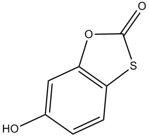 噻克索酮
噻克索酮
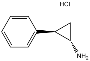 反苯环丙胺
反苯环丙胺
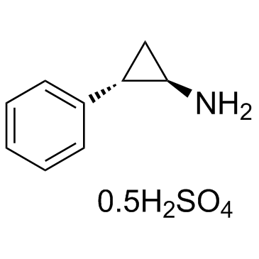 硫酸反苯环丙胺
 Benzenesulfonamide (Benzenesulphonamide, Benzosulfonamide)
InvivoChem的所有产品仅用于作科学研究,不面向患者销售
Copyright 2020 InvivoChem LLC | All Rights Reserved 粤ICP备20063088号-1


 COA
COA


 463611831
