| 规格 | 价格 | 库存 | 数量 |
|---|---|---|---|
| 100mg |
|
||
| Other Sizes |
|

| 体外研究 (In Vitro) |
当暴露于 DBAA (5–40 μM) 时,体外胸腺细胞增殖显着降低[1]。 DBAA 处理 (5–40 μM) 诱导细胞周期停滞 24 小时。根据数据[1],用不同浓度的 DBAA 处理的胸腺细胞在 G0 /G1 期至少增加 40%,在 S 期减少 50%。在 24 小时内,DBAA (5–40 μM) 会增加 Fas/FasL 的表达并减少 Bcl-2 的表达[1]。
|
|---|---|
| 体内研究 (In Vivo) |
基于雄性大鼠恶性间皮瘤的发生率较高,有一些迹象表明二溴乙酸具有致癌潜力。在雄性大鼠中观察到的单核细胞白血病发病率升高可能与接触二溴乙酸有关[2]。雌性大鼠中的二溴乙酸活性是基于单核细胞白血病的阳性趋势和较高的频率[2]。根据肝细胞肿瘤和肝母细胞瘤(仅限雄性)发生率的升高,二溴乙酸对雄性和雌性小鼠均具有明显的致癌作用。雄性小鼠的肺部肿瘤发生率也较高,这被认为与暴露有关[2]。
|
| 细胞实验 |
细胞增殖测定[1]
细胞类型: BALB/c 小鼠的胸腺细胞 测试浓度: 0、5、10、20 和 40 μM 孵育持续时间:6、12、24、48 和 72 小时 实验结果:导致细胞对 T- 的增殖反应显着减弱细胞有丝分裂原6小时或更长时间。 6 小时时,仅在 40 μM 浓度下观察到显着抑制,而在 24、48 和 72 小时时,所有浓度均观察到显着抑制。 蛋白质印迹分析[1] 细胞类型:胸腺细胞 测试浓度:0、5、10、20 和 40 μM 孵育时间:24小时 实验结果:Fas/FasL的表达量从10 μM开始急剧增加,Bcl-2的表达量增加所有浓度均减弱。 |
| 动物实验 |
动物/疾病模型:雄性和雌性F344/N大鼠和B6C3F1小鼠[2]
剂量:每组5只雄性和5只雌性大鼠/小鼠分别暴露于浓度为0、125、250、500、1000或2000 mg/L的二溴乙酸饮用水中,持续2周。每组10只雄性和10只雌性大鼠/小鼠分别暴露于浓度为0、125、250、500、1000或2000 mg/L的二溴乙酸饮用水中,持续3个月。每组50只雄性和50只雌性大鼠/小鼠分别暴露于浓度为0、50、500和1000 mg/L的二溴乙酸饮用水中,持续2年。 给药途径给药方式:饮用水中添加二溴乙酸(纯度大于99%),持续2周、3个月或2年。 实验结果:雄性大鼠暴露于二溴乙酸2年后,肝脏囊性变性发生率增加;雌性大鼠肺泡上皮增生和肾病发生率增加;雄性小鼠脾脏造血发生率增加。 |
| 药代性质 (ADME/PK) |
吸收、分布和排泄
在初步生殖/发育毒性研究(大鼠)中,二溴乙酸 (DBA) 被添加到饮用水中。研究内容包括 DBA 的吸收和生物分布,包括其进入胎盘、羊水、胎儿或乳汁的情况。DBA 的初步生殖/发育毒性研究每组均包含 50 只 Sprague-Dawley 大鼠(每性别每组)。DBA(0、125、250、500 或 1000 ppm)在交配前 14 天至妊娠和哺乳期(63 至 70 天)期间添加到饮用水中。 ……卫星组(每组每项研究6只雄性大鼠,17只雌性大鼠)……用于生物分析采样。大鼠……由于对DBA的明显味觉厌恶,导致其饮水量减少,尤其是在两个最高暴露水平(500和1000 ppm DBA)下的亲代动物中。雌性大鼠的DBA摄入量(mg/kg/天)略高于雄性大鼠,尤其是在妊娠和哺乳期;断奶大鼠的摄入量(mg/kg/天)最高。DBA在血浆、胎盘、羊水和乳汁中均达到可检测和可定量的浓度。血浆样本证实,大鼠主要在黑暗中饮水;这种饮水模式,而非蓄积,导致血浆中DBA浓度持续18至24小时…… 对连续五天灌胃给予250 mg/kg体重的DBA的雄性Sprague-Dawley大鼠的睾丸间质液进行了DBA浓度测定。体重……。睾丸液中二溴乙酸的浓度在末次给药后30分钟达到峰值,为79 μg/mL(约370 μM),半衰期约为1.5小时。 在Sprague-Dawley大鼠的饮用水中添加浓度范围为125至1000 ppm(mg/L)的二溴乙酸,从同居前14天开始,持续到妊娠和哺乳期……在亲代和胎儿血浆、胎盘组织、羊水和乳汁中均检测到了可定量的二溴乙酸。因此,二溴乙酸可以穿过胎盘并被胎儿组织吸收。 据报道,二溴乙酸在雄性F344/N大鼠中的口服生物利用度为30%……。与二氯乙酸相比,二溴乙酸的生物利用度较低是由于其……二溴乙酸在肝脏的首过代谢。 生物半衰期 对连续五天灌胃给予250 mg/kg体重二溴乙酸的雄性Sprague-Dawley大鼠,测定其睾丸间质液中的二溴乙酸含量……。半衰期约为1.5小时。 |
| 毒性/毒理 (Toxicokinetics/TK) |
相互作用
研究发现,消毒副产物二溴乙酸 (DBA) 可提高雌性大鼠体内循环雌二醇 (E2) 和雌酮 (E1) 的浓度。这种效应显然至少部分是由于肝脏分解代谢受到抑制所致。本研究旨在探讨 DBA 是否能够通过提高性激素水平,增强下丘脑上调以触发黄体生成素 (LH) 激增,或者影响神经毒物二甲基二硫代氨基甲酸钠 (DMDC) 阻断 LH 激增的能力。Sprague-Dawley 大鼠连续 14 天灌胃给予 DBA(0-150 mg/kg),并在给药第 11 天进行卵巢切除术,同时植入雌二醇胶囊以诱导每日 LH 激增。在第14天13:00注射0.1 mM/kg DMDC,并在当天下午采集血样。DBA诱导总雌激素水平呈剂量依赖性升高。对于已识别的LH峰值,LH曲线下面积分为两组,分别对应两个低剂量组(0和37.5 mg/kg DBA)和两个高剂量组(75和150 mg/kg DBA)。因此,对低剂量组和高剂量组进行了比较,发现两组间存在显著差异。在150 mg DBA/0.1 mM DMDC组中,可识别的LH峰值出现时间与未接受DMDC处理的雌性小鼠相当,而37.5 mg DBA/0.1 mM DMDC组的峰值出现时间则有所延迟。单独使用DBA处理未观察到显著效应。结果表明,暴露于DBA会导致总雌激素浓度呈剂量依赖性升高,同时DMDC对LH峰值的阻断作用减弱。这种效应似乎归因于雌激素相关的脑机制上调增强,从而刺激LH峰值的产生。饮用水氯化会产生消毒副产物(DBPs),研究表明,高剂量DBPs会破坏啮齿动物的精子发生,提示DBPs可能对男性生殖构成风险。……一项队列研究旨在评估DBPs暴露情况明确的男性精液质量。……本研究结果不支持DBPs暴露水平接近监管限值与不良精子结局之间存在关联,尽管总有机卤化物与精子浓度之间存在关联。 ……总有机卤化物暴露与精子浓度的唯一关联可能支持以下研究结果:总有机卤化物比任何受监管的消毒副产物 (DBP) 类或种类都更能导致不良妊娠结局,并且总有机卤化物的毒性大于单个或亚类消毒副产物的毒性。……/消毒副产物/ 非人类毒性值 大鼠口服 LD50 1737 mg/kg |
| 参考文献 |
[1]. Shu-Ying Gao, et al. Dibromoacetic Acid Induces Thymocyte Apoptosis by Blocking Cell Cycle Progression, Increasing Intracellular Calcium, and the Fas/FasL Pathway in Vitro.Toxicol Pathol. 2016 Jan;44(1):88-97.
[2]. National Toxicology Program. Toxicology and carcinogenesis studies of dibromoacetic acid (Cas No. 631-64-1) in F344/N rats and B6C3F1 mice (drinking water studies). Natl Toxicol Program Tech Rep Ser. 2007 Apr;(537):1-320. |
| 其他信息 |
根据美国国家毒理学计划(NTP)的数据,二溴乙酸可能致癌。
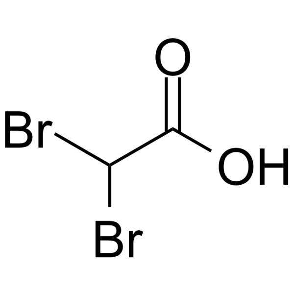
二溴乙酸是一种单羧酸,是乙酸分子中两个甲基氢被溴原子取代后形成的。它是一种海洋代谢物,具有诱导细胞凋亡和延缓衰老的作用。它是一种单羧酸,也是一种2-溴羧酸。其功能与乙酸相关。 已有报道称,在海蓬子(Asparagopsis taxiformis)中发现了二溴乙酸,并有相关数据。 作用机制 ……研究了二溴乙酸(DBA)引起DNA低甲基化、糖原积累和过氧化物酶体增殖的能力……分别给雌性B6C3F1小鼠和雄性Fischer 344大鼠饮用浓度为0、1000和2000 mg/L的DBA溶液。动物分别在暴露2、4、7和28天后实施安乐死。二溴乙酸导致DNA中5-甲基胞嘧啶含量呈剂量依赖性和时间依赖性下降20%至46%。小鼠暴露于二溴乙酸7天后,观察到c-myc基因的低甲基化。胰岛素样生长因子2 (IGF-II) 基因中24个CpG位点的甲基化水平在2000 mg/L二溴乙酸处理28天后,从80.2% ± 9.2%降低至18.8% ± 12.9%。二溴乙酸可增加小鼠肝脏中c-myc和IGF-II基因的mRNA表达。在大鼠中也观察到c-myc基因mRNA表达的剂量依赖性增加。在小鼠和大鼠中,DBA均能引起剂量依赖性的糖原积累和过氧化物酶体月桂酰辅酶A氧化酶活性的增加。因此,DBA与二氯乙酸和三氯乙酸类似,可诱导DNA以及c-myc和IGF-II基因的低甲基化,增加这两个基因的mRNA表达,并导致过氧化物酶体增殖。同样,DBA也能诱导糖原积累。这些结果表明,DBA与二氯乙酸和/或三氯乙酸具有共同的生化和分子活性,提示其也可能是一种肝癌致癌物。 卤乙酸(HA)是饮用水中常见的胚胎毒性污染物。HA的胚胎毒性机制可能部分是通过抑制蛋白激酶C(PKC)介导的。本研究旨在评估透明质酸(HA)胚胎毒性的发病机制,并将这些数据与特异性(Bis I)和非特异性(星形孢菌素)蛋白激酶C(PKC)抑制剂的数据进行比较。将胚胎与多种HA、Bis I、星形孢菌素或Bis V(阴性对照)孵育不同时间。采用碘化丙啶(PI)染色后,通过流式细胞术进行细胞周期分析;采用LysoTracker染色后,通过荧光显微镜评估细胞凋亡。在产生100%胚胎毒性但不致死的浓度下,只有星形孢菌素会扰乱细胞周期。然而,流式细胞术分析显示,溴氯乙酸、二氯乙酸和星形孢菌素处理后,亚G1期事件(一种凋亡指标)随时间推移而积累,而二溴乙酸、Bis I 或 Bis V 处理后则未观察到此现象。亚G1期事件在头部区域尤为显著,而在心脏中则维持在对照水平。溶酶体示踪剂染色证实了完整胚胎中类似的凋亡模式;BCA 和 DCA 在前脑中产生强烈的染色,而在心脏中几乎没有染色。这些数据表明,虽然细胞周期紊乱可能并非 HA 胚胎毒性的致病机制,但这些药物确实会诱导胚胎细胞凋亡。此外,Bis I 未诱导细胞凋亡表明 PKC 抑制不太可能是 HA 胚胎毒性的唯一介质。 HA 致癌作用的相关机制包括 DCA 和 TCA 的致癌机制。显然,导致此类化合物产生效应的机制不止一种,而且这些机制对该类化合物中不同成员活性的重要性也各不相同。部分机制上的差异可能与诱导的肿瘤表型差异有关。一种表型似乎与先前对过氧化物酶体增殖剂诱导的肿瘤的特征描述相关,并且是由三氯乙酸(TCA)诱导的。第二种表型涉及糖原含量低的肿瘤,这些肿瘤对c-Jun和c-Fos抗体有强烈的染色反应。这种表型是由二氯乙酸(DCA)产生的。这些效应可能是由于选择性地导致细胞信号通路中存在不同缺陷的病变所致,这些缺陷控制着细胞分裂和细胞死亡的过程。溴代杂环胺(HAAs)诱导点突变的能力比其氯代类似物强约10倍。这并不能证明它们在体内通过诱变机制诱发癌症,但随着其致癌活性数据的日益完善,这种活性必须被纳入考虑。 杂环胺类化合物诱导氧化应激和提高肝脏核DNA中8-羟基脱氧鸟苷(8-OH-dG)含量的能力差异很大。溴代化合物的这种特性尤为明显。值得注意的是,溴代类似物诱导肝脏肿瘤的能力并不比相应的氯代杂环胺类化合物更强。因此,这种机制是否是决定这种效应的最重要因素尚存疑问。 |
| 分子式 |
C2H2BR2O2
|
|---|---|
| 分子量 |
217.84
|
| 精确质量 |
215.842
|
| CAS号 |
631-64-1
|
| PubChem CID |
12433
|
| 外观&性状 |
Hygroscopic crystals
|
| 密度 |
2.382 g/mL at 25ºC(lit.)
|
| 沸点 |
128-130ºC16 mm Hg(lit.)
|
| 熔点 |
32-38ºC(lit.)
|
| 闪点 |
>230 °F
|
| 折射率 |
1.598
|
| LogP |
1.186
|
| tPSA |
37.3
|
| 氢键供体(HBD)数目 |
1
|
| 氢键受体(HBA)数目 |
2
|
| 可旋转键数目(RBC) |
1
|
| 重原子数目 |
6
|
| 分子复杂度/Complexity |
60.6
|
| 定义原子立体中心数目 |
0
|
| SMILES |
C(C(=O)O)(Br)Br
|
| InChi Key |
SIEILFNCEFEENQ-UHFFFAOYSA-N
|
| InChi Code |
InChI=1S/C2H2Br2O2/c3-1(4)2(5)6/h1H,(H,5,6)
|
| 化学名 |
2,2-dibromoacetic acid
|
| HS Tariff Code |
2934.99.9001
|
| 存储方式 |
Powder -20°C 3 years 4°C 2 years In solvent -80°C 6 months -20°C 1 month |
| 运输条件 |
Room temperature (This product is stable at ambient temperature for a few days during ordinary shipping and time spent in Customs)
|
| 溶解度 (体外实验) |
DMSO : 100 mg/mL (459.05 mM)
|
|---|---|
| 溶解度 (体内实验) |
配方 1 中的溶解度: 2.5 mg/mL (11.48 mM) in 10% DMSO + 90% (20% SBE-β-CD in Saline) (这些助溶剂从左到右依次添加,逐一添加), 澄清溶液; 超声助溶。
例如,若需制备1 mL的工作液,可将100 μL 25.0 mg/mL澄清DMSO储备液加入900 μL 20% SBE-β-CD生理盐水溶液中,混匀。 *20% SBE-β-CD 生理盐水溶液的制备(4°C,1 周):将 2 g SBE-β-CD 溶解于 10 mL 生理盐水中,得到澄清溶液。 配方 2 中的溶解度: ≥ 2.5 mg/mL (11.48 mM) (饱和度未知) in 10% DMSO + 90% Corn Oil (这些助溶剂从左到右依次添加,逐一添加), 澄清溶液。 例如,若需制备1 mL的工作液,可将 100 μL 25.0 mg/mL 澄清 DMSO 储备液添加到 900 μL 玉米油中并混合均匀。 请根据您的实验动物和给药方式选择适当的溶解配方/方案: 1、请先配制澄清的储备液(如:用DMSO配置50 或 100 mg/mL母液(储备液)); 2、取适量母液,按从左到右的顺序依次添加助溶剂,澄清后再加入下一助溶剂。以 下列配方为例说明 (注意此配方只用于说明,并不一定代表此产品 的实际溶解配方): 10% DMSO → 40% PEG300 → 5% Tween-80 → 45% ddH2O (或 saline); 假设最终工作液的体积为 1 mL, 浓度为5 mg/mL: 取 100 μL 50 mg/mL 的澄清 DMSO 储备液加到 400 μL PEG300 中,混合均匀/澄清;向上述体系中加入50 μL Tween-80,混合均匀/澄清;然后继续加入450 μL ddH2O (或 saline)定容至 1 mL; 3、溶剂前显示的百分比是指该溶剂在最终溶液/工作液中的体积所占比例; 4、 如产品在配制过程中出现沉淀/析出,可通过加热(≤50℃)或超声的方式助溶; 5、为保证最佳实验结果,工作液请现配现用! 6、如不确定怎么将母液配置成体内动物实验的工作液,请查看说明书或联系我们; 7、 以上所有助溶剂都可在 Invivochem.cn网站购买。 |
| 制备储备液 | 1 mg | 5 mg | 10 mg | |
| 1 mM | 4.5905 mL | 22.9526 mL | 45.9053 mL | |
| 5 mM | 0.9181 mL | 4.5905 mL | 9.1811 mL | |
| 10 mM | 0.4591 mL | 2.2953 mL | 4.5905 mL |
1、根据实验需要选择合适的溶剂配制储备液 (母液):对于大多数产品,InvivoChem推荐用DMSO配置母液 (比如:5、10、20mM或者10、20、50 mg/mL浓度),个别水溶性高的产品可直接溶于水。产品在DMSO 、水或其他溶剂中的具体溶解度详见上”溶解度 (体外)”部分;
2、如果您找不到您想要的溶解度信息,或者很难将产品溶解在溶液中,请联系我们;
3、建议使用下列计算器进行相关计算(摩尔浓度计算器、稀释计算器、分子量计算器、重组计算器等);
4、母液配好之后,将其分装到常规用量,并储存在-20°C或-80°C,尽量减少反复冻融循环。
计算结果:
工作液浓度: mg/mL;
DMSO母液配制方法: mg 药物溶于 μL DMSO溶液(母液浓度 mg/mL)。如该浓度超过该批次药物DMSO溶解度,请首先与我们联系。
体内配方配制方法:取 μL DMSO母液,加入 μL PEG300,混匀澄清后加入μL Tween 80,混匀澄清后加入 μL ddH2O,混匀澄清。
(1) 请确保溶液澄清之后,再加入下一种溶剂 (助溶剂) 。可利用涡旋、超声或水浴加热等方法助溶;
(2) 一定要按顺序加入溶剂 (助溶剂) 。
 BBO-11818
 RMC-5127
 Degarelix acetate hydrate
 DDO-2728
InvivoChem的所有产品仅用于作科学研究,不面向患者销售
Copyright 2020 InvivoChem LLC | All Rights Reserved 粤ICP备20063088号-1


 463611831
