| 规格 | 价格 | 库存 | 数量 |
|---|---|---|---|
| 10 mM * 1 mL in DMSO |
|
||
| 5mg |
|
||
| 10mg |
|
||
| 50mg |
|
||
| 100mg |
|
||
| 250mg |
|
||
| 500mg |
|
||
| 1g |
|
||
| 5g |
|
||
| 10g |
|
||
| Other Sizes |
|

| 靶点 |
ENaC[1]; uTPA[2]; polycystin-2 (TRPP2)[3]
Amiloride HCl (MK-870) targets delta epithelial sodium channel (δENaC) with an IC50 of 1.8 μM [1] It also targets canonical epithelial sodium channel (ENaC, α/β/γ subunits) with an IC50 of 0.9 μM [2] |
|---|---|
| 体外研究 (In Vitro) |
阿米洛利的 IC50 为 2.6 μM,阻断 δβγ 通道。与αβγ通道(αβγENaC为0.1μM)相比,盐酸阿米洛利对于δHnENaC的Ki大26倍。与阻断 αβγ 通道相比,盐酸阿米洛利更依赖于电压来阻断 δβγ ENaC。根据αβη和δβγ通道的Ki[1],盐酸阿米洛利对δαβγ通道的Ki在-120和+80 mV下分别为920和13.7 μM。阿米洛利的 IC50(实现离子通道抑制 50% 所需的浓度)在 0.1 至 0.5 μM 的浓度范围内,是一种相对选择性的上皮钠通道 (ENaC) 抑制剂。在低外部 [Na+] 存在下,IC50 低至 3 μM,在高 [Na+] 存在下,IC50 高达 1 mM,阿米洛利是 Na+/H+ 交换器 (NHE) 的相对较差的抑制剂。阿米洛利的 IC50 为 1 mM,使其成为 Na+/Ca2+ 交换器 (NCX) 较弱的抑制剂。通过抑制 ENaC 蛋白的活性,阿米洛利 (1 μM) 和亚微摩尔剂量的苯扎米尔 (30 nM)(已知可抑制 ENaC)可防止对灌注压升高的肌源性血管收缩反应。在血管平滑肌细胞 (VSMC) 中,阿米洛利以已知对 ENaC 相对特异性的剂量 (1.5 μM) 完全抑制 Na+ 内流 [2]。
在稳定表达人δENaC的HEK293细胞中,Amiloride HCl(0.1-10 μM)剂量依赖性抑制钠通道电流:1.8 μM时实现50%抑制(IC50),10 μM时在-60 mV膜电位下抑制92%的电流 [1] - 在大鼠原代肺泡上皮细胞中,Amiloride HCl(0.5-5 μM)抑制ENaC介导的钠重吸收:2 μM处理30分钟后,放射性钠摄取实验显示钠内流减少65% [2] - 在人血管内皮细胞中,Amiloride HCl(1-10 μM)抑制ENaC依赖的细胞增殖:5 μM处理72小时后细胞活力降低40%,且不影响细胞凋亡(膜联蛋白V阳性细胞<5%)[2] - 膜片钳分析显示,1 μM Amiloride HCl 使δENaC激活曲线向更正电位偏移15 mV,通道开放概率从0.42降至0.18 [1] |
| 体内研究 (In Vivo) |
研究发现,给 DOCA 盐高血压大鼠皮下注射阿米洛利(1 毫克/公斤/天)将逆转胶原蛋白沉积的最初上升并阻止任何额外的增加。在易发生中风、饮用盐水的自发性高血压大鼠(SHRSP)中,与对照组相比,阿米洛利改善了肾脏和大脑组织学评分,并推迟了蛋白尿的发生。在患有盐依赖性高血压的动物中,盐酸阿米洛利抵消或抑制醛固酮在这些细胞以及心血管和肾脏器官中的作用[2]。
在自发性高血压大鼠(SHR)中,口服 Amiloride HCl(10 mg/kg/天,连续14天)使收缩压降低28 mmHg(从185±10降至157±8 mmHg),舒张压降低16 mmHg(从122±8降至106±6 mmHg)[2] - 在盐负荷高血压大鼠中,腹腔注射 Amiloride HCl(5 mg/kg/天,连续7天)抑制肾脏ENaC活性,尿钠排泄减少35%,尿量较溶媒组增加22% [2] - 在小鼠肺水肿模型中,气管内给予 Amiloride HCl(0.5 mg/kg)后4小时,通过抑制肺泡上皮δENaC,使肺泡液清除率降低48% [1] |
| 酶活实验 |
δENaC电流记录实验:培养表达δENaC的HEK293细胞,采用全细胞膜片钳技术。向浴液中加入系列浓度的 Amiloride HCl(0.1-10 μM),在-60 mV钳制电位下记录钠电流。测量电流幅度,从电流抑制的剂量-反应曲线计算IC50值 [1]
- ENaC介导的钠摄取实验:大鼠原代肺泡上皮细胞接种于24孔板,用 Amiloride HCl(0.5-5 μM)处理30分钟。向培养基中加入放射性22Na+,摄取10分钟后洗涤细胞并裂解,计数放射性强度以量化钠内流抑制率 [2] |
| 细胞实验 |
MR和ENaC已在成纤维细胞、VSMC和内皮细胞内的mRNA和蛋白质水平上得到证实。Kornel等人发现,用生理剂量醛固酮(5 nmol/L)治疗7至10天的兔子主动脉VSMC的Na+内流显著增加。此外,在已知对ENaC相对特异的剂量(1.5μmol/L)下,阿米洛利几乎完全抑制了Na+内流,但NHE特异性阿米洛利类似物乙基异丙基阿米洛利或NCX特异性二氯苯甲酰胺都没有。在另一项研究中,用醛固酮(2mg/天)治疗兔子4周。随后,从主动脉中分离VSMC,并使用ENaC特异性[3H]阿米洛利结合对Na+通道进行定量。醛固酮治疗的动物VSMC Na+通道数量增加了一倍,表明VSMC内醛固酮和ENaC之间可能存在关系。[2]
Golestaneh等人研究了从人脐带获得的内皮细胞是否也有类似的发现。共聚焦显微镜记录双标免疫荧光后,发现MR和ENaC在内皮细胞中共定位。免疫细胞化学定位显示,细胞内MR主要标记为具有核周偏好的细胞质蛋白,而膜结合的ENaC显示为细胞质内核周模式的弥漫颗粒。这两项研究分别研究了ENaC与醛固酮水平为10μmol/L和1nmol/L的关系。在各自的研究中,通过RT-PCR和免疫细胞化学定位发现醛固酮显著增加了ENaC的水平。这些数据表明,内皮细胞中醛固酮介导的信号传导与上皮细胞中的信号传导相当。[2] Oberleithner等人在醛固酮和低剂量阿米洛利(1μmol/L)存在和不存在的情况下对人脐内皮细胞进行了两项生理学研究,为内皮细胞中MR和ENaC的存在提供了间接证据。首先,通过原子力显微镜评估内皮细胞对醛固酮(0.1μmol/L)暴露20分钟的体积反应,原子力显微镜是细胞体积和体积变化(细胞质到细胞核)的测量方法。醛固酮在10分钟时产生18%的最大细胞体积增加,在5分钟时产生核肿胀,15分钟后出现核收缩。螺内酯(1μmol/L)和阿米洛利(1μmol/L)阻止了细胞质和核体积的这些变化。相比之下,尽管使用的剂量高出10倍(10μmol/L),但NHE阻断剂cariporide无效。在第二项研究中,在72小时内评估了暴露于10nmol/L醛固酮浓度的内皮细胞的内皮细胞体积反应。在此浓度下,醛固酮在此期间使细胞体积增加了18%。螺内酯(100nmol/L)可防止醛固酮诱导的肿胀。当在醛固酮存在的情况下将阿米洛利(1μmol/L)加入内皮细胞时,细胞体积急剧减少。相比之下,缺乏醛固酮的细胞对阿米洛利没有反应。20作者得出结论,醛固酮反应性ENaC在一定程度上导致了这些观察到的细胞和核体积的变化。这些数据支持醛固酮对心血管系统具有重要生理/病理生理作用的观点,低剂量阿米洛利可能会拮抗醛固酮对心血管的不良影响。然而,尽管细胞研究有助于识别某些机制数据,但人们只能通过使用动物模型来评估潜在的临床相关疗效。 血管内皮细胞增殖实验:人血管内皮细胞接种于96孔板(2×10³个细胞/孔),用 Amiloride HCl(1-10 μM)处理72小时。MTT法评估细胞活力,计算增殖抑制率 [2] - δENaC激活曲线实验:对表达δENaC的HEK293细胞进行膜片钳记录。在存在或不存在1 μM Amiloride HCl 的条件下,测量不同膜电位(-100至+40 mV)下的钠电流。采用玻尔兹曼方程拟合激活曲线,分析半最大激活电位的偏移 [1] - 凋亡实验:人血管内皮细胞用10 μM Amiloride HCl 处理72小时,用膜联蛋白V-FITC/碘化丙啶染色,流式细胞术分析凋亡率 [2] |
| 动物实验 |
Campbell等人研究了高盐/醛固酮状态下的心肌纤维化。他们将单侧肾切除的Sprague-Dawley大鼠置于1%氯化钠饮用水中,并分别给予以下四种处理之一,持续8周:1)皮下注射醛固酮0.75 μg/h;2)皮下注射阿米洛利1 mg/kg/天;3)皮下注射醛固酮+阿米洛利;或4)溶剂对照。醛固酮显著升高血压,而阿米洛利可减弱这种作用。在接受醛固酮治疗的动物中,可见微观瘢痕形成,这是一种修复性纤维化,提示心肌细胞丢失。然而,当阿米洛利与醛固酮同时给药时,左心室和右心室的微观瘢痕形成均被完全抑制。非高血压右心室瘢痕的发现表明,阿米洛利的益处并非仅仅源于其降压作用。作者推测,原发性醛固酮增多症中的心肌细胞坏死可能是由于钾排泄增加所致,而阿米洛利可以预防这种增加。尽管这仍然是一种可能性,但并未进行尿钠/钾比值和血清钾水平的测定。此外,后续研究提示了其他可能的获益机制(详见后文)[2]。Mirkovic 等人利用 DOCA 盐诱导的高血压大鼠模型,研究了阿米洛利在另一种高盐皮质激素状态下对心肌纤维化的抑制作用。在给予 1% NaCl 饮用水和每 4 天皮下注射 25 mg 醋酸脱氧皮质酮 (DOCA) 的 Wistar 大鼠中,发现 2 周后间质胶原沉积显著增加,4 周后左心室瘢痕进一步加重。研究发现,皮下注射1 mg/kg/天的阿米洛利可逆转胶原沉积的初始增加,并阻止其进一步增加。这些益处是在收缩压无显著变化的情况下实现的。由于既往使用血管紧张素转换酶(ACE)抑制剂、血管紧张素受体阻滞剂和盐皮质激素受体拮抗剂(MRA)的研究表明,即使血压没有显著降低或心脏肥大没有减少,心脏纤维化也可以逆转,因此本研究将阿米洛利添加到似乎能够预防或减轻实验性高血压动物模型中心脏纤维化的药物列表中。作者推测,阿米洛利预防瘢痕形成可能与维持心肌钾浓度有关。然而,他们并未测量血液或组织中阿米洛利的浓度,也未测定细胞内钾浓度或其对钾转运的影响。事实上,他们的研究表明,DOCA-盐组和随后DOCA-盐+阿米洛利治疗组的大鼠血浆钾水平无显著差异。[2]
Sepehrdad等人对饮用生理盐水的易卒中自发性高血压大鼠(SHRSP)进行了阿米洛利给药的生存分析。首先,作者证实,急性给予递增剂量的阿米洛利(1至30 mg/kg/天)不会改变尿量、尿钠/钾比值或体重。此外,SHRSP的平均动脉压升高仅在最高剂量(即30 mg/kg/天)的阿米洛利下受到影响。在对8.5周龄SHRSP大鼠的生存分析中,给予阿米洛利(1 mg/kg/天)和1%氯化钠饮用水,与未治疗的大鼠相比,未观察到收缩压下降、尿钠/钾排泄或尿量变化。所有对照组SHRSP大鼠在16.4周时全部死亡,而接受阿米洛利治疗的SHRSP大鼠在20周的研究结束时仍有75%存活。所有在研究结束时存活的阿米洛利治疗组SHRSP大鼠均未出现中风迹象,而所有对照组大鼠在死亡前均出现中风的神经系统症状。此外,尽管阿米洛利平均延长了6周的生存期,但与对照组相比,它延缓了蛋白尿的发生,并改善了脑和肾脏的组织学评分。23 这项研究表明,在饮用生理盐水的SHRSP模型中,阿米洛利与ACE抑制剂43、血管紧张素转换酶1型拮抗剂和盐皮质激素受体拮抗剂(MRA)类似,可在不降低血压的情况下显著减少卒中、蛋白尿和血管损伤。[2] 为了确定抑制ENaC或NHE在提高饮用生理盐水的SHRSP大鼠的生存率方面发挥更重要的作用,Sepehrdad进行了第二项生存研究。将皮下注射0.7 mg/kg/天的苯扎米(一种ENaC特异性阿米洛利类似物)与皮下注射0.7 mg/kg/天的二甲基阿米洛利(一种选择性NHE抑制剂阿米洛利类似物)以及对照组大鼠的数值进行了比较。二甲基阿米洛利治疗组大鼠的平均存活时间为14.7周龄,显著长于对照组大鼠(12.7周龄时存活率为0%,P < 0.005)。然而,苯扎米治疗组SHRSP大鼠的平均存活时间为16.1周龄,显著长于二甲基阿米洛利治疗组大鼠(P < 0.05)。所有大鼠的血压均显著升高,且各组间在整个研究期间血压无显著差异。此外,与二甲基阿米洛利相比,苯扎米延迟了蛋白尿的发生。急性给予苯扎米(1 mg/kg)或二甲基阿米洛利(1 mg/kg)均未改变血浆钠浓度或体重。与对照组相比,两组大鼠在给药后4小时血浆钾浓度均出现轻微但显著的升高(24小时时未见升高)(出乎意料的是,二甲基阿米洛利组也出现了升高)。然而,所给剂量大于生存分析中使用的剂量,并且采用的是单次推注给药,而非全天持续输注。尽管饮用生理盐水的SHRSP大鼠对苯扎米不表现出高钾血症反应的原因尚不清楚,但低钾饮食、钠摄入量增加或这些大鼠的钾转运基因缺陷(或所有这些因素)可能是原因所在。 高血压大鼠模型(SHR):将12周龄的SHR大鼠随机分组(每组n=8),并分别接受以下治疗:(1)口服赋形剂(0.9%生理盐水);(2)口服盐酸阿米洛利 10 mg/kg/天。采用尾套式血压计每3天测量一次血压,持续14天[2] - 高盐高血压大鼠模型:Wistar大鼠喂食高盐饮食(8% NaCl)2周以诱导高血压,然后随机分组(n=8/组),并分别接受以下治疗:(1)腹腔注射溶剂;(2)腹腔注射盐酸阿米洛利 5 mg/kg/天,持续7天。每日收集尿液样本,以测量钠排泄量和尿量[2] - 肺水肿小鼠模型:C57BL/6小鼠经静脉注射油酸(0.1 mL/kg)以诱导肺水肿,然后随机分组(n=6/组),并分别接受以下治疗:(1)气管内注射溶剂(生理盐水);(2)气管内注射盐酸阿米洛利 0.5 mg/kg。给药后 4 小时,通过向肺部注入荧光标记的白蛋白来测量肺泡液清除率 [1] - 盐酸阿米洛利 溶解于 0.9% 无菌生理盐水中,用于所有动物给药 [1][2] |
| 药代性质 (ADME/PK) |
盐酸阿米洛利的口服生物利用度约为50%,口服10 mg后2小时达到血浆峰浓度(Cmax)0.8 μg/mL [2]
- 其在人体内的终末半衰期(t1/2)为6-9小时,在大鼠体内为4-6小时 [2] - 盐酸阿米洛利在肝脏中的代谢极少(≤10%的剂量),80%以原形经尿液排出,10%经粪便排出 [2] - 治疗浓度下,盐酸阿米洛利的血浆蛋白结合率为40-50% [2] |
| 毒性/毒理 (Toxicokinetics/TK) |
人体毒性:主要不良反应是高钾血症(治疗剂量下15-20%的患者报告出现),其特征是血清钾水平>5.5 mmol/L;其他轻微副作用包括恶心(8%)、头晕(6%)和疲劳(4%)[2]
- 动物毒性:用盐酸阿米洛利(20 mg/kg/天,口服,持续21天)治疗的大鼠血清钾水平从4.2 mmol/L升高至5.8 mmol/L,肝脏、肾脏或心脏未见明显的组织病理学异常,体重减轻<3%[2] |
| 参考文献 |
[1]. Ji, H.L., et al. delta ENaC: a novel divergent amiloride-inhibitable sodium channel. Am J Physiol Lung Cell Mol Physiol, 2012. 303(12): p. L1013-26.
[2]. Teiwes J, et al. Epithelial sodium channel inhibition in cardiovascular disease. A potential role for amiloride. Am J Hypertens. 2007 Jan;20(1):109-17. [3]. Giamarchi A, et al. A polycystin-2 (TRPP2) dimerization domain essential for the function of heteromeric polycystin complexes. EMBO J. 2010 Apr 7;29(7):1176-91. |
| 其他信息 |
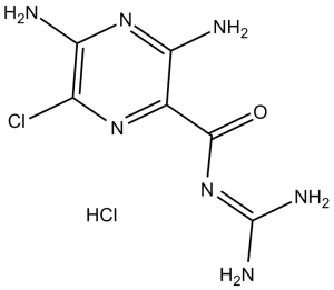
盐酸阿米洛利呈结晶固体或浅黄色粉末状。(NTP, 1992)
盐酸阿米洛利是由阿米洛利与等摩尔量的盐酸反应制得的盐酸盐。它是一种利尿剂和钠通道阻滞剂。它含有阿米洛利(1+)离子。 盐酸阿米洛利是阿米洛利的盐酸盐,阿米洛利是一种合成的吡嗪衍生物,具有利尿和抗利尿作用。阿米洛利抑制位于肾脏远曲小管和集合管中的钠通道,从而阻止钠的吸收,并增加钠与水的排泄,产生利尿作用。为应对肾脏高钠血症,血浆膜发生超极化,电化学作用力减弱,从而阻止钾离子和氢离子排泄到管腔。 阿米洛利是一种吡嗪类化合物,可抑制肾上皮细胞通过钠通道对钠的重吸收。这种抑制作用会在远曲小管和集合管的主细胞管腔膜上产生负电位。负电位会减少钾离子和氢离子的分泌。阿米洛利与利尿剂联合使用,以减少钾的流失。 (摘自 Gilman 等,《古德曼和吉尔曼药理学基础》,第 9 版,第 705 页) 盐酸阿米洛利 是一种保钾利尿剂和选择性 ENaC 抑制剂 [1][2] 其核心作用机制是可逆性阻断上皮细胞中的 ENaC(包括 δENaC 和经典的 α/β/γ ENaC),从而抑制钠重吸收并促进钠排泄,这使其在临床上用于治疗高血压、水肿和心力衰竭 [1][2] 除了心血管应用外,它还能抑制肺泡上皮细胞中的 δENaC 以减少肺泡液清除,这可能在肺水肿的治疗中具有潜在价值 [1] 治疗剂量下毒性较低,但需要监测血清钾水平以避免高钾血症,尤其是在肾功能不全的患者中。 [2] |
| 分子式 |
C6H8CLN7O.HCL
|
|
|---|---|---|
| 分子量 |
266.09
|
|
| 精确质量 |
265.024
|
|
| 元素分析 |
C, 27.08; H, 3.41; Cl, 26.65; N, 36.85; O, 6.01
|
|
| CAS号 |
2016-88-8
|
|
| 相关CAS号 |
Amiloride hydrochloride dihydrate;17440-83-4;Amiloride;2609-46-3;Amiloride hydrochloride (Standard);2016-88-8;Amiloride-15N3 hydrochloride;1216796-18-7
|
|
| PubChem CID |
16230
|
|
| 外观&性状 |
Typically exists as Light yellow to yellow solids at room temperature
|
|
| 密度 |
2.11 g/cm3
|
|
| 沸点 |
628.1ºC at 760 mmHg
|
|
| 熔点 |
560.3 °F (NTP, 1992)
|
|
| 闪点 |
333.7ºC
|
|
| 蒸汽压 |
1.08E-15mmHg at 25°C
|
|
| LogP |
2.073
|
|
| tPSA |
156.79
|
|
| 氢键供体(HBD)数目 |
5
|
|
| 氢键受体(HBA)数目 |
5
|
|
| 可旋转键数目(RBC) |
1
|
|
| 重原子数目 |
16
|
|
| 分子复杂度/Complexity |
279
|
|
| 定义原子立体中心数目 |
0
|
|
| SMILES |
N=C(NC(=O)C1C(N)=NC(=C(N=1)Cl)N)N.Cl
|
|
| InChi Key |
ACHKKGDWZVCSNH-UHFFFAOYSA-N
|
|
| InChi Code |
InChI=1S/C6H8ClN7O.ClH/c7-2-4(9)13-3(8)1(12-2)5(15)14-6(10)11;/h(H4,8,9,13)(H4,10,11,14,15);1H
|
|
| 化学名 |
3,5-Diamino-N-(aminoiminomethyl)-6-chloropyrazinecarboxamide hydrochloride
|
|
| 别名 |
|
|
| HS Tariff Code |
2934.99.9001
|
|
| 存储方式 |
Powder -20°C 3 years 4°C 2 years In solvent -80°C 6 months -20°C 1 month 注意: 请将本产品存放在密封且受保护的环境中,避免吸湿/受潮。 |
|
| 运输条件 |
Room temperature (This product is stable at ambient temperature for a few days during ordinary shipping and time spent in Customs)
|
| 溶解度 (体外实验) |
|
|||
|---|---|---|---|---|
| 溶解度 (体内实验) |
配方 1 中的溶解度: ≥ 2.5 mg/mL (9.40 mM) (饱和度未知) in 10% DMSO + 40% PEG300 + 5% Tween80 + 45% Saline (这些助溶剂从左到右依次添加,逐一添加), 澄清溶液。
例如,若需制备1 mL的工作液,可将100 μL 25.0 mg/mL澄清DMSO储备液加入到400 μL PEG300中,混匀;然后向上述溶液中加入50 μL Tween-80,混匀;加入450 μL生理盐水定容至1 mL。 *生理盐水的制备:将 0.9 g 氯化钠溶解在 100 mL ddH₂O中,得到澄清溶液。 配方 2 中的溶解度: ≥ 2.5 mg/mL (9.40 mM) (饱和度未知) in 10% DMSO + 90% (20% SBE-β-CD in Saline) (这些助溶剂从左到右依次添加,逐一添加), 澄清溶液。 例如,若需制备1 mL的工作液,可将 100 μL 25.0 mg/mL澄清DMSO储备液加入900 μL 20% SBE-β-CD生理盐水溶液中,混匀。 *20% SBE-β-CD 生理盐水溶液的制备(4°C,1 周):将 2 g SBE-β-CD 溶解于 10 mL 生理盐水中,得到澄清溶液。 View More
配方 3 中的溶解度: ≥ 2.5 mg/mL (9.40 mM) (饱和度未知) in 10% DMSO + 90% Corn Oil (这些助溶剂从左到右依次添加,逐一添加), 澄清溶液。 1、请先配制澄清的储备液(如:用DMSO配置50 或 100 mg/mL母液(储备液)); 2、取适量母液,按从左到右的顺序依次添加助溶剂,澄清后再加入下一助溶剂。以 下列配方为例说明 (注意此配方只用于说明,并不一定代表此产品 的实际溶解配方): 10% DMSO → 40% PEG300 → 5% Tween-80 → 45% ddH2O (或 saline); 假设最终工作液的体积为 1 mL, 浓度为5 mg/mL: 取 100 μL 50 mg/mL 的澄清 DMSO 储备液加到 400 μL PEG300 中,混合均匀/澄清;向上述体系中加入50 μL Tween-80,混合均匀/澄清;然后继续加入450 μL ddH2O (或 saline)定容至 1 mL; 3、溶剂前显示的百分比是指该溶剂在最终溶液/工作液中的体积所占比例; 4、 如产品在配制过程中出现沉淀/析出,可通过加热(≤50℃)或超声的方式助溶; 5、为保证最佳实验结果,工作液请现配现用! 6、如不确定怎么将母液配置成体内动物实验的工作液,请查看说明书或联系我们; 7、 以上所有助溶剂都可在 Invivochem.cn网站购买。 |
| 制备储备液 | 1 mg | 5 mg | 10 mg | |
| 1 mM | 3.7581 mL | 18.7906 mL | 37.5813 mL | |
| 5 mM | 0.7516 mL | 3.7581 mL | 7.5163 mL | |
| 10 mM | 0.3758 mL | 1.8791 mL | 3.7581 mL |
1、根据实验需要选择合适的溶剂配制储备液 (母液):对于大多数产品,InvivoChem推荐用DMSO配置母液 (比如:5、10、20mM或者10、20、50 mg/mL浓度),个别水溶性高的产品可直接溶于水。产品在DMSO 、水或其他溶剂中的具体溶解度详见上”溶解度 (体外)”部分;
2、如果您找不到您想要的溶解度信息,或者很难将产品溶解在溶液中,请联系我们;
3、建议使用下列计算器进行相关计算(摩尔浓度计算器、稀释计算器、分子量计算器、重组计算器等);
4、母液配好之后,将其分装到常规用量,并储存在-20°C或-80°C,尽量减少反复冻融循环。
计算结果:
工作液浓度: mg/mL;
DMSO母液配制方法: mg 药物溶于 μL DMSO溶液(母液浓度 mg/mL)。如该浓度超过该批次药物DMSO溶解度,请首先与我们联系。
体内配方配制方法:取 μL DMSO母液,加入 μL PEG300,混匀澄清后加入μL Tween 80,混匀澄清后加入 μL ddH2O,混匀澄清。
(1) 请确保溶液澄清之后,再加入下一种溶剂 (助溶剂) 。可利用涡旋、超声或水浴加热等方法助溶;
(2) 一定要按顺序加入溶剂 (助溶剂) 。
InvivoChem的所有产品仅用于作科学研究,不面向患者销售
Copyright 2020 InvivoChem LLC | All Rights Reserved 粤ICP备20063088号-1


 COA
COA
 463611831
