| 规格 | 价格 | 库存 | 数量 |
|---|---|---|---|
| 10 mM * 1 mL in DMSO |
|
||
| 5mg |
|
||
| 10mg |
|
||
| 25mg |
|
||
| 50mg |
|
||
| 100mg |
|
||
| 250mg |
|
||
| 500mg |
|
||
| Other Sizes |
|

| 靶点 |
Calpain II (ID50 = 34 nM); Calpain I (ID50 = 40 nM); Calpain I (ID50 = 52 nM); Papainb (ID50 = 138 nM)
Calpeptin is a specific inhibitor of calpain (a calcium-dependent cysteine protease), with Ki values of 0.5 nM for μ-calpain and 6 nM for m-calpain [1] |
|---|---|
| 体外研究 (In Vitro) |
Calpeptin 以剂量相关的方式抑制凝血酶、离子霉素或胶原蛋白刺激的血小板中的 20K 磷酸化。在分化 PC12 细胞时,Calpeptin 通过抑制 Calpain 活性来促进神经突伸长。在大鼠视网膜神经节细胞中,Calpeptin 可减弱细胞凋亡,维持正常的全细胞膜电位,从而提供功能性神经保护激酶测定:Calpeptin 是一种有效的细胞穿透性钙蛋白酶抑制剂,在人血小板细胞测定中 Calpain I 的 ID50 为 40 nM :钙肽素减少肺成纤维细胞生成 TGF-b1、IL-6、血管生成素-1 和胶原蛋白合成。 Calpeptin 还可以减少血管生成素 1 依赖性细胞迁移和 IL-6 依赖性细胞增殖,这可能是 Calpeptin 预防肺纤维化作用的潜在机制。
在使用纯化μ-钙蛋白酶和m-钙蛋白酶的酶活实验中,Calpeptin 以剂量依赖性方式抑制酶活性;浓度为10 nM时,对μ-钙蛋白酶活性的抑制率超过90%,对m-钙蛋白酶活性的抑制率约为70% [1] - 在暴露于100 μM谷氨酸(诱导兴奋性毒性)的原代培养大鼠皮质神经元中,10 μM Calpeptin 预处理可将神经元存活率从对照组的约30%提升至约60%(通过MTT法检测)[2] - 在经历氧糖剥夺(OGD,模拟缺血)的原代培养小鼠海马神经元中,1 μM Calpeptin 可使OGD诱导的神经元凋亡率降低约40%(通过TUNEL染色检测),并减少caspase-3剪切体的表达(通过Western blot检测)[3] - 在暴露于100 μM H₂O₂(诱导氧化应激)的原代培养新生大鼠心肌细胞中,10 μM Calpeptin 可抑制钙蛋白酶介导的肌钙蛋白I降解(抑制率约50%,通过Western blot检测),并减少心肌细胞凋亡(乳酸脱氢酶(LDH)释放量降低约35%)[4] - 在C₂C₁₂小鼠骨骼肌成肌细胞中,5 μM Calpeptin 可促进肌管形成:分化7天后,多核肌管(含≥3个细胞核)数量较对照组增加约30%,肌分化标志物肌球蛋白重链(MHC)的表达量上调约2倍(通过Western blot检测)[5] |
| 体内研究 (In Vivo) |
在猫右心室 (RV) PO (RVPO) 模型中,calpeptin(0.6 mg/kg,静脉注射)可阻断钙蛋白酶和 caspase-3 的激活、其底物的裂解以及心肌细胞程序性细胞死亡。在大鼠局灶性脑缺血再灌注损伤模型中,Calpeptin 通过抑制 Caspase-3 的表达来减少海马 CA1 区的神经元凋亡。
钙蛋白酶激活与几种细胞骨架蛋白的切割有关,可能是心脏压力超负荷(PO)期间心肌细胞损失和收缩功能障碍的重要因素。我们使用猫右心室(RV)PO模型,分析了心肌肥大早期代偿期钙蛋白酶的激活情况。在24至48小时PO心肌中观察到钙蛋白酶富集及其活性增加,钙蛋白酶抑制剂水平降低,这些变化在PO 1周后恢复到基础水平。24小时PO心肌的组织化学研究显示,存在TdT介导的dUTP缺口末端标记(TUNEL)阳性心肌细胞,其表现出钙蛋白酶和凝溶胶蛋白的富集。生化研究表明,组蛋白H2B磷酸化和凝溶胶蛋白的细胞骨架结合和切割增加,这表明心肌细胞程序性死亡。为了测试钙蛋白酶抑制是否可以预防这些变化,我们在24小时PO诱导前15分钟和诱导后6小时两次推注钙蛋白酶(0.6 mg/kg iv)。Calpeptin阻断了以下PO诱导的变化:钙蛋白酶富集和激活、钙蛋白酶抑制剂水平降低、半胱氨酸天冬氨酸蛋白酶-3激活、凝溶胶蛋白富集和切割、TUNEL染色和组蛋白H2B磷酸化。尽管类似地施用胱天蛋白酶抑制剂N-苯甲酰羰基-Val-Ala-Asp-氟甲基酮(Z-VD-fmk)阻断了胱天蛋白酶-3的激活,但它并没有缓解上述其他变化。这些结果表明,心肌细胞死亡的生化标志物,如肌节紊乱、凝溶胶蛋白切割和TUNEL阳性核,至少部分是由钙蛋白酶介导的,钙蛋白酶可能是一种潜在的治疗剂,可以预防心肌细胞损失,并在心肌肥大期间保护心肌结构和功能。[4] 证明钙蛋白酶抑制剂Calpeptin对大鼠局灶性脑缺血再灌注损伤的保护作用,并探讨其可能机制。96只大鼠随机分为4组。采用大脑中动脉闭塞模型研究局灶性脑缺血。利用该动物模型,研究了钙肽对大鼠局灶性脑缺血再灌注损伤后神经功能、脑梗死体积和梗死体积百分比、海马CA1区Caspase-3表达和神经元凋亡的影响。目前的研究结果证实,Calpeptin作为钙蛋白酶抑制剂可能在针对局灶性脑缺血再灌注损伤的神经保护中发挥重要作用。Calpeptin可减少大鼠局灶性脑缺血再灌注时海马CA1区神经元的凋亡,其潜在机制可能与Calpeptin-3抑制Caspase-3的表达有关。然而,Calpeptin在这一过程中抑制Caspase-3激活的确切机制尚不清楚。因此,未来需要进一步的研究来揭示潜在的机制[5]。 在大鼠创伤性脑损伤(TBI,通过可控皮质撞击法建立)模型中,于TBI后30分钟侧脑室注射1 nmol Calpeptin,可使损伤皮质区的TUNEL阳性神经元数量减少约50%(TBI后24小时检测),并改善大鼠运动功能(通过平衡木行走实验评估)[2] - 在小鼠短暂性大脑中动脉阻塞(tMCAO,诱导局灶性脑缺血)模型中,再灌注后1小时腹腔注射3 mg/kg Calpeptin(每日1次,连续3天),可使脑梗死体积减少约40%(再灌注后7天通过TTC染色检测),并降低神经功能缺损评分[3] - 在大鼠心肌梗死(MI,通过结扎左冠状动脉前降支建立)模型中,结扎后立即静脉注射5 mg/kg Calpeptin,可使心肌凋亡细胞数量减少约35%(通过TUNEL检测),并在MI后2周将左心室射血分数从对照组的约40%提升至约55%[4] - 在小鼠去神经诱导肌萎缩(通过切断坐骨神经建立)模型中,术后第1天开始皮下注射2 mg/kg Calpeptin(每周2次,连续4周),可使腓肠肌重量较溶剂对照组增加约25%,并减轻肌纤维萎缩(通过苏木精-伊红染色检测)[5] |
| 酶活实验 |
Calpeptin(0.6 mg/kg,静脉注射)可抑制猫右心室 (RV) PO (RVPO) 模型中钙蛋白酶和 caspase-3 的激活、其底物的裂解以及心肌细胞程序性细胞死亡。 [4] Calpeptin 通过抑制 Caspase-3 表达,降低大鼠局灶性脑缺血再灌注损伤模型中海马 CA1 区神经元凋亡。 [5]
修饰Leu norleucinal或Leu methoninal的N端,以获得针对钙蛋白酶的细胞渗透性肽抑制剂。苄氧羰基(Z)衍生物对木瓜蛋白酶的活性低于苯丁酰基衍生物和亮肽。Z-Leu-nLeu-H(calpeptin)对钙蛋白酶I的敏感性高于Z-Leu-Met-H和leupeptin。在合成的抑制剂中,Calpeptin在防止Ca2+离子载体诱导的完整血小板中肌动蛋白结合蛋白和P235的降解方面最有效。与完整血小板孵育30分钟后,钙蛋白酶完全消除了血小板中的钙蛋白酶活性,但在亮肽的情况下没有观察到任何影响。Calpeptin还抑制凝血酶、离子霉素或胶原蛋白刺激的血小板20K磷酸化。因此,发现钙蛋白酶是一种有用的细胞渗透性钙蛋白酶抑制剂。[1] 钙蛋白酶活性检测流程:将纯化的μ-钙蛋白酶或m-钙蛋白酶与钙蛋白酶特异性荧光肽底物混合于含5 mM CaCl₂的反应缓冲液中,加入不同浓度(0.1 nM~100 nM)的Calpeptin,在37°C下孵育30分钟。检测荧光强度(激发波长380 nm,发射波长460 nm)以计算酶活性,通过与溶剂对照组比较确定抑制率,并采用Lineweaver-Burk双倒数作图法计算Ki值[1] |
| 细胞实验 |
Calpeptin 抑制肺成纤维细胞产生 TGF-b1、IL-6、angiopoietin-1 和胶原蛋白的能力。 Calpeptin 还抑制依赖于 angiopoietin-1 的细胞迁移和依赖于 IL-6 的细胞增殖。这可能是 Calpeptin 对抗肺纤维化的保护作用的基本机制。
视网膜神经节细胞(RGCs)的凋亡损害青光眼患者的视力。RGCs在多发性硬化症(MS)中也会退化,导致MS患者视觉感知丧失。我们研究了在暴露于250 nM离子霉素(IMN)或300单位/ml干扰素-γ(IFN-γ)24小时后,钙蛋白酶和胱天蛋白酶级联在大鼠视网膜神经节细胞系RGC-5凋亡中的作用,然后评估了2微M钙蛋白酶(CP,钙蛋白酶特异性抑制剂)的功能性神经保护作用。在暴露于IMN或IFN-γ后,在RGC-5细胞中检测到凋亡的形态学和生化特征。Fura-2测定确定,暴露于IMN或IFN-γ后,细胞内游离[Ca2+]显著增加。用CP预处理1小时可防止RGC-5细胞中的Ca2+内流、蛋白水解活性和凋亡。Western blot分析显示钙蛋白酶和胱天蛋白酶-12的活性增加,Bax:Bcl-2比值上调,线粒体释放细胞色素c,凋亡过程中胱天蛋白酶-9和胱天酶-3活性增加。比色分析也证实了半胱氨酸天冬氨酸蛋白酶-3活性的增加。暴露于IFN-γ后,RGC-5细胞中胱天蛋白酶-8的激活和Bid向tBid的切割表明了凋亡的外在和内在途径之间的合作。膜片钳记录显示,CP预处理可减轻细胞凋亡,并保持正常的全细胞膜电位,表明具有功能性神经保护作用。综上所述,我们的结果表明,Ca2+超载可能是钙蛋白酶和胱天蛋白酶级联激活的原因,导致RGC-5细胞凋亡死亡,CP提供了功能性神经保护[3]。 血小板聚集实验:从新鲜血液中分离人血小板,重悬于Tyrode缓冲液中。将Calpeptin(1 μM~100 μM)与血小板预孵育10分钟后,加入10 μM ADP(血小板激活剂),通过浊度计监测5分钟内的血小板聚集情况并计算聚集率。浓度为10 μM时,Calpeptin 对ADP诱导的血小板聚集抑制率约为80%[1] - 皮质神经元兴奋性毒性实验:培养E18大鼠胚胎皮质神经元7天(使用神经基础培养基),用Calpeptin(1 μM~100 μM)预处理神经元1小时后,暴露于100 μM谷氨酸24小时。通过MTT法(检测570 nm吸光度)测定细胞活力,并在相差显微镜下观察神经元形态[2] - 海马神经元缺氧实验:培养P0小鼠海马神经元10天,将神经元置于氧糖剥夺环境(95% N₂+5% CO₂,无糖培养基)中2小时,复氧时加入Calpeptin(0.1 μM~10 μM)。24小时后,通过TUNEL染色(计数每视野TUNEL阳性细胞)检测凋亡,通过Western blot(使用抗剪切型caspase-3抗体)分析caspase-3剪切情况[3] - 心肌细胞氧化应激实验:培养P1~P3新生大鼠心肌细胞(使用含10%胎牛血清的DMEM培养基),用Calpeptin(1 μM~100 μM)处理细胞1小时后,暴露于100 μM H₂O₂ 6小时。通过比色法(检测490 nm吸光度)测定LDH释放量(细胞死亡标志物),通过Western blot(使用抗肌钙蛋白I抗体)分析肌钙蛋白I降解情况[4] - 骨骼肌成肌细胞分化实验:C₂C₁₂细胞在含10%胎牛血清的DMEM培养基(生长培养基)中培养至汇合,随后换用含Calpeptin(1 μM~10 μM)的分化培养基(含2%马血清的DMEM)。7天后,在显微镜下观察肌管形成(计数含≥3个细胞核的多核肌管),通过Western blot(使用抗MHC抗体)检测MHC表达[5] |
| 动物实验 |
C57BL/6雌性小鼠(8周龄)[3]
0.04 mg/只。 腹腔注射,每周三次,持续28天(与博来霉素联用)。 体内给药。[4] 在24小时口服(PO)猫模型中进行了钙蛋白酶和半胱天冬酶抑制剂的研究。Calpeptin(25 mg)溶于1 ml DMSO中,并用生理盐水进一步稀释至250 μg/ml。Z-VD-fmk溶于0.05 M Tris·HCl(pH 8.5,10 mg/ml)。给药前将药物溶液的pH值调整至7.2。每种药物均通过静脉推注给药两次,分别在PO诱导前15分钟和诱导后6小时进行。钙蛋白酶抑制剂Calpeptin的初始剂量和最终剂量均为0.6 mg/kg。Z-VD-fmk的首剂剂量为20 mg/kg,后续剂量为10 mg/kg。 分组[5] 将96只健康成年SD大鼠随机分为4组,每组24只。MCAO组(n = 24):左侧大脑中动脉闭塞2小时后进行再灌注;Calpeptin组(n = 24):在左侧大脑中动脉闭塞30分钟前,脑室内注射50 μg钙蛋白酶抑制剂Calpeptin(溶于5 μl DMSO); DMSO组(n = 24):在左侧大脑中动脉闭塞30分钟前,脑室内注射5 μl二甲基亚砜;假手术组(n = 24):在建立动物模型时未插入闭塞线,但其他步骤与实验组相似。左侧大脑中动脉闭塞2小时后,分别进行12、24或48小时的再灌注。 大鼠创伤性脑损伤(TBI)模型:雄性Sprague-Dawley大鼠(250-300 g)用异氟烷麻醉。通过控制性皮层冲击(冲击速度:5 m/s,深度:2 mm)诱导TBI。 TBI后30分钟,将Calpeptin(1 nmol)溶于5 μL生理盐水中,以1 μL/min的速率注射到侧脑室(立体定位坐标:距前囟AP -0.8 mm,ML ±1.5 mm,DV -3.5 mm)。对照组大鼠注射5 μL生理盐水。注射24小时后,处死大鼠,收集脑组织进行TUNEL染色和神经元计数[2] - 小鼠脑缺血模型:雄性C57BL/6小鼠(20-25 g)用氯胺酮/甲苯噻嗪麻醉。使用6-0尼龙缝线(距颈外动脉8-10 mm处插入)进行大脑中动脉闭塞(MCAO)诱导局灶性脑缺血。闭塞60分钟后,移除缝线进行再灌注。再灌注1小时后,将Calpeptin(3 mg/kg)溶解于0.1 mL溶剂(5% DMSO + 95%生理盐水)中,并通过腹腔注射给药。连续3天,每天注射一次。溶剂对照组小鼠注射0.1 mL溶剂。再灌注7天后,处死小鼠,并用2,3,5-氯化三苯基四氮唑(TTC)染色脑组织以测量梗死体积;在处死前评估神经功能缺损评分(0-5分)[3] - 大鼠心肌梗死(MI)模型:雄性Wistar大鼠(300-350 g)用戊巴比妥钠麻醉。通过结扎左前降支冠状动脉(左心耳下方 2 mm)诱导心肌梗死 (MI)。结扎后立即将 Calpeptin(5 mg/kg)溶于 0.2 mL 生理盐水中,经尾静脉注射给药。对照组大鼠注射 0.2 mL 生理盐水。MI 两周后,进行超声心动图检查以测量左心室射血分数;随后处死大鼠,并收集心肌组织进行 TUNEL 染色 [4] - 小鼠肌肉萎缩模型:雄性 BALB/c 小鼠(18-22 g)用异氟烷麻醉。切断右侧坐骨神经(距腘窝 1 cm)以诱导神经支配丧失。术后第1天,将Calpeptin(2 mg/kg)溶于0.1 mL PBS中,经皮下注射(背部)给药。每周注射两次,持续4周。对照组小鼠注射0.1 mL PBS。术后4周,处死小鼠,称量右侧腓肠肌重量;肌肉组织经苏木精-伊红染色后测量肌纤维横截面积[5] |
| 毒性/毒理 (Toxicokinetics/TK) |
在大鼠心肌梗死模型中,静脉注射5 mg/kg Calpeptin(单次)2周后,与载体对照组相比,血清丙氨酸氨基转移酶(ALT)或天冬氨酸氨基转移酶(AST)(肝毒性标志物)水平未发生显著变化[4]。在小鼠肌肉萎缩模型中,皮下注射2 mg/kg Calpeptin(每周两次,持续4周)未影响体重,也未引起白细胞计数(WBC)或红细胞计数(RBC)(全身毒性标志物)的异常变化[5]。
|
| 参考文献 | |
| 其他信息 |
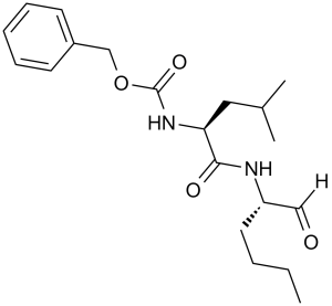
钙肽是一种氨基酸酰胺。
对亮氨酸正亮氨酸醛或亮氨酸甲硫氨酸醛的N端进行修饰,得到一种可穿透细胞的钙蛋白酶抑制剂。苄氧羰基(Z)衍生物对木瓜蛋白酶的活性低于苯基丁酰基衍生物和亮肽。Z-亮氨酸正亮氨酸-H(钙肽)对钙蛋白酶I的敏感性高于Z-亮氨酸甲硫氨酸-H和亮肽。在合成的抑制剂中,钙肽抑制完整血小板中Ca2+离子载体诱导的肌动蛋白结合蛋白和P235降解的能力最强。与完整血小板孵育30分钟后,钙肽完全抑制了血小板中的钙蛋白酶活性,而亮肽则无此作用。钙蛋白酶抑制剂calpeptin还能抑制凝血酶、离子霉素或胶原蛋白刺激的血小板中20K的磷酸化。因此,calpeptin被发现是一种有效的细胞穿透性钙蛋白酶抑制剂。[1] 神经生长因子(NGF)诱导的大鼠嗜铬细胞瘤(PC12)细胞的钙蛋白酶活性在分化早期阶段出现短暂下降。细胞渗透性钙蛋白酶抑制剂calpeptin可进一步降低钙蛋白酶活性,并通过刺激神经突延伸增强NGF的作用。calpeptin不会增加单个细胞产生的神经突数量。本文讨论了钙蛋白酶抑制在PC12细胞早期分化过程中的可能作用。 [2] Calpeptin 是一种合成的、可渗透细胞的肽醛抑制剂,特异性靶向钙蛋白酶,广泛用作研究工具,用于研究钙蛋白酶在各种生理和病理过程(例如细胞凋亡、组织损伤)中的作用。[1] - 在神经生物学研究中,Calpeptin 通过抑制钙蛋白酶介导的神经元死亡发挥神经保护作用,提示其在治疗神经退行性疾病或脑损伤方面具有潜在应用价值。[2,3] - 在心血管研究中,Calpeptin 可减少心肌细胞凋亡并改善心肌梗死模型中的心脏功能,表明其具有作为心血管疾病治疗候选药物的潜力。[4] - 在骨骼肌研究中,Calpeptin 通过抑制钙蛋白酶介导的肌肉蛋白降解来减轻去神经支配引起的肌肉萎缩,为……提供了理论基础。肌肉萎缩症的治疗[5] |
| 分子式 |
C20H30N2O4
|
|
|---|---|---|
| 分子量 |
362.46
|
|
| 精确质量 |
362.22
|
|
| 元素分析 |
C, 66.27; H, 8.34; N, 7.73; O, 17.66
|
|
| CAS号 |
117591-20-5
|
|
| 相关CAS号 |
|
|
| PubChem CID |
73364
|
|
| 外观&性状 |
White to off-white solid powder
|
|
| 密度 |
1.1±0.1 g/cm3
|
|
| 沸点 |
550.7±45.0 °C at 760 mmHg
|
|
| 熔点 |
60-75 °C
|
|
| 闪点 |
286.8±28.7 °C
|
|
| 蒸汽压 |
0.0±1.5 mmHg at 25°C
|
|
| 折射率 |
1.508
|
|
| LogP |
4.37
|
|
| tPSA |
84.5
|
|
| 氢键供体(HBD)数目 |
2
|
|
| 氢键受体(HBA)数目 |
4
|
|
| 可旋转键数目(RBC) |
12
|
|
| 重原子数目 |
26
|
|
| 分子复杂度/Complexity |
434
|
|
| 定义原子立体中心数目 |
2
|
|
| SMILES |
O=C(OCC1=CC=CC=C1)N[C@H](C(N[C@H](C=O)CCCC)=O)CC(C)C
|
|
| InChi Key |
PGGUOGKHUUUWAF-ROUUACIJSA-N
|
|
| InChi Code |
InChI=1S/C20H30N2O4/c1-4-5-11-17(13-23)21-19(24)18(12-15(2)3)22-20(25)26-14-16-9-7-6-8-10-16/h6-10,13,15,17-18H,4-5,11-12,14H2,1-3H3,(H,21,24)(H,22,25)/t17-,18-/m0/s1
|
|
| 化学名 |
benzyl N-[(2S)-4-methyl-1-oxo-1-[[(2S)-1-oxohexan-2-yl]amino]pentan-2-yl]carbamate
|
|
| 别名 |
Calpain; N-Cbz-leu-nleu-al; calpeptin; 117591-20-5; N-Cbz-leu-nleu-al; Benzylcarbonyl-leu-nleu-H; UNII-18X9FR245W; 18X9FR245W; N-Benzyloxycarbonyl-L-leucylnorleucinal; CHEMBL92708; Benzylcarbonyl-leu-nleu-H
|
|
| HS Tariff Code |
2934.99.9001
|
|
| 存储方式 |
Powder -20°C 3 years 4°C 2 years In solvent -80°C 6 months -20°C 1 month |
|
| 运输条件 |
Room temperature (This product is stable at ambient temperature for a few days during ordinary shipping and time spent in Customs)
|
| 溶解度 (体外实验) |
|
|||
|---|---|---|---|---|
| 溶解度 (体内实验) |
配方 1 中的溶解度: ≥ 2.5 mg/mL (6.90 mM) (饱和度未知) in 10% DMSO + 40% PEG300 + 5% Tween80 + 45% Saline (这些助溶剂从左到右依次添加,逐一添加), 澄清溶液。
例如,若需制备1 mL的工作液,可将100 μL 25.0 mg/mL澄清DMSO储备液加入到400 μL PEG300中,混匀;然后向上述溶液中加入50 μL Tween-80,混匀;加入450 μL生理盐水定容至1 mL。 *生理盐水的制备:将 0.9 g 氯化钠溶解在 100 mL ddH₂O中,得到澄清溶液。 配方 2 中的溶解度: ≥ 2.5 mg/mL (6.90 mM) (饱和度未知) in 10% DMSO + 90% Corn Oil (这些助溶剂从左到右依次添加,逐一添加), 澄清溶液。 例如,若需制备1 mL的工作液,可将 100 μL 25.0 mg/mL 澄清 DMSO 储备液加入到 900 μL 玉米油中并混合均匀。 请根据您的实验动物和给药方式选择适当的溶解配方/方案: 1、请先配制澄清的储备液(如:用DMSO配置50 或 100 mg/mL母液(储备液)); 2、取适量母液,按从左到右的顺序依次添加助溶剂,澄清后再加入下一助溶剂。以 下列配方为例说明 (注意此配方只用于说明,并不一定代表此产品 的实际溶解配方): 10% DMSO → 40% PEG300 → 5% Tween-80 → 45% ddH2O (或 saline); 假设最终工作液的体积为 1 mL, 浓度为5 mg/mL: 取 100 μL 50 mg/mL 的澄清 DMSO 储备液加到 400 μL PEG300 中,混合均匀/澄清;向上述体系中加入50 μL Tween-80,混合均匀/澄清;然后继续加入450 μL ddH2O (或 saline)定容至 1 mL; 3、溶剂前显示的百分比是指该溶剂在最终溶液/工作液中的体积所占比例; 4、 如产品在配制过程中出现沉淀/析出,可通过加热(≤50℃)或超声的方式助溶; 5、为保证最佳实验结果,工作液请现配现用! 6、如不确定怎么将母液配置成体内动物实验的工作液,请查看说明书或联系我们; 7、 以上所有助溶剂都可在 Invivochem.cn网站购买。 |
| 制备储备液 | 1 mg | 5 mg | 10 mg | |
| 1 mM | 2.7589 mL | 13.7946 mL | 27.5893 mL | |
| 5 mM | 0.5518 mL | 2.7589 mL | 5.5179 mL | |
| 10 mM | 0.2759 mL | 1.3795 mL | 2.7589 mL |
1、根据实验需要选择合适的溶剂配制储备液 (母液):对于大多数产品,InvivoChem推荐用DMSO配置母液 (比如:5、10、20mM或者10、20、50 mg/mL浓度),个别水溶性高的产品可直接溶于水。产品在DMSO 、水或其他溶剂中的具体溶解度详见上”溶解度 (体外)”部分;
2、如果您找不到您想要的溶解度信息,或者很难将产品溶解在溶液中,请联系我们;
3、建议使用下列计算器进行相关计算(摩尔浓度计算器、稀释计算器、分子量计算器、重组计算器等);
4、母液配好之后,将其分装到常规用量,并储存在-20°C或-80°C,尽量减少反复冻融循环。
计算结果:
工作液浓度: mg/mL;
DMSO母液配制方法: mg 药物溶于 μL DMSO溶液(母液浓度 mg/mL)。如该浓度超过该批次药物DMSO溶解度,请首先与我们联系。
体内配方配制方法:取 μL DMSO母液,加入 μL PEG300,混匀澄清后加入μL Tween 80,混匀澄清后加入 μL ddH2O,混匀澄清。
(1) 请确保溶液澄清之后,再加入下一种溶剂 (助溶剂) 。可利用涡旋、超声或水浴加热等方法助溶;
(2) 一定要按顺序加入溶剂 (助溶剂) 。
 |
|---|
 |
 |
 |
|---|
 |
InvivoChem的所有产品仅用于作科学研究,不面向患者销售
Copyright 2020 InvivoChem LLC | All Rights Reserved 粤ICP备20063088号-1


 COA
COA
 463611831
