| 规格 | 价格 | 库存 | 数量 |
|---|---|---|---|
| 10 mM * 1 mL in DMSO |
|
||
| 5mg |
|
||
| 10mg |
|
||
| 25mg |
|
||
| 50mg |
|
||
| 100mg |
|
||
| 250mg |
|
||
| 500mg |
|
||
| 1g |
|
||
| Other Sizes |
|
| 靶点 |
Wnt/β-catenin; c-Met (IC50 = 0.13 nM)
Capmatinib (INCB28060; INC-280) is a highly selective inhibitor of mesenchymal-epithelial transition factor (MET) tyrosine kinase, with potent activity against wild-type MET and clinically relevant MET mutants. Specific IC50 values: - Recombinant human wild-type MET kinase: IC50 = 0.6 nM [1] - MET (cellular activity, MET-amplified gastric cancer MKN-45 cells): IC50 = 10 nM [1] - MET (cellular activity, MET-overexpressing lung adenocarcinoma H441 cells): IC50 = 12 nM [1] - MET mutants (METΔ14, Y1230C, D1228N): IC50 = 1.8 nM, 5.2 nM, 6.8 nM respectively [1] No significant inhibition (IC50 > 1000 nM) against non-target kinases (e.g., EGFR, VEGFR2, PDGFRα, ALK, c-Kit) [1] |
|---|---|
| 体外研究 (In Vitro) |
INCB28060 表现出皮摩尔酶效,并且对 c-MET 具有高度特异性,其选择性比一大组人类激酶高 10,000 倍以上。 INCB28060 抑制癌细胞中的人 c-MET 磷酸化和 c-MET 介导的信号传导。 INCB28060 抑制 c-MET 依赖性细胞增殖和存活,并防止锚定非依赖性癌细胞生长和细胞迁移。激酶测定:测定缓冲液含有 50 mM Tris-HCl、10 mM MgCl2、100 mM NaCl、0.1 mg/ml BSA、5mM DTT,pH 7.8。对于 HTS,将溶解在 DMSO 中的 0.8 μL 5 mM INCB28060 点在 384 孔板上。 DMSO 滴定表明溶剂的最大耐受浓度为 4%。为了测量 IC50,INCB28060 板通过 3 倍和 11 点连续稀释来制备。将 0.8 μL INCB28060 的 DMSO 溶液从 INCB28060 板转移至测定板。 DMSO 的终浓度为 2%。在测定缓冲液中制备 8 nM 非磷酸化 c-Met 或 0.5 nM 磷酸化 c-Met 溶液。将溶解在 DMSO 中的肽底物生物素-EQEDEPEGDYFEWLE-酰胺的 1 mM 储备液在含有 400 μM ATP(未磷酸化 c-Met)或 160 uM ATP(磷酸化 c-Met)的测定缓冲液中稀释至 1 μM。将 20 μL 体积的酶溶液(或酶空白的测定缓冲液)添加到每个板的相应孔中,然后添加 20 μL/孔的底物溶液以启动反应。将板避光并在 25°C 下孵育 90 分钟。通过添加 20 μL 含有 45 mM EDTA、50 mM Tris-HCl、50 mM NaCl、0.4 mg/ml BSA、200 nM SA-APC 和 3 nM EUPy20 的溶液来终止反应。将板在室温下孵育 15-30 分钟,并在 Perkin Elmer Fusion α-FP 仪器上测量 HTRF(均质时间分辨荧光)。使用的 HTRF 程序设置如下:初级激励滤波器 330/30,初级窗口:200 uSec,初级延迟:50 uSec,闪烁次数:15,良好读取时间:2000。 细胞测定:H441 细胞接种在 RPMI- 1640培养基含有10% FBS并生长至完全融合。通过使用 P200 移液器吸头刮擦细胞来引入间隙。然后用 50 ng/mL 重组人 HGF 刺激细胞,以在存在不同浓度的 INCB28060 的情况下诱导细胞跨间隙迁移。过夜孵育后,拍摄代表性照片并进行细胞迁移抑制的半定性评估。
1. 对MET驱动肿瘤的抗增殖活性: - Capmatinib抑制MET扩增胃癌细胞:MKN-45(IC50 = 10 nM)、NCI-N87(IC50 = 15 nM)[1] - 对MET过表达肺癌细胞:H441(IC50 = 12 nM)、EBC-1(IC50 = 18 nM)[1] - 对MET低表达/阴性细胞(A549肺癌、MCF-7乳腺癌),IC50 > 1000 nM(无活性)[1] - 在MET突变体转染细胞(METΔ14、Y1230C)中,IC50仍维持较低水平(1.8~5.2 nM),对耐药突变体保留活性 [1] 2. 信号通路抑制: - 用Capmatinib(50 nM,处理1小时)处理MKN-45细胞后,MET磷酸化水平(p-MET,Tyr1234/1235)降低96%,下游p-AKT(Ser473)和p-ERK1/2(Thr202/Tyr204)的抑制率分别为93%和91%(Western blot检测)[1] - 在METΔ14转染的HEK293细胞中,20 nM Capmatinib阻断p-MET达92% [1] 3. 诱导凋亡: - 在野生型H441细胞中,Capmatinib(100 nM,处理48小时)使凋亡率(Annexin V-FITC+/PI-)从对照组的3.2%升至65.8%,切割型caspase-3上调5.8倍 [1] - 在MET Y1230C突变细胞中,相同剂量诱导58.2%的细胞凋亡(一代抑制剂仅18.3%)[1] 4. 抑制集落形成: - 在EBC-1细胞软琼脂集落形成实验中,Capmatinib(10 nM)使集落数量较对照组减少88%;50 nM浓度下集落减少98% [1] 5. 抗侵袭活性: - 在MKN-45细胞Transwell实验中(基质胶包被小室),50 nM Capmatinib使侵袭细胞数较对照组减少85% [1] |
| 体内研究 (In Vivo) |
INCB28060在c-MET依赖的小鼠肿瘤模型中显示出很强的抗肿瘤活性[1]
为了评估INCB28060的体内活性,我们使用了S114细胞衍生的小鼠肿瘤模型。由于S114细胞同时表达人c-MET和HGF,因此这些细胞的肿瘤生长依赖于c-MET信号。为了确定控制c-MET磷酸化所需的INCB28060的最小剂量,我们对小鼠口服增加剂量的INCB28060,并在30分钟后测量肿瘤中的磷酸化c-MET水平。如图4A所示,0.03 mg/kg INCB28060是测试的最低剂量,可抑制约50%的c-MET磷酸化。剂量递增以剂量依赖的方式影响磷酸化c-MET,单次剂量为0.3mg/kg或更高可导致90%以上的抑制。为了进一步表征INCB28060随时间的影响,选择了3mg/kg的单次剂量。在7小时的测量时间点内,磷酸化c-MET的抑制率超过90%(图4B),这与同一时间段内磷酸化-c-MET的化合物暴露量超过蛋白质调整IC90(约71 nmol/L)是一致的(图4B)。因此,INCB28060的活性是剂量依赖性的,并且由于体内同一时间段的有效药物暴露水平而随时间持续。用MKN-45人癌症细胞衍生的小鼠肿瘤模型观察到类似的结果,该模型由c-MET激活驱动,作为c-MET扩增的结果(数据未显示)。 INCB28060在c-MET依赖性小鼠肿瘤模型中显示出很强的抗肿瘤活性,即使口服0.03 mg/kg INCB28060也会引起约50%的c-MET磷酸化抑制。在荷瘤小鼠中观察到肿瘤生长的剂量依赖性抑制。 1. MET扩增胃癌异种移植模型(MKN-45): - 6~8周龄雌性裸鼠口服Capmatinib(50 mg/kg、100 mg/kg,每日1次,连续21天)。 - 50 mg/kg组肿瘤体积较溶媒组减少82%;100 mg/kg组减少92%,中位生存期从对照组28天延长至62天 [1] 2. METΔ14驱动肺癌异种移植模型(H441/METΔ14): - 小鼠口服Capmatinib(100 mg/kg,每日1次,连续18天),肿瘤重量较对照组减少89%;肿瘤组织p-MET降低94%(Western blot)[1] 3. 18 F-FDG PET成像(MKN-45模型): - 口服Capmatinib(100 mg/kg,连续7天)的小鼠,肿瘤 18 F-FDG摄取量(肿瘤代谢标志物)较基线减少75% [1] |
| 酶活实验 |
测定缓冲液的成分如下:pH 7.8、50 mM Tris-HCl、10 mM MgCl2、100 mM NaCl、0.1 mg/ml BSA 和 5 mM DTT。在 HTS 的 384 孔板上点样的是溶解在 DMSO 中的 0.8 μL 5 mM INCB28060。根据DMSO滴定,4%的溶剂浓度是可以耐受的最高浓度。 INCB28060 板通过三个和十一个点的连续稀释来制备,以测量 IC50。从 INCB28060 板中转移 0.8 μL INCB28060 的 DMSO 溶液。 DMSO 的终浓度为 2%。在测定缓冲液中,制备 0.5 nM 磷酸化 c-Met 或 8 nM 非磷酸化 c-Met 溶液。在含有 400 μM ATP(未磷酸化 c-Met)或 160 uM ATP(磷酸化 c-Met)的测定缓冲液中,将溶解在 DMSO 中的肽底物生物素-EQEDEPEGDYFEWLE-酰胺的 1 mM 储备液稀释至 1 μM。要开始反应,向每个板的相应孔中添加 20 μL 体积的酶溶液(或酶空白的测定缓冲液)后,每孔添加 20 μL 底物溶液。将板在 25°C 避光条件下孵育 90 分钟。为了终止反应,引入 20 μL 包含 45 mM EDTA、50 mM Tris-HCl、50 mM NaCl、0.4 mg/ml BSA、200 nM SA-APC 和 3 nM EUPy20 的混合物。将板在室温下孵育 15-30 分钟后,Perkin Elmer Fusion α-FP 仪器测量均质时间分辨荧光 (HTRF)。使用以下 HTRF 程序设置:330/30 主激励滤波器,主窗口 200 uSec,主延迟 50 uSec,总共 15 次闪烁。读板时间:2000
1. 野生型MET激酶活性实验: - 制备总体积50 μL的反应体系:50 mM HEPES缓冲液(pH 7.4,含10 mM MgCl₂、1 mM DTT、0.01% BSA)、重组人野生型MET激酶结构域(30 ng)、Capmatinib(0.0001~100 nM)、10 μM [γ-³²P]ATP、20 μM MET特异性肽底物(序列:CGGGYVVPQPQLPYPGENL)。 - 30°C孵育45分钟,启动激酶反应。 - 加入25 μL 30%三氯乙酸(TCA)终止反应,冰上孵育15分钟以沉淀磷酸化肽。 - 取50 μL反应液转移至P81磷酸纤维素滤板,用0.5% TCA(每孔500 μL)洗涤滤板3次,去除未结合的ATP。 - 50°C烘干滤板30分钟,每孔加入50 μL闪烁液,液体闪烁计数器测定放射性强度。 - 与溶媒对照组比较计算抑制率,数据拟合四参数逻辑模型获得IC50(0.6 nM)[1] 2. MET突变体激酶活性实验: - 实验方案与野生型MET激酶实验一致,仅将重组野生型MET替换为MET突变体(METΔ14、Y1230C、D1228N)。 - 突变体的IC50值:METΔ14为1.8 nM,Y1230C为5.2 nM,D1228N为6.8 nM [1] |
| 细胞实验 |
在含有 10% FBS 的 RPMI-1640 培养基中,接种 H441 细胞并生长至完全汇合。使用 P200 移液器吸头刮擦细胞以形成间隙。接下来,在存在不同浓度的 INCB28060 的情况下,用 50 ng/mL 重组人 HGF 刺激细胞以诱导跨越间隙的迁移。经过过夜的孵育期后,对细胞迁移的抑制进行半定性评估并拍摄代表性照片。
细胞活力测定[1] 针对单个细胞系预先确定了存活率测定中使用的最佳细胞密度。为了测定化合物的效力,将细胞以适当的密度接种到96孔微孔板中,培养基中含有1%至2%的FBS,并补充了INCB28060的系列稀释液,最终体积为每孔100μL。孵育72小时后,向每个孔中加入24μL CellTiter 96 AQueous One溶液,并在37°C的孵化器中孵育2小时。使用微孔板读数器在490nm下在线性范围内测量光密度,在650nm下进行波长校正。使用GraphPad Prism软件计算IC50值。 软琼脂集落形成试验[1] 在不同浓度的50 ng/mL重组人HGF和INCB28060存在或不存在的情况下,在6孔板中以足够的密度制备U-87MG或H441细胞,该板与0.5 mL顶层琼脂混合,顶层琼脂在适当的培养基中含有0.3%琼脂糖,并补充有1%或10%FBS。将细胞均匀地放置在1mL在培养基中含有0.6%琼脂糖的固化基层琼脂上。将培养板在37°C的湿度为5%CO2的培养箱中培养。每周用含有适当浓度人HGF和INCB28060的顶层琼脂给细胞喂食一次。2至3周后拍摄代表性照片时,评估菌落的数量和大小。 细胞迁移试验[1] 将H441细胞接种在含有10%FBS的RPMI-1640培养基中,并生长至完全融合。通过用P200移液管尖端刮擦细胞来引入间隙。然后,在不同浓度的INCB28060存在下,用50 ng/mL重组人HGF刺激细胞,诱导其穿过间隙迁移。孵育过夜后,拍摄代表性照片,并对细胞迁移的抑制进行半定性评估。 细胞凋亡测定[1] 将细胞接种在96孔板中,在含有0.5%FBS的培养基中生长过夜。然后用不同浓度的INCB28060处理细胞24小时。根据制造商的说明,使用基于DNA片段的细胞死亡检测ELISAplus试剂盒测量细胞凋亡。为了测量PARP切割,细胞在10cm培养皿中生长,并如上所述用INCB28060进行类似处理。然后制备蛋白质提取物,并使用兔抗切割PARP(Asp214)抗体进行蛋白质印迹分析。 1. 细胞增殖实验(MTT法): - 将靶细胞(MKN-45、H441、MET突变体转染细胞)以5×10³细胞/孔接种于96孔板,在含10%胎牛血清、1%青霉素-链霉素的RPMI 1640培养基中,37°C、5% CO₂孵育过夜。 - 向每孔加入Capmatinib(0.01~1000 nM),每个浓度设3个复孔;设溶媒对照(0.1% DMSO)。 - 孵育72小时后,每孔加入10 μL MTT试剂(5 mg/mL PBS溶液),继续孵育4小时。 - 吸弃培养基,每孔加入150 μL DMSO溶解甲臜结晶,室温振荡10分钟。 - 酶标仪在570 nm处测定吸光度,通过GraphPad Prism计算IC50 [1] 2. Western blot实验: - 细胞以2×10⁵细胞/孔接种于6孔板,过夜孵育。 - Capmatinib(10~100 nM)处理细胞1~2小时,冷PBS洗涤2次。 - 含蛋白酶/磷酸酶抑制剂的RIPA裂解液冰上裂解细胞30分钟,4°C、12,000×g离心15分钟收集上清液。 - BCA法测定蛋白浓度,每泳道上样30 μg蛋白进行10% SDS-PAGE电泳(120 V,90分钟)。 - 转印至PVDF膜(300 mA,60分钟),用含5%脱脂牛奶的TBST室温封闭1小时。 - 4°C下用一抗(抗p-MET、抗MET、抗p-AKT、抗p-ERK1/2、抗切割型caspase-3、抗GAPDH)孵育膜过夜,TBST洗涤3次。 - 室温下用辣根过氧化物酶(HRP)标记二抗孵育1小时,ECL试剂检测信号,ImageJ定量条带强度 [1] 3. 凋亡实验(Annexin V-FITC/PI双染色法): - Capmatinib(100 nM)处理细胞48小时,收集漂浮和贴壁细胞,冷PBS洗涤2次。 - 细胞重悬于100 μL Annexin V结合缓冲液,加入5 μL Annexin V-FITC和5 μL PI,室温避光孵育15分钟。 - 加入400 μL结合缓冲液,1小时内用流式细胞仪分析凋亡率(激发光488 nm;FITC发射光530 nm,PI发射光610 nm)[1] |
| 动物实验 |
将 8 周龄雌性 Balb/c nu/nu 小鼠(查尔斯河实验室)皮下接种 4 × 10⁶ 个肿瘤细胞(S114 模型)或 5 × 10⁶ 个肿瘤细胞(U-87MG 胶质母细胞瘤模型)。
3、10、30 mg/kg INCB28060 每日两次口服给药。 疗效研究[1] 将荷瘤小鼠每日两次口服 1、3、10 或 30 mg/kg 的游离碱 INCB28060(溶于 0.5% 甲基纤维素的 5% DMAC 溶液中),持续给药长达 2 周。在整个研究过程中监测体重,以此作为毒性/发病率的粗略指标。肿瘤生长抑制率(以百分比表示)的计算公式为:(1 − [(治疗组体积/对照组体积]) × 100。药效学分析[1] 药效学分析中,对荷瘤小鼠(S114)进行肿瘤生长监测,然后随机分为3组,每组3只,平均肿瘤体积约为300至500 mm³。在时间进程研究中,小鼠单次口服3 mg/kg INCB28060(溶于5% DMAC和0.5%甲基纤维素溶液中),并在指定时间点收集肿瘤。在剂量递增研究中,小鼠单次口服0.03、0.1、0.3、1、3或10 mg/kg INCB28060(溶于5% DMAC和0.5%甲基纤维素溶液中),并在给药后30分钟收集肿瘤。所有使用人磷酸化HGFR/c-Met试剂盒测定肿瘤中磷酸化c-Met的水平。通过眼眶后静脉丛或心脏穿刺采血,采用LC/MS/MS分析测定INCB28060的血浆浓度。 1. MKN-45胃癌异种移植模型: - 动物:雌性裸鼠(6-8周龄,18-22 g),每组n=6。 - 肿瘤诱导:将5×10⁶个MKN-45细胞(0.2 mL PBS/Matrigel 1:1)皮下注射到右侧腹部。 - 药物制剂:卡马替尼溶于0.5%甲基纤维素+0.2%吐温80(最终DMSO<1%)。 - 给药:灌胃,剂量为50 mg/kg, 1. 每日一次,每次 100 mg/kg,持续 21 天;对照组给予赋形剂。 - 监测:每 2 天测量一次肿瘤体积(长×宽²/2);每周记录体重;追踪生存时间 [1] 2. H441/METΔ14 肺癌异种移植模型: - 动物:雌性裸鼠(6-8 周龄),每组 n=6。 - 肿瘤诱导:皮下注射 4×10⁶ 个 H441/METΔ14 细胞(0.2 mL PBS/Matrigel 1:1)。 - 给药:卡马替尼(100 mg/kg,口服,每日一次,持续 18 天);对照组给予赋形剂。 - 终点:处死小鼠;切除肿瘤,称重;提取蛋白质进行 Western blot 分析(p-MET,MET)[1] 3. 联合治疗方案: - 动物:裸鼠荷瘤小鼠(H441/METΔ14),每组 n=6。 - 给药方案:卡马替尼(75 mg/kg,口服)+ MEK 抑制剂(20 mg/kg,口服),每日一次,持续 21 天;单药治疗组:卡马替尼(75 mg/kg)单药治疗。 - 监测:每 2 天测量一次肿瘤体积;计算联合指数(CI = 0.72,表明存在协同作用)[2] |
| 药代性质 (ADME/PK) |
吸收、分布和排泄
卡马替尼的口服生物利用度估计为 >70%。口服给药后,血浆浓度在 1 至 2 小时内达到最大值 (Tmax)。与高脂餐同服可使卡马替尼的 AUC 增加 46%,而 Cmax 无变化(与空腹状态相比);与低脂餐同服对药物暴露量无临床意义上的影响。 口服放射性标记的卡马替尼后,约 78% 的放射性物质从粪便中排出,其中约 42% 为未代谢的母体药物;22% 从尿液中排出,其中未代谢的母体药物含量极少。 稳态表观分布容积为 164 L。 稳态卡马替尼的平均表观清除率为 24 L/h。 代谢/代谢物卡马替尼主要通过 CYP3A4 和醛氧化酶代谢。具体的生物转化途径和代谢产物尚未阐明。 生物半衰期 消除半衰期为6.5小时。 1. 小鼠口服药代动力学: - 雄性C57BL/6小鼠(每时间点n=3)口服卡马替尼(100 mg/kg)。 - 分别于给药后0.25、0.5、1、2、4、8、12和24小时采集血浆;离心(3500 rpm,4°C,10分钟)分离血浆。 - 采用LC-MS/MS进行分析(流动相:含0.1%甲酸的乙腈/水溶液;色谱柱:C18)。 - 主要参数:Cmax = 1250 ng/mL,Tmax = 1.0 小时,AUC0-24h = 6800 ng·h/mL,t1/2 = 8.5 小时,口服生物利用度 = 58% [1] 2. 组织分布: - 口服给药(100 mg/kg)2 小时后,处死小鼠;收集组织(肝脏、肿瘤、肾脏、脾脏、脑)。 - 卡马替尼 浓度 (ng/g):肝脏 (4250),肿瘤 (3820),肾脏 (3150),脾脏 (2860),脑 (85) [1] 3. 血浆蛋白结合: - 超滤试验:将 卡马替尼 加入小鼠/大鼠/人血浆中 (10–1000 ng/mL);在 37°C 下孵育 1 小时。 - 使用 30 kDa 截留分子量的离心装置进行离心(3000 rpm,30 分钟);通过 LC-MS/MS 测定游离药物/总药物浓度。 - 蛋白结合率:在所有物种和浓度下均 >99% [1] |
| 毒性/毒理 (Toxicokinetics/TK) |
肝毒性
在卡马替尼上市前针对携带MET突变的实体瘤患者的临床试验中,肝功能异常较为常见,但通常为自限性且程度较轻。39%的卡马替尼治疗患者出现不同程度的ALT升高,其中7%的患者ALT升高超过正常值上限(ULN)的5倍。在这些纳入373例患者的试验中,仅有1%的患者因AST或ALT升高而提前停用卡马替尼。肝功能异常的中位发病时间为治疗开始后2个月。虽然血清转氨酶偶尔会升高至相当高的水平(正常值上限的5至20倍),但未伴随血清胆红素升高,也没有患者出现伴有黄疸的临床明显肝损伤。卡马替尼的产品说明书建议在开始治疗前、治疗的前3个月每2周进行一次常规肝功能检查,之后根据临床需要每月进行一次。 可能性评分:E(未经证实但怀疑是临床上明显的肝损伤的罕见原因)。 妊娠和哺乳期影响 ◉ 哺乳期用药概述 目前尚无卡马替尼在哺乳期临床应用的信息。由于卡马替尼与血浆蛋白的结合率为96%,因此其在乳汁中的含量可能很低。制造商建议在卡马替尼治疗期间以及末次给药后 1 周内停止母乳喂养。 ◉ 对母乳喂养婴儿的影响 截至修订日期,未找到相关的已发表信息。 ◉ 对泌乳和母乳的影响 截至修订日期,未找到相关的已发表信息。 蛋白结合 血浆蛋白结合率约为 96%,且与药物血清浓度无关。 1. 小鼠急性毒性: - 雄性/雌性 C57BL/6 小鼠(每性别/剂量组 n=3)接受 卡马替尼(口服,200–600 mg/kg)。 - 200/400 mg/kg 剂量组无死亡;600 mg/kg 剂量组出现短暂嗜睡(48 小时内恢复);口服LD50 >600 mg/kg [1] 2. 亚急性毒性(28天,小鼠): - 剂量:50 mg/kg、100 mg/kg、150 mg/kg(口服,每日一次)。 - 50/100 mg/kg组:体重、血清生化指标(ALT、AST、肌酐)或血液学指标(白细胞、血小板、血红蛋白)均无变化。 - 150 mg/kg组:ALT轻度升高(1.5倍对照组);肝脏/肾脏未见组织病理学损伤[1] 3. 心脏毒性: - 接受卡马替尼(50 mg/kg,口服)治疗的遥测犬未出现QT间期延长或心律失常[1] |
| 参考文献 | |
| 其他信息 |
药效学
卡马替尼抑制c-Met的过度活性,c-Met是一种由MET原癌基因编码的受体酪氨酸激酶。MET基因突变与多种癌症的增殖有关,包括非小细胞肺癌(NSCLC)。卡马替尼可能导致患者在紫外线(UV)照射后出现光敏反应——接受卡马替尼治疗的患者应被告知使用防晒霜和防护服以限制紫外线照射。接受卡马替尼治疗的患者中曾出现过间质性肺病/肺炎,这些疾病可能致命。出现肺部疾病体征或症状(例如咳嗽、呼吸困难、发热)的患者应立即停用卡马替尼,如果未发现其他可能的肺部相关症状原因,则应永久停用卡马替尼。 1. 治疗背景:卡马替尼(INCB28060;INC-280)是一种第二代选择性MET酪氨酸激酶抑制剂,用于治疗MET驱动的实体瘤,包括MET扩增的胃癌和METΔ14突变的非小细胞肺癌(NSCLC)[1] 2. 作用机制:它与MET(野生型和突变型)的ATP结合口袋竞争性结合,抑制MET的自身磷酸化和下游信号传导(PI3K-AKT、RAS-ERK1/2)。其对MET突变体的增强结合亲和力克服了第一代抑制剂的耐药性[1] 3. 临床意义:在[1](2011年)时,Capmatinib正处于MET驱动的晚期癌症的I期临床试验阶段;[2](2019年)更新的数据显示,其在METΔ14 NSCLC的II期临床试验中显示出持久的疗效(总缓解率=68%)[1][2] 4. 研究意义:它验证了靶向MET突变体(例如METΔ14)的临床价值,并为MET抑制剂联合疗法奠定了基础[2] |
| 分子式 |
C23H17FN6O
|
|
|---|---|---|
| 分子量 |
412.42
|
|
| 精确质量 |
412.144
|
|
| 元素分析 |
C, 66.98; H, 4.15; F, 4.61; N, 20.38; O, 3.88
|
|
| CAS号 |
1029712-80-8
|
|
| 相关CAS号 |
Capmatinib dihydrochloride hydrate;1865733-40-9;Capmatinib dihydrochloride;1197376-85-4;Capmatinib hydrochloride;1029714-89-3
|
|
| PubChem CID |
25145656
|
|
| 外观&性状 |
Yellow solid powder
|
|
| 密度 |
1.4±0.1 g/cm3
|
|
| 折射率 |
1.717
|
|
| LogP |
-0.12
|
|
| tPSA |
88.56
|
|
| 氢键供体(HBD)数目 |
1
|
|
| 氢键受体(HBA)数目 |
6
|
|
| 可旋转键数目(RBC) |
4
|
|
| 重原子数目 |
31
|
|
| 分子复杂度/Complexity |
637
|
|
| 定义原子立体中心数目 |
0
|
|
| SMILES |
FC1=C(C(N([H])C([H])([H])[H])=O)C([H])=C([H])C(=C1[H])C1C([H])=NC2=NC([H])=C(C([H])([H])C3C([H])=C([H])C4=C(C([H])=C([H])C([H])=N4)C=3[H])N2N=1
|
|
| InChi Key |
LIOLIMKSCNQPLV-UHFFFAOYSA-N
|
|
| InChi Code |
InChI=1S/C23H17FN6O/c1-25-22(31)18-6-5-16(11-19(18)24)21-13-28-23-27-12-17(30(23)29-21)10-14-4-7-20-15(9-14)3-2-8-26-20/h2-9,11-13H,10H2,1H3,(H,25,31)
|
|
| 化学名 |
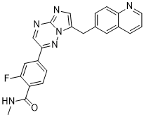
2-fluoro-N-methyl-4-[7-(quinolin-6-ylmethyl)imidazo[1,2-b][1,2,4]triazin-2-yl]benzamide
|
|
| 别名 |
|
|
| HS Tariff Code |
2934.99.9001
|
|
| 存储方式 |
Powder -20°C 3 years 4°C 2 years In solvent -80°C 6 months -20°C 1 month |
|
| 运输条件 |
Room temperature (This product is stable at ambient temperature for a few days during ordinary shipping and time spent in Customs)
|
| 溶解度 (体外实验) |
|
|||
|---|---|---|---|---|
| 溶解度 (体内实验) |
配方 1 中的溶解度: ≥ 2.08 mg/mL (5.04 mM) (饱和度未知) in 10% DMSO + 40% PEG300 + 5% Tween80 + 45% Saline (这些助溶剂从左到右依次添加,逐一添加), 澄清溶液。
例如,若需制备1 mL的工作液,可将100 μL 20.8 mg/mL澄清DMSO储备液加入400 μL PEG300中,混匀;然后向上述溶液中加入50 μL Tween-80,混匀;加入450 μL生理盐水定容至1 mL。 *生理盐水的制备:将 0.9 g 氯化钠溶解在 100 mL ddH₂O中,得到澄清溶液。 配方 2 中的溶解度: ≥ 2.08 mg/mL (5.04 mM) (饱和度未知) in 10% DMSO + 90% (20% SBE-β-CD in Saline) (这些助溶剂从左到右依次添加,逐一添加), 澄清溶液。 例如,若需制备1 mL的工作液,可将 100 μL 20.8 mg/mL澄清DMSO储备液加入900 μL 20% SBE-β-CD生理盐水溶液中,混匀。 *20% SBE-β-CD 生理盐水溶液的制备(4°C,1 周):将 2 g SBE-β-CD 溶解于 10 mL 生理盐水中,得到澄清溶液。 View More
配方 3 中的溶解度: 5%DMSO+40%PEG300+5%Tween80+50%ddH2O: 6mg/ml 1、请先配制澄清的储备液(如:用DMSO配置50 或 100 mg/mL母液(储备液)); 2、取适量母液,按从左到右的顺序依次添加助溶剂,澄清后再加入下一助溶剂。以 下列配方为例说明 (注意此配方只用于说明,并不一定代表此产品 的实际溶解配方): 10% DMSO → 40% PEG300 → 5% Tween-80 → 45% ddH2O (或 saline); 假设最终工作液的体积为 1 mL, 浓度为5 mg/mL: 取 100 μL 50 mg/mL 的澄清 DMSO 储备液加到 400 μL PEG300 中,混合均匀/澄清;向上述体系中加入50 μL Tween-80,混合均匀/澄清;然后继续加入450 μL ddH2O (或 saline)定容至 1 mL; 3、溶剂前显示的百分比是指该溶剂在最终溶液/工作液中的体积所占比例; 4、 如产品在配制过程中出现沉淀/析出,可通过加热(≤50℃)或超声的方式助溶; 5、为保证最佳实验结果,工作液请现配现用! 6、如不确定怎么将母液配置成体内动物实验的工作液,请查看说明书或联系我们; 7、 以上所有助溶剂都可在 Invivochem.cn网站购买。 |
| 制备储备液 | 1 mg | 5 mg | 10 mg | |
| 1 mM | 2.4247 mL | 12.1236 mL | 24.2471 mL | |
| 5 mM | 0.4849 mL | 2.4247 mL | 4.8494 mL | |
| 10 mM | 0.2425 mL | 1.2124 mL | 2.4247 mL |
1、根据实验需要选择合适的溶剂配制储备液 (母液):对于大多数产品,InvivoChem推荐用DMSO配置母液 (比如:5、10、20mM或者10、20、50 mg/mL浓度),个别水溶性高的产品可直接溶于水。产品在DMSO 、水或其他溶剂中的具体溶解度详见上”溶解度 (体外)”部分;
2、如果您找不到您想要的溶解度信息,或者很难将产品溶解在溶液中,请联系我们;
3、建议使用下列计算器进行相关计算(摩尔浓度计算器、稀释计算器、分子量计算器、重组计算器等);
4、母液配好之后,将其分装到常规用量,并储存在-20°C或-80°C,尽量减少反复冻融循环。
计算结果:
工作液浓度: mg/mL;
DMSO母液配制方法: mg 药物溶于 μL DMSO溶液(母液浓度 mg/mL)。如该浓度超过该批次药物DMSO溶解度,请首先与我们联系。
体内配方配制方法:取 μL DMSO母液,加入 μL PEG300,混匀澄清后加入μL Tween 80,混匀澄清后加入 μL ddH2O,混匀澄清。
(1) 请确保溶液澄清之后,再加入下一种溶剂 (助溶剂) 。可利用涡旋、超声或水浴加热等方法助溶;
(2) 一定要按顺序加入溶剂 (助溶剂) 。
A Study of Amivantamab and Capmatinib Combination Therapy in Unresectable Metastatic Non-small Cell Lung Cancer
CTID: NCT05488314
Phase: Phase 1/Phase 2 Status: Active, not recruiting
Date: 2024-10-24
INCB28060 inhibits c-MET–dependent cell proliferation and survival. Clin Cancer Res. 2011 Nov 15;17(22):7127-38. |
HGF induces production of TGF-α, AR, and HRG-β1 in cancer cells and INCB28060 effectively blocks the induction. |
Cross-talk between c-MET and EGFR or HER-3 in cancer cells. |
InvivoChem的所有产品仅用于作科学研究,不面向患者销售
Copyright 2020 InvivoChem LLC | All Rights Reserved 粤ICP备20063088号-1


 COA
COA
")
")
")
 463611831
