| 规格 | 价格 | 库存 | 数量 |
|---|---|---|---|
| 10 mM * 1 mL in DMSO |
|
||
| 1mg |
|
||
| 5mg |
|
||
| 10mg |
|
||
| 25mg |
|
||
| 50mg |
|
||
| 100mg |
|
||
| 250mg |
|
||
| 500mg |
|
||
| 1g |
|
||
| Other Sizes |
|

| 靶点 |
Aurora A (Ki = 8 nM); Aurora B (Ki = 9.2 nM)
Dual inhibitor of Aurora A kinase and Aurora B kinase with potent activity. For recombinant Aurora A: IC₅₀ = 1.2 nM (kinase activity assay); for recombinant Aurora B: IC₅₀ = 3.5 nM. It showed high selectivity over other kinases, with IC₅₀ > 1000 nM for CDK1/cyclin B, EGFR, and VEGFR2, confirming >800-fold selectivity for Aurora kinases over non-Aurora kinases [1] - In HCT116 colorectal cancer cells, inhibition of Aurora A-mediated TPX2 phosphorylation showed an EC₅₀ = 4.8 nM, and inhibition of Aurora B-mediated histone H3 (Ser10) phosphorylation showed an EC₅₀ = 6.2 nM [1] |
|---|---|
| 体外研究 (In Vitro) |
此外,CYC-116 分别以 44、82、280 和 44 nM 的浓度抑制 FLT3、Src、Lck 和 VEGFR2。 CYC-116 可能具有广谱抗癌作用。 MCF7、HeLa、Co 的 IC50 为 0.599、0.59、0.241、0.34、0.725、1.375、0.471、0.034、0.372、0.681、0.151、1.626、0.775、0.308、0.110 和 0.09 lo 205、HCT-116、HT29、K562 、CCRF-CEM、MV4-11、HL60、NCI-H460、A2780、BxPC3、HuPT4、Mia-Paca-2、Saos-2 和 Messa 细胞,CYC-116 对癌细胞系表现出强大的抗增殖活性。用 1.25 μM CYC-116 处理 7 小时后,HeLa 细胞裂解物中的组蛋白 H3 磷酸化被完全抑制[1]。
对人癌细胞系的抗增殖活性:CYC116对多种实体瘤细胞系表现出广谱强效抗增殖作用,IC₅₀值范围为15 nM至30 nM。具体示例包括: - HCT116(结直肠癌):IC₅₀=18 nM - MCF-7(乳腺癌):IC₅₀=25 nM - SK-OV-3(卵巢癌):IC₅₀=15 nM - A549(肺癌):IC₅₀=30 nM[1] - 诱导G2/M期细胞周期停滞:用CYC116(20 nM)处理HCT116细胞24小时,通过碘化丙啶(PI)染色和流式细胞术检测显示,G2/M期细胞比例从溶剂对照组的14%显著升高至处理组的58%。这种停滞与有丝分裂纺锤体形态异常相关,通过α-微管蛋白免疫荧光可观察到65%的处理细胞出现异常纺锤体[1] - 抑制Aurora底物磷酸化:对经CYC116(5-50 nM)处理6小时的HCT116细胞进行蛋白质印迹(western blot)分析,发现底物磷酸化呈剂量依赖性降低: - Aurora A介导的TPX2磷酸化:20 nM剂量下较对照组降低80% - Aurora B介导的组蛋白H3(Ser10)磷酸化:30 nM剂量下较对照组降低70% 总TPX2和总组蛋白H3水平无显著变化[1] - 诱导癌细胞凋亡:用CYC116(30 nM)处理MCF-7细胞48小时,膜联蛋白V阳性凋亡细胞(早期+晚期凋亡)比例较溶剂对照组增加38%。Western blot结果显示,凋亡标志物切割型caspase-3增加3.0倍,切割型PARP增加2.6倍[1] |
| 体内研究 (In Vivo) |
口服 CYC-116 剂量水平分别为 75 和 100 mg/kg qd,分别导致肿瘤生长延迟 2.3 天和 5.8 天。这些肿瘤生长延迟转化为 0.32 和 0.81 的特定生长延迟。在试验期间,接受两种剂量水平的 CYC-116 的小鼠的平均相对肿瘤体积小于接受赋形剂的小鼠的相对肿瘤体积。在第 6 天和第 9 天,每天口服 100 mg/kg 时,生长下降具有统计学意义[1]。
HCT116结直肠癌裸鼠异种移植模型:对携带HCT116异种移植瘤的雌性裸鼠(6-7周龄,每组8只),以50 mg/kg剂量口服CYC116,每日1次,连续14天。该处理相较于溶剂对照组实现72%的肿瘤生长抑制率(TGI)。实验结束时,处理组肿瘤体积为200±28 mm³,对照组为710±42 mm³(p<0.001)。处理组小鼠未出现显著体重下降(<5%)[1] - SK-OV-3卵巢癌裸鼠异种移植模型:对携带SK-OV-3异种移植瘤的裸鼠,口服CYC116(60 mg/kg/日)18天,TGI达68%。对剥离肿瘤的免疫组化分析显示: - 磷酸化组蛋白H3(Ser10,Aurora B活性标志物)染色降低85% - 磷酸化TPX2(Aurora A活性标志物)染色降低75%[1] |
| 酶活实验 |
激酶测定[1]
如前所述进行这些操作。使用XLfit软件(IDBS)测定IC50值。使用Cheng和Prussoff的方法,根据每种激酶的IC50值和适当的Km(ATP)值计算表观抑制常数(Ki)。重组人极光A和B激酶购自Upstate Discovery。使用25μL反应体积(25mMβ-甘油磷酸,20mM Tris/HCl,pH 7.5,5mM EGTA,1mM DTT,1mM Na3VO4,10μg keptide(肽底物))进行Aurora A激酶测定,并将重组Aurora激酶在20mM Tris/HCl中稀释,pH 8,含有0.5mg/mL BSA,2.5%甘油和0.006%Brij-35。通过添加5μL Mg/ATP混合物(15 mM MgCl2,100μM ATP,每孔18.5 kBqγ-32P-ATP)开始反应,并在30°C下孵育30分钟,然后通过添加25μL 75 mM H3PO4终止反应。Aurora B激酶测定与Aurora A相同,只是在使用前,Aurora B在30°C下与内着丝粒蛋白的单独反应中活化60分钟。 Aurora A激酶活性实验(HTRF格式):将重组人Aurora A激酶(与TPX2结合以增强催化活性)与CYC116(系列浓度:0.01 nM至500 nM)、ATP(10 μM)及生物素化TPX2衍生肽底物(含Ser466位Aurora A磷酸化位点)在激酶缓冲液(50 mM Tris-HCl、10 mM MgCl₂、1 mM DTT、0.01% BSA,pH 7.5)中于30°C孵育60分钟。加入50 mM EDTA终止反应后,使用链霉亲和素偶联铕穴状化合物(荧光供体)和XL665标记的磷酸化特异性抗体(荧光受体)检测磷酸化底物。通过酶标仪测量荧光共振能量转移(FRET)信号,将剂量-反应曲线拟合至四参数逻辑模型计算IC₅₀值[1] - Aurora B激酶活性实验:将重组人Aurora B激酶(与INCENP结合)与CYC116(0.01 nM至500 nM)、ATP(10 μM)及生物素化组蛋白H3(Ser10)肽底物在上述相同激酶缓冲液中孵育。30°C孵育60分钟后,加入50 mM EDTA终止反应,采用与Aurora A实验相同的HTRF方法检测磷酸化底物,从剂量-反应曲线中确定IC₅₀值[1] |
| 细胞实验 |
流式细胞术的细胞周期分析[1]
为了使早期S期的细胞同步,对它们进行双胸苷阻断。将HeLa细胞以每10 cm培养皿5×10⁵个细胞的速度接种,并在37°C下孵育16−18小时。加入胸苷(2 mM),将细胞孵育18小时。通过在5 mL PBS中洗涤3次,将细胞从块中释放。加入新鲜培养基,将细胞培养8小时。然后再次加入2 mM胸苷,持续第二个16小时。再次释放细胞,并加入新鲜培养基以及适当的测试化合物稀释液 为了使A549细胞在M期同步,将其与40 ng/mL诺可唑(Sigma)孵育18小时。如有说明,还用50μM MG132蛋白酶体抑制剂处理细胞孵育的最后2小时。将通过摇动和重复洗涤从平板上分离的圆形有丝分裂细胞制成丸粒并在PBS中洗涤以从诺可唑嵌段释放。将细胞用合适的测试化合物稀释液在新鲜培养基中重新接种。 结合TUNEL染色或细胞周期蛋白B分析对细胞进行细胞周期分析。因此,收获所有细胞,在PBS中洗涤,用0.5%PFA固定,并在−20°C的80%乙醇中储存。末端脱氧核苷酸转移酶介导的缺口末端标记(TUNEL)测定按照制造商的说明进行。流式细胞术期间的细胞周期蛋白B分析使用在400μL 0.5%BSA PBS中的细胞周期素B抗体的1:1000稀释液,用1:400 FITC山羊多克隆至小鼠二次,然后用PI孵育用于DNA染色。使用BD FACSCalibur流式细胞仪分析每个样品的20000个单细胞事件。 组蛋白H3磷酸化的蛋白质印迹分析[1] 为了检测磷酸化组蛋白H3,用化合物处理HeLa细胞7小时。然后通过酸提取制备提取物。简言之,将细胞从板上刮下,成丸,在PBS中洗涤一次,然后重悬于含有罗氏完全蛋白酶抑制剂混合物的裂解缓冲液(10mM Tris/HCl,pH 8.0,1.5mM MgCl2,10mM KCl,0.5mM DTT)中。将HCl和H2SO4添加到0.2M的最终浓度,并将裂解物在冰上孵育1小时。通过离心将不溶性材料制成颗粒,将酸溶解的上清液添加到1mL Me2CO中,并在−20°C下储存24小时。通过离心机将沉淀的蛋白质制成颗粒,短暂风干,并重悬于SDS−PAGE负载缓冲液中。样品在15%SDS-聚丙烯酰胺凝胶上分离,并通过电印迹转移到硝化纤维膜上。用兔抗磷酸组蛋白H3抗体在膜上检测磷酸化组蛋白H3.用小鼠抗组蛋白H3:检测总组蛋白H3,然后用适当的二抗和化学发光检测。 抗增殖实验(CellTiter-Glo法):将人癌细胞系(HCT116、MCF-7、SK-OV-3、A549)以2×10³个细胞/孔接种于96孔板,37°C(5% CO₂)过夜孵育。加入系列浓度(1 nM至200 nM)的CYC116,继续培养72小时。向每孔加入CellTiter-Glo试剂(其产生的发光强度与活细胞ATP含量成正比),室温孵育10分钟后测量发光强度。使用GraphPad Prism软件计算抑制50%活细胞的CYC116浓度(IC₅₀)[1] - 细胞周期分析(PI染色):将HCT116细胞以5×10⁵个细胞/孔接种于6孔板,用CYC116(20 nM)或溶剂处理24小时。胰酶消化收集细胞,冷PBS洗涤后,在-20°C下用70%乙醇固定过夜。固定细胞再次用PBS洗涤,重悬于PI染色液(50 μg/mL PI、100 μg/mL RNase A、0.1% Triton X-100溶于PBS),37°C孵育30分钟。通过流式细胞仪分析细胞周期分布(G0/G1、S、G2/M期),使用ModFit软件定量各时期细胞百分比[1] - 凋亡实验(膜联蛋白V-FITC/PI双染法):用CYC116(30 nM)或溶剂处理MCF-7细胞48小时。收集细胞,冷PBS洗涤后,重悬于膜联蛋白V结合缓冲液。向细胞悬液中加入膜联蛋白V-FITC和PI,室温避光孵育15分钟。通过流式细胞仪检测并计数凋亡细胞(早期凋亡:膜联蛋白V阳性/PI阴性;晚期凋亡:膜联蛋白V阳性/PI阳性)[1] - Aurora底物Western blot实验:用CYC116(5、10、20、30、50 nM)处理HCT116细胞6小时,随后用含蛋白酶和磷酸酶抑制剂的RIPA缓冲液裂解细胞。将蛋白提取物(每泳道30 μg)通过10% SDS-PAGE分离,转移至PVDF膜。膜用5%脱脂牛奶-TBST封闭1小时,然后用抗磷酸化TPX2(Ser466)、抗磷酸化组蛋白H3(Ser10)、抗总TPX2、抗总组蛋白H3及抗β-肌动蛋白(内参)一抗在4°C下孵育过夜。TBST洗涤后,膜与辣根过氧化物酶偶联的二抗在室温下孵育1小时。使用增强化学发光(ECL)试剂检测信号,通过ImageJ软件定量条带强度[1] |
| 动物实验 |
将化合物溶于DMSO,然后用水稀释;剂量为75、100 mg/kg;采用灌胃法给药。将NCI-H460细胞腹腔注射到小鼠体内。大鼠药代动力学研究。[1]
在雄性Wistar大鼠中测定了测试化合物的药代动力学参数。每种化合物分别通过静脉推注或灌胃给药,每组3只大鼠。灌胃给药的剂量为10 mL/kg,静脉给药的剂量为12 mL/kg(1 mL/min)。静脉给药后,分别于0、5、15和30分钟,1、2、4和6小时,通过颈静脉插管从每只大鼠采集3份血清样本;灌胃给药后,分别于0、0.5、1、2、4、6、8和24小时采集血清样本。所有血液样本采集后立即进行离心。收集血浆并储存于–20 °C直至分析。采用LC-MS/MS方法分析样本。使用WinNonlin 5.2软件,通过非房室模型方法推导药代动力学参数。口服生物利用度(%F)通过口服与静脉给药剂量标准化AUC值的比值计算得出。 小鼠P388/D1白血病模型。[1] 雌性Balb/c × DBA/2J F1小鼠于第0天腹腔注射2.1 × 10⁵个P388/D1白血病细胞。从第1天开始,在第1-3天和第7-9天,每天两次通过灌胃给予动物化合物18,剂量为0.1 mL/10 g体重。通过比较各治疗组小鼠与载体对照组小鼠接种后中位生存期(ILS)来评估治疗效果。治疗组与对照组的ILS值比值以百分比(% ILS)表示,并用作相对疗效的指标。 NCI-H460异种移植瘤。[1] 从体外培养的亚融合培养物中收集人NCI-H460非小细胞肺癌细胞,并测定活细胞数量。然后将细胞重悬于无菌PBS中,浓度约为7 × 10⁷个细胞/mL。将约7 × 10⁶个细胞皮下注射到裸鼠(无胸腺)右侧腹部。当肿瘤体积达到可测量范围(80-100 mm³)时,将动物随机分为治疗组和对照组,每组10只。每周至少测量两次肿瘤大小。研究期间,若肿瘤体积过大或出现任何不良反应,则终止动物实验。治疗方案为灌胃给药,每日一次,从第1天开始,持续5天。对照组动物灌胃给予赋形剂,每日一次,从第1天开始,持续5天。记录研究期间测量的肿瘤尺寸。计算相对肿瘤体积并绘制平均肿瘤生长曲线图(图S8)。采用单因素方差分析 (ANOVA) 比较各组的相对肿瘤体积数据,并使用 Dunnett's t 检验确定统计学显著性。 HCT116 结直肠癌异种移植模型:将 5×10⁶ 个 HCT116 细胞(悬浮于 PBS 和 Matrigel 的 1:1 混合物中)皮下注射到 6-7 周龄雌性裸鼠的右侧腹部。当肿瘤体积达到 100-150 mm³ 时,将小鼠随机分为两组(每组 n=8):载体对照组(0.5% 羧甲基纤维素钠 + 0.1% Tween 80 的蒸馏水)和 CYC116 治疗组。将CYC116溶解于溶剂中,浓度为10 mg/mL,并以50 mg/kg的剂量每日一次经口灌胃给药,连续14天。每2天使用游标卡尺测量肿瘤体积,计算公式为(长×宽²)/2。同时每2天测量小鼠体重,以监测潜在的毒性[1] - SK-OV-3卵巢癌异种移植模型:将1×10⁷个SK-OV-3细胞(与Matrigel混合)皮下植入雌性裸鼠体内。当肿瘤体积达到约120 mm³时,将小鼠分组(每组n=8)。将CYC116以12 mg/mL的浓度溶于上述溶剂中,并以60 mg/kg的剂量每日一次经口给药,连续18天。研究结束时,切除肿瘤组织,称重,并用10%中性缓冲福尔马林固定,用于磷酸化组蛋白H3(Ser10)和磷酸化TPX2的免疫组织化学分析[1] |
| 药代性质 (ADME/PK) |
口服生物利用度:在雄性 Sprague-Dawley 大鼠中,口服 CYC116 (20 mg/kg) 的口服生物利用度为 30%。血浆浓度-时间曲线显示,给药后 1.8 小时达到血浆峰浓度 (Cmax) 为 1.1 μg/mL,末端半衰期 (t₁/₂) 为 4.5 小时 [1]
- 静脉注射药代动力学(大鼠):大鼠静脉注射 CYC116 (5 mg/kg) 后,清除率 (CL) 为 15 mL/min/kg,稳态分布容积 (Vss) 为 5.2 L/kg,t₁/₂ 为 4.2 小时 [1] - 血浆蛋白结合率:通过平衡透析法测定,CYC116 在人 (95%)、大鼠 (94%) 和小鼠 (93%) 血浆中均表现出较高的血浆蛋白结合率。在37°C下,使用10 kDa分子量截留膜进行4小时透析,血浆中CYC116浓度为1 μg/mL [1] - 代谢稳定性:在人肝微粒体中,CYC116的半衰期为3.8小时(中等代谢稳定性);在大鼠肝微粒体中,t₁/₂为4.3小时。液相色谱-串联质谱(LC-MS/MS)鉴定出主要代谢物为单羟基化衍生物(占总代谢物的55%),主要通过CYP3A4介导的氧化作用生成[1] |
| 毒性/毒理 (Toxicokinetics/TK) |
蛋白质结合
CYC116 是一种口服有效的 Aurora 激酶 A 和 B 以及 VEGFR2 抑制剂,用于治疗晚期实体瘤患者。 急性口服毒性(小鼠):雌性 CD-1 小鼠单次口服剂量高达 2000 mg/kg 的 CYC116 未导致死亡。剂量 ≥1500 mg/kg 时,小鼠出现短暂的运动活性降低,但在 24 小时内恢复。剂量 ≤1000 mg/kg 时,未观察到体重的显著变化 [1] - 慢性口服毒性(大鼠):雄性 Sprague-Dawley 大鼠接受 CYC116(50 mg/kg,每日一次,口服)治疗 28 天。观察到轻度骨髓抑制:与载体对照组相比,白细胞计数下降了18%,而红细胞计数和血小板计数保持在正常范围内。肝功能指标(ALT、AST)和肾功能指标(BUN、肌酐)的血清水平与对照组无显著差异,肝脏、肾脏和心脏组织的病理学检查未发现与治疗相关的病变[1] |
| 参考文献 | |
| 其他信息 |
CYC116 是一种新型抗癌化合物,具有独特的靶点谱,涉及细胞周期和血管生成抑制机制。在临床前研究中,CYC116 已在实体瘤和血液肿瘤中均显示出抗肿瘤活性。CYC116 是 Cyclacel 公司研发的第三款口服药物,其抗癌活性机制与 Aurora 激酶抑制相符。
适应症 晚期实体瘤 作用机制 Aurora 激酶是一种帮助分裂细胞在两个子细胞之间共享物质的酶。在许多癌症患者中,Aurora 激酶功能异常,导致细胞分裂的正常调控丧失,从而引起异常生长。 CYC116 抑制 Aurora 激酶,可能减缓癌细胞的生长并导致其凋亡。 化学类别和设计:CYC116 属于 N-苯基-4-(噻唑-5-基)嘧啶-2-胺衍生物类化合物,该类化合物的化学骨架经过优化,可同时抑制 Aurora A 和 B 激酶。其设计平衡了对两种 Aurora 亚型的抑制效力,同时最大限度地减少了脱靶激酶活性,从而克服了单一亚型抑制剂的局限性(单一亚型抑制剂可能允许肿瘤细胞通过补偿性 Aurora 活性逃避免疫抑制)[1]。 - 作用机制:CYC116 通过同时抑制 Aurora A 和 B 激酶(有丝分裂进程的关键调节因子)发挥抗肿瘤活性。抑制 Aurora A 会破坏有丝分裂纺锤体的组装和中心体的成熟,而抑制 Aurora B 则会损害染色体分离和胞质分裂。这种联合作用会导致癌细胞(尤其是 Aurora 激酶表达升高的癌细胞,这是结直肠癌、乳腺癌和卵巢癌的常见特征)发生 G2/M 期细胞周期阻滞、有丝分裂灾难和随后的细胞凋亡 [1]。 - 临床前治疗潜力:CYC116 在 HCT116 和 SK-OV-3 细胞系中与标准化疗药物(例如 5-氟尿嘧啶、紫杉醇)无交叉耐药性,支持其治疗化疗难治性实体瘤的潜力。此外,其高口服生物利用度和良好的药代动力学特征使其适合在临床环境中口服给药 [1]。 |
| 分子式 |
C18H20N6OS
|
|
|---|---|---|
| 分子量 |
368.46
|
|
| 精确质量 |
368.141
|
|
| 元素分析 |
C, 58.68; H, 5.47; N, 22.81; O, 4.34; S, 8.70
|
|
| CAS号 |
693228-63-6
|
|
| 相关CAS号 |
|
|
| PubChem CID |
6420138
|
|
| 外观&性状 |
Typically exists as light yellow to yellow solids at room temperature
|
|
| 密度 |
1.4±0.1 g/cm3
|
|
| 沸点 |
648.8±65.0 °C at 760 mmHg
|
|
| 闪点 |
346.2±34.3 °C
|
|
| 蒸汽压 |
0.0±1.9 mmHg at 25°C
|
|
| 折射率 |
1.689
|
|
| LogP |
1.44
|
|
| tPSA |
118.16
|
|
| 氢键供体(HBD)数目 |
2
|
|
| 氢键受体(HBA)数目 |
8
|
|
| 可旋转键数目(RBC) |
4
|
|
| 重原子数目 |
26
|
|
| 分子复杂度/Complexity |
443
|
|
| 定义原子立体中心数目 |
0
|
|
| SMILES |
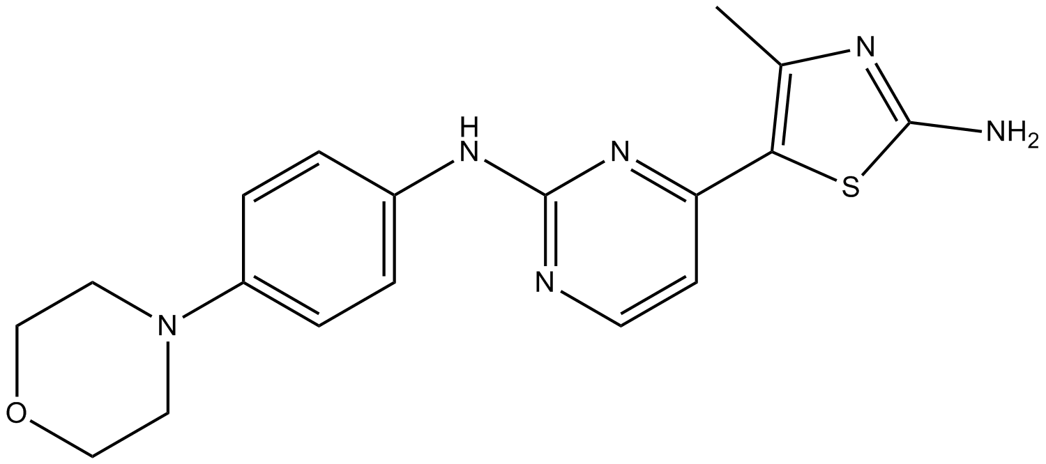
S1C(N([H])[H])=NC(C([H])([H])[H])=C1C1C([H])=C([H])N=C(N=1)N([H])C1C([H])=C([H])C(=C([H])C=1[H])N1C([H])([H])C([H])([H])OC([H])([H])C1([H])[H]
|
|
| InChi Key |
GPSZYOIFQZPWEJ-UHFFFAOYSA-N
|
|
| InChi Code |
InChI=1S/C18H20N6OS/c1-12-16(26-17(19)21-12)15-6-7-20-18(23-15)22-13-2-4-14(5-3-13)24-8-10-25-11-9-24/h2-7H,8-11H2,1H3,(H2,19,21)(H,20,22,23)
|
|
| 化学名 |
4-methyl-5-(2-(4-morpholinophenylamino)pyrimidin-4-yl)thiazol-2-amine
|
|
| 别名 |
CYC 116; CYC-116; CYC116.
|
|
| HS Tariff Code |
2934.99.9001
|
|
| 存储方式 |
Powder -20°C 3 years 4°C 2 years In solvent -80°C 6 months -20°C 1 month |
|
| 运输条件 |
Room temperature (This product is stable at ambient temperature for a few days during ordinary shipping and time spent in Customs)
|
| 溶解度 (体外实验) |
|
|||
|---|---|---|---|---|
| 溶解度 (体内实验) |
配方 1 中的溶解度: 1.5 mg/mL (4.07 mM) in 10% DMSO + 40% PEG300 + 5% Tween80 + 45% Saline (这些助溶剂从左到右依次添加,逐一添加), 悬浮液;超声助溶。
例如,若需制备1 mL的工作液,可将100 μL 15.0 mg/mL澄清DMSO储备液加入到400 μL PEG300中,混匀;然后向上述溶液中加入50 μL Tween-80,混匀;加入450 μL生理盐水定容至1 mL。 *生理盐水的制备:将 0.9 g 氯化钠溶解在 100 mL ddH₂O中,得到澄清溶液。 配方 2 中的溶解度: ≥ 1.5 mg/mL (4.07 mM) (饱和度未知) in 10% DMSO + 90% (20% SBE-β-CD in Saline) (这些助溶剂从左到右依次添加,逐一添加), 澄清溶液。 例如,若需制备1 mL的工作液,可将 100 μL 15.0 mg/mL澄清DMSO储备液加入900 μL 20% SBE-β-CD生理盐水溶液中,混匀。 *20% SBE-β-CD 生理盐水溶液的制备(4°C,1 周):将 2 g SBE-β-CD 溶解于 10 mL 生理盐水中,得到澄清溶液。 View More
配方 3 中的溶解度: 1% DMSO+30% polyethylene glycol+1% Tween 80:30mg/mL 1、请先配制澄清的储备液(如:用DMSO配置50 或 100 mg/mL母液(储备液)); 2、取适量母液,按从左到右的顺序依次添加助溶剂,澄清后再加入下一助溶剂。以 下列配方为例说明 (注意此配方只用于说明,并不一定代表此产品 的实际溶解配方): 10% DMSO → 40% PEG300 → 5% Tween-80 → 45% ddH2O (或 saline); 假设最终工作液的体积为 1 mL, 浓度为5 mg/mL: 取 100 μL 50 mg/mL 的澄清 DMSO 储备液加到 400 μL PEG300 中,混合均匀/澄清;向上述体系中加入50 μL Tween-80,混合均匀/澄清;然后继续加入450 μL ddH2O (或 saline)定容至 1 mL; 3、溶剂前显示的百分比是指该溶剂在最终溶液/工作液中的体积所占比例; 4、 如产品在配制过程中出现沉淀/析出,可通过加热(≤50℃)或超声的方式助溶; 5、为保证最佳实验结果,工作液请现配现用! 6、如不确定怎么将母液配置成体内动物实验的工作液,请查看说明书或联系我们; 7、 以上所有助溶剂都可在 Invivochem.cn网站购买。 |
| 制备储备液 | 1 mg | 5 mg | 10 mg | |
| 1 mM | 2.7140 mL | 13.5700 mL | 27.1400 mL | |
| 5 mM | 0.5428 mL | 2.7140 mL | 5.4280 mL | |
| 10 mM | 0.2714 mL | 1.3570 mL | 2.7140 mL |
1、根据实验需要选择合适的溶剂配制储备液 (母液):对于大多数产品,InvivoChem推荐用DMSO配置母液 (比如:5、10、20mM或者10、20、50 mg/mL浓度),个别水溶性高的产品可直接溶于水。产品在DMSO 、水或其他溶剂中的具体溶解度详见上”溶解度 (体外)”部分;
2、如果您找不到您想要的溶解度信息,或者很难将产品溶解在溶液中,请联系我们;
3、建议使用下列计算器进行相关计算(摩尔浓度计算器、稀释计算器、分子量计算器、重组计算器等);
4、母液配好之后,将其分装到常规用量,并储存在-20°C或-80°C,尽量减少反复冻融循环。
计算结果:
工作液浓度: mg/mL;
DMSO母液配制方法: mg 药物溶于 μL DMSO溶液(母液浓度 mg/mL)。如该浓度超过该批次药物DMSO溶解度,请首先与我们联系。
体内配方配制方法:取 μL DMSO母液,加入 μL PEG300,混匀澄清后加入μL Tween 80,混匀澄清后加入 μL ddH2O,混匀澄清。
(1) 请确保溶液澄清之后,再加入下一种溶剂 (助溶剂) 。可利用涡旋、超声或水浴加热等方法助溶;
(2) 一定要按顺序加入溶剂 (助溶剂) 。
| NCT Number | Recruitment | interventions | Conditions | Sponsor/Collaborators | Start Date | Phases |
| NCT00560716 | Terminated | Drug: CYC116 | Solid Tumors | Cyclacel Pharmaceuticals, Inc. | June 2007 | Phase 1 |
|
InvivoChem的所有产品仅用于作科学研究,不面向患者销售
Copyright 2020 InvivoChem LLC | All Rights Reserved 粤ICP备20063088号-1


 COA
COA

 463611831
