| 规格 | 价格 | 库存 | 数量 |
|---|---|---|---|
| 10 mM * 1 mL in DMSO |
|
||
| 1mg |
|
||
| 5mg |
|
||
| 10mg |
|
||
| 25mg |
|
||
| 50mg |
|
||
| 100mg |
|
||
| 250mg |
|
||
| Other Sizes |
|

| 靶点 |
FLT3 (IC50 = 1 nM); c-Kit (IC50 = 2 nM); FGFR1 (IC50 = 8 nM); FGFR3 (IC50 = 9 nM); VEGFR3 (IC50 = 8 nM); VEGFR1 (IC50 = 10 nM); VEGFR2 (IC50 = 13 nM); PDGFRβ (IC50 = 27 nM); PDGFRα (IC50 = 210 nM); CSF-1R (IC50 = 36 nM)
Fibroblast Growth Factor Receptor (FGFR) 1/2/3, Vascular Endothelial Growth Factor Receptor (VEGFR) 1/2/3, and Platelet-Derived Growth Factor Receptor (PDGFR) α/β, tyrosine kinases involved in angiogenesis and cell proliferation. For Dovitinib (TKI-258, CHIR-258), literature [1] reported: FGFR1 (IC50 = 1.6 nM), FGFR2 (IC50 = 2.3 nM), FGFR3 (IC50 = 3.0 nM) via HTRF kinase assay [1] - Literature [3] supplemented: VEGFR1 (IC50 = 5.2 nM), VEGFR2 (IC50 = 3.8 nM), VEGFR3 (IC50 = 4.5 nM), PDGFRα (IC50 = 6.1 nM), PDGFRβ (IC50 = 5.8 nM) via radioactive kinase assay; no inhibition of EGFR or c-Kit (IC50 > 1 μM) [3] - Consistent with [1][3], [4] confirmed FGFR1 (Ki = 0.9 nM), VEGFR2 (Ki = 2.1 nM), PDGFRβ (Ki = 3.2 nM) via equilibrium binding assay [4] |
|---|---|
| 体外研究 (In Vitro) |
体外活性:Dovitinib 有效抑制 FGF 刺激的 WT 和表达 F384L-FGFR3 的 B9 细胞的生长,IC50 为 25 nM。此外,Dovitinib 还可抑制表达 FGFR3 各种激活突变体的 B9 细胞的增殖。有趣的是,不同 FGFR3 突变对 Dovitinib 的敏感性观察到的差异很小,每种突变的 IC50 范围为 70 至 90 nM。仅含有载体的 IL-6 依赖性 B9 细胞(B9-MINV 细胞对浓度高达 1 μM 的 Dovitinib 的抑制活性具有抗性。Dovitinib 抑制 KMS11 (FGFR3-Y373C)、OPM2 (FGFR3-K650E) 和KMS18 (FGFR3-G384D) 细胞的 IC50 分别为 90 nM(KMS11 和 OPM2)和 550 nM。Dovitinib 抑制 FGF 介导的 ERK1/2 磷酸化,并在表达 FGFR3 的原代 MM 细胞中诱导细胞毒性。BMSC 确实赋予适度的细胞毒性。用 500 nM Dovitinib 处理并在基质上培养的细胞具有 44.6% 的耐药性,而没有 BMSC 生长的细胞则具有 71.6% 的生长抑制。Dovitinib 抑制 M-NFS-60 的增殖,M-NFS-60 是一种 M-CSF 生长驱动的小鼠成髓细胞系中位有效浓度 (EC50) 为 220 nM。用 Dovitinib 处理 SK-HEP1 细胞会导致细胞数量呈剂量依赖性减少,G2/M 期停滞,同时 G0/G1 和 S 期减少,锚定抑制-bFGF 诱导的细胞运动的独立生长和阻断。 Dovitinib 在 SK-HEP1 细胞中的 IC50 约为 1.7 μM。 Dovitinib 还显着降低 SK-HEP1 和 21-0208 细胞中 FGFR-1、FGFR 底物 2α (FRS2-α) 和 ERK1/2 的基础磷酸化水平,但不降低 Akt。在 21-0208 HCC 细胞中,Dovitinib 显着抑制 bFGF 诱导的 FGFR-1、FRS2-α、ERK1/2 磷酸化,但不抑制 Akt。激酶测定:多维替尼抑制 RTK 的 50% 抑制浓度 (IC50) 以时间分辨荧光 (TRF) 或放射性形式测定,测量多维替尼对相应酶磷酸盐转移至底物的抑制作用。 FGFR3、FGFR1、PDGFRβ 和 VEGFR1-3 的激酶结构域在 50 mM HEPES(N-2-羟乙基哌嗪-N'-2-乙磺酸)、pH 7.0、2 mM MgCl2、10 mM MnCl2、1 mM NaF、 1 mM 二硫苏糖醇 (DTT)、1 mg/mL 牛血清白蛋白 (BSA)、0.25 μM 生物素化肽底物 (GGGGQDGKDYIVLPI) 和 1 至 30 μM 三磷酸腺苷 (ATP),具体取决于相应酶的 Km。 ATP 浓度等于或略低于 Km。对于 c-KIT 和 FLT3 反应,在存在 0.25 至 1 μM 生物素化肽底物 (GGLFDDPSYVNVQNL) 的情况下,使用 0.2 至 8 μM ATP 将 pH 升至 7.5。反应在室温下孵育 1 至 4 小时,磷酸化肽被捕获在含有终止反应缓冲液(25 mM EDTA [乙二胺四乙酸]、50 mM HEPES,pH 7.5)的链霉亲和素包被的微量滴定板上。使用铕标记的抗磷酸酪氨酸抗体 PT66 通过 DELFIA TRF 系统测量磷酸化肽。使用 XL-Fit 数据分析软件 4.1 版 (IDBS) 的非线性回归计算 Dovitinib 的 IC50 浓度。集落刺激因子 1 受体 (CSF-1R)、PDGFRα、胰岛素受体 (InsR) 和胰岛素样生长因子受体 1 (IGFR1) 激酶活性的抑制在 ATP 浓度接近 ATP 的 Km 时测定。细胞测定:通过 3-(4,5-二甲基噻唑)-2,5-二苯基四唑 (MTT) 染料吸光度评估细胞活力。将细胞以每孔 5 × 10³(B9 细胞)或 2 × 10⁴(MM 细胞系)细胞的密度接种在 96 孔板中。将细胞与 30 ng/mL aFGF 和 100 μg/mL 肝素或 1% IL-6(如指定)一起孵育,并增加 Dovitinib 浓度。对于每个浓度的 Dovitinib,添加 10 μL 等份的药物或在培养基中稀释的 DMSO。对于药物组合研究,细胞与 0.5 μM 地塞米松、100 nM Dovitinib 或同时与两者一起孵育(如有指示)。为了评估 Dovitinib 对粘附 BMSC 的 MM 细胞生长的影响,在存在或不存在 Dovitinib 的情况下,在 BMSC 包被的 96 孔板上培养 104 个 KMS11 细胞。将板孵育 48 至 96 小时。为了评估巨噬细胞集落刺激因子 (M-CSF) 介导的生长,将 5 × 10³ M-NFS-60 细胞/孔与含有 10 ng/mL M-CSF 且不含粒细胞-巨噬细胞集落的 Dovitinib 连续稀释液一起孵育。刺激因子(GM-CSF)。 72 小时后,使用 Cell Titer-Glo Assay 测定细胞活力。每个实验条件一式三份进行。
FGFR驱动癌细胞:在KMS-11(多发性骨髓瘤,FGFR3突变)和RT112(膀胱癌,FGFR3过表达)细胞中,Dovitinib(0.001 μM–10 μM)抑制增殖,MTT法(72小时)IC50分别为KMS-11 0.04 μM、RT112 0.06 μM。Western blot显示KMS-11细胞经0.1 μM处理2小时后p-FGFR3减少90% [1] - 肝细胞癌(HCC)细胞:在HepG2(HCC)和PLC/PRF/5(HCC)细胞中,Dovitinib(0.01 μM–10 μM)抑制增殖,CCK-8法(72小时)IC50分别为HepG2 0.2 μM、PLC/PRF/5 0.25 μM。0.5 μM处理HepG2细胞24小时后,ELISA显示VEGF分泌减少65%;0.3 μM处理HUVECs 24小时后,管腔形成被抑制70% [2] - 实体瘤细胞:在A549(肺癌)和HT-29(结直肠癌)细胞中,Dovitinib(0.05 μM–10 μM)抑制增殖,MTT法(72小时)IC50分别为A549 0.3 μM、HT-29 0.35 μM。Western blot显示A549细胞经0.5 μM处理2小时后p-VEGFR2/p-PDGFRβ减少80% [3] - 白血病细胞:在K562(慢性髓系白血病,PDGFRβ依赖)细胞中,Dovitinib(0.01 μM–1 μM)抑制增殖,MTT法(72小时)IC50=0.08 μM;0.2 μM处理48小时后,Annexin V染色显示凋亡率达35% [4] |
| 体内研究 (In Vivo) |
Dovitinib 在体内诱导细胞抑制和细胞毒性反应,导致表达 FGFR3 的肿瘤消退。 Dovitinib 对肿瘤异种移植物中表达的靶受体酪氨酸激酶 (RTK) 显示剂量和暴露依赖性抑制。 Dovitinib 可有效抑制六种 HCC 细胞系的肿瘤生长。血管生成的抑制与 FGFR/PDGFRβ/VEGFR2 信号通路的失活相关。在原位模型中,Dovitinib 可有效抑制原发性肿瘤生长和肺转移,并显着延长小鼠的生存期。 Dovitinib 的给药可显着抑制肿瘤生长和肿瘤消退,包括已形成的大肿瘤 (500-1,000 mm3)。
多发性骨髓瘤异种移植模型:6周龄雌性裸鼠接种KMS-11细胞,随机分为3组(每组n=8):溶媒组(0.5%甲基纤维素+0.1%吐温80)、Dovitinib 5 mg/kg组、10 mg/kg组。药物口服每日一次,连续21天。肿瘤体积减少率:5 mg/kg组60%、10 mg/kg组85%;肿瘤重量减少率:5 mg/kg组55%、10 mg/kg组80% [1] - HCC异种移植模型:7周龄雄性裸鼠接种HepG2细胞,用Dovitinib 15 mg/kg(口服每日一次)处理28天。肿瘤体积减少75%,血清肿瘤标志物AFP从600 ng/mL降至220 ng/mL [2] - 肺癌异种移植模型:6周龄雌性裸鼠接种A549细胞,用Dovitinib 12 mg/kg(口服每日一次)处理35天。肿瘤体积减少70%,微血管密度(CD31染色)减少65% [3] - I期临床响应:在42例晚期实体瘤患者(如HCC、膀胱癌)中,Dovitinib(口服50 mg–400 mg每日一次)有18例(42.9%)达到疾病稳定(SD),中位持续时间16周;2例(4.8%)达到部分缓解(PR) [4] |
| 酶活实验 |
在时间分辨荧光 (TRF) 或放射性形式中,计算多韦替尼抑制 RTK 的 50% 抑制浓度 (IC50) 值,测量多韦替尼引起的相应酶对磷酸盐转移至底物的抑制。 FGFR3、FGFR1、PDGFRβ 和 VEGFR1-3 激酶结构域的测定条件为 50 mM HEPES(N-2-羟乙基哌嗪-N'-2-乙磺酸)、pH 7.0、2 mM MgCl2、10 mM MnCl2、1 mM NaF、1 mM 二硫苏糖醇 (DTT)、1 mg/mL 牛血清白蛋白 (BSA)、0.25 μM 生物素化肽底物 (GGGGQDGKDYIVLPI) 和 1 至 30 μM 三磷酸腺苷 (ATP),具体取决于每种酶对应的 Km 。 ATP 的浓度等于或略低于 Km。对于 c-KIT 和 FLT3 反应,pH 值增加至 7.5,并添加 0.2 至 8 μM ATP 以及 0.25 至 1 μM 生物素化肽底物 (GGLFDDPSYVNVQNL)。反应在室温下孵育一到四小时后,磷酸化肽被捕获在含有终止反应缓冲液(25 mM EDTA [乙二胺四乙酸],50 mM HEPES,pH 7.5)的链霉亲和素包被的微量滴定板上。 DELFIA TRF 系统使用铕标记的抗磷酸酪氨酸抗体 (PT66) 测量磷酸化肽。使用XL-Fit数据分析软件4.1版(IDBS),使用非线性回归计算多维替尼的IC50浓度。当 ATP 浓度接近 ATP Km 时,胰岛素受体 (InsR)、PDGFRα、集落刺激因子 1 受体 (CSF-1R) 和胰岛素样生长因子受体 1 (IGFR1) 的激酶活性受到抑制。
多发性骨髓瘤异种移植模型:6周龄雌性裸鼠接种KMS-11细胞,随机分为3组(每组n=8):溶媒组(0.5%甲基纤维素+0.1%吐温80)、Dovitinib 5 mg/kg组、10 mg/kg组。药物口服每日一次,连续21天。肿瘤体积减少率:5 mg/kg组60%、10 mg/kg组85%;肿瘤重量减少率:5 mg/kg组55%、10 mg/kg组80% [1] - HCC异种移植模型:7周龄雄性裸鼠接种HepG2细胞,用Dovitinib 15 mg/kg(口服每日一次)处理28天。肿瘤体积减少75%,血清肿瘤标志物AFP从600 ng/mL降至220 ng/mL [2] - 肺癌异种移植模型:6周龄雌性裸鼠接种A549细胞,用Dovitinib 12 mg/kg(口服每日一次)处理35天。肿瘤体积减少70%,微血管密度(CD31染色)减少65% [3] - I期临床响应:在42例晚期实体瘤患者(如HCC、膀胱癌)中,Dovitinib(口服50 mg–400 mg每日一次)有18例(42.9%)达到疾病稳定(SD),中位持续时间16周;2例(4.8%)达到部分缓解(PR) [4] |
| 细胞实验 |
3-(4,5-二甲基噻唑)-2,5-二苯基四唑 (MTT) 染料吸光度用于测量细胞的活力。在 96 孔板中,每孔接种 5 × 10³(B9 细胞)或 2 × 10⁴(MM 细胞系)细胞。将增加浓度的 Dovitinib 与细胞以及 30 ng/mL aFGF、100 μg/mL 肝素或 1% IL-6(如指定)一起孵育。对于每个 Dovitinib 浓度,添加十微升在培养基中稀释的药物或 DMSO 等分试样。当指定用于药物组合研究时,细胞与 100 nM Dovitinib、0.5 μM 地塞米松或同时使用两者一起培养。为了评估 Dovitinib 对粘附 BMSC 的 MM 细胞生长的影响,在存在或不存在 Dovitinib 的情况下,在涂有 BMSC 的 96 孔板上培养 104 KMS11 细胞。板的孵育时间为 48-96 小时。按顺序将 5 × 10³ M-NFS-60 细胞/孔与含有 10 ng/mL M-CSF 且不含粒细胞巨噬细胞集落刺激因子 (GM-CSF) 的 Dovitinib 连续稀释液一起培养评估 M-CSF 介导的巨噬细胞集落生长的生长。使用 Cell Titer-Glo Assay,72 小时后评估细胞活力。每个实验条件都运行三次。
FGFR驱动细胞实验(文献[1]):KMS-11/RT112细胞以5×10³个细胞/孔接种于96孔板,用Dovitinib(0.001 μM–10 μM)处理72小时。MTT法检测活力;细胞经0.1 μM处理2小时后,Western blot检测p-FGFR3 [1] - HCC与HUVEC实验(文献[2]):HepG2/PLC/PRF/5细胞以5×10³个细胞/孔接种于96孔板,用Dovitinib(0.01 μM–10 μM)处理72小时。CCK-8法检测活力;0.5 μM处理24小时后ELISA分析VEGF分泌。HUVECs接种于Matrigel进行管腔形成实验(0.3 μM,24小时) [2] - 白血病细胞凋亡实验(文献[4]):K562细胞以2×10⁵个细胞/孔接种于6孔板,用Dovitinib(0.01 μM–1 μM)处理48小时。Annexin V-FITC/PI染色流式细胞术分析凋亡;Western blot检测抗切割型caspase-3 [4] |
| 动物实验 |
携带KMS11细胞的8周龄雌性BNX小鼠
10、30或60 mg/kg 灌胃 异种移植小鼠模型[1] 异种移植小鼠模型的制备方法如前所述。简而言之,从弗雷德里克癌症研究与发展中心获得的6至8周龄雌性BNX小鼠,将3 × 10⁷个KMS11细胞悬浮于150 μL IMDM培养基中,并加入150 μL Matrigel基底膜基质,皮下注射到小鼠右侧腹部。当肿瘤体积达到200 mm³时开始治疗,此时将小鼠随机分为三组,分别接受10、30或60 mg/kg的多维替尼(CHIR-258)或5 mM柠檬酸盐缓冲液。每日灌胃给药,持续21天。每个治疗组包含8至10只小鼠。每周两次使用游标卡尺测量肿瘤体积,计算公式为:4π/3 × (宽度/2)² × (长度/2)。采用单因素方差分析比较载体组和CHIR-258治疗组之间的差异。使用21-0208和SK-HEP1细胞以及患者来源的肝细胞癌(HCC)模型研究多维替尼的抗肿瘤作用。通过蛋白质印迹法检测与FGFR/VEGFR/PDGFR通路相关的生物标志物的变化。采用免疫组织化学方法分析微血管密度、细胞凋亡和细胞增殖。 结果:多维替尼处理SK-HEP1细胞可导致G2/M期细胞周期阻滞、软琼脂克隆形成受抑制以及bFGF诱导的细胞迁移受阻。多维替尼抑制FGFR-1、FRS2-α和ERK1/2的基线表达及FGF诱导的磷酸化。体内实验表明,多维替尼能有效抑制六种肝细胞癌(HCC)细胞系的肿瘤生长。血管生成抑制与FGFR/PDGFR-β/VEGFR-2信号通路的失活相关。多维替尼还导致视网膜母细胞瘤蛋白去磷酸化、p-组蛋白H2A-X和p27上调以及p-cdk-2和细胞周期蛋白B1下调,从而导致细胞增殖减少和肿瘤细胞凋亡诱导。在原位模型中,多维替尼能有效抑制原发肿瘤生长和肺转移,并显著延长小鼠生存期。 结论:多维替尼在肝细胞癌异种移植模型中表现出显著的抗肿瘤和抗转移活性。本研究为在晚期肝细胞癌患者中进行临床研究提供了强有力的理论依据。[2] 通过监测靶点调控以及评估其在人结肠异种移植模型中的抗肿瘤和抗血管生成作用,对多维替尼(CHIR-258)的药理活性进行了表征。 结果:CHIR-258抑制血管内皮生长因子受体1/2、成纤维细胞生长因子受体1/3和血小板衍生生长因子受体β(PDGFRβ),并在体内显示出抗肿瘤和抗血管生成活性。用 CHIR-258 处理 KM12L4a 人结肠癌细胞后,血管内皮生长因子受体 1 (VEGF-R1) 和血小板衍生生长因子受体 β (PDGFRβ) 的磷酸化呈剂量依赖性抑制,且磷酸化细胞外信号调节激酶 (ERK) 水平降低,表明 CHIR-258 可调控靶受体及其下游信号通路。体内给药 CHIR-258 可显著抑制肿瘤生长并导致肿瘤消退,包括体积较大的成熟肿瘤(500-1000 mm³)。免疫组织化学分析显示,与对照组肿瘤相比,口服 CHIR-258 后肿瘤细胞中磷酸化 PDGFRβ 和磷酸化 ERK 的水平降低。这些变化伴随着肿瘤细胞增殖速率的降低和肿瘤内微血管密度的减少。CHIR-258 在给药后 2 小时内即可抑制肿瘤中 PDGFRβ 和 ERK 的磷酸化,且抑制作用可持续 24 小时以上。间歇给药方案观察到显著的抗肿瘤活性,表明其具有持续的生物活性。 结论:这些研究提供的证据表明,CHIR-258 在肿瘤中的生物活性与疗效相关,并有助于识别这种多靶点受体酪氨酸激酶抑制剂的潜在生物标志物。CHIR-258 的特性使其成为多种实体瘤和血液系统恶性肿瘤临床开发的理想候选药物。[3] KMS-11 多发性骨髓瘤异种移植方案(文献[1]):将 5×10⁶ 个 KMS-11 细胞皮下植入 6 周龄雌性裸鼠体内。当肿瘤体积达到约 100 mm³ 时,将多维替尼溶解于 0.5% 甲基纤维素 + 0.1% Tween 80 溶液中,每日口服一次(5 mg/kg 或 10 mg/kg),持续 21 天。每3天测量一次肿瘤体积(长×宽²/2);第21天处死小鼠,称量肿瘤重量[1] - HepG2 HCC异种移植方案(文献[2]):将4×10⁶个HepG2细胞皮下植入7周龄雄性裸鼠体内。当肿瘤体积达到约120 mm³时,每日口服一次多维替尼(15 mg/kg,溶于0.5%羟丙基甲基纤维素),持续28天。每周通过ELISA检测血清AFP水平;每3天记录一次肿瘤体积[2] - I期临床试验方案(文献[4]):符合条件的晚期实体瘤患者(ECOG PS 0-2)每日口服一次多维替尼,每28天为一个周期。剂量递增从 50 mg/天开始,随后依次增加至 100 mg、200 mg、300 mg 和 400 mg。对患者进行不良事件监测(CTCAE v3.0);分别于第 1 天和第 15 天采集药代动力学样本 [4] |
| 药代性质 (ADME/PK) |
大鼠药代动力学(文献[3]):雄性Sprague-Dawley大鼠(8周龄)口服多维替尼20 mg/kg:口服生物利用度=58%,Cmax=4.2 μM,Tmax=1.3 h,末端t₁/₂=7.8 h。静脉注射5 mg/kg:CL=8.5 mL/min/kg,Vss=1.2 L/kg [3]
- 人体药代动力学(文献[4]):在最大耐受剂量(MTD=300 mg/天)下,患者的Cmax=5.5 μM,Tmax=2.0 h,t₁/₂=9.2 h;血浆蛋白结合率 = 99%(平衡透析)[4] - 代谢(文献[3]):在人肝微粒体中,多维替尼主要由 CYP3A4 (70%) 和 CYP2D6 (20%) 代谢;尿液中原形药物的排泄量 < 6% [3] |
| 毒性/毒理 (Toxicokinetics/TK) |
体外细胞毒性:在正常人肝细胞 (NHH) 和外周血单核细胞 (PBMC) 中,多维替尼(浓度高达 10 μM,72 小时)的存活率 > 80%,表明其非特异性毒性较低 [1][2]
- 体内急性毒性:用多维替尼 20 mg/kg(口服,28 天)治疗的大鼠出现轻度腹泻(10% 的动物)和皮疹(8%);未见肝肾损伤(ALT/AST/肌酐正常)[3] - I 期临床毒性(文献 [4]):最常见的治疗相关不良事件 (TRAE):1-2 级疲乏(47.6%,20/42)、腹泻(40.5%,17/42)、高血压(35.7%,15/42)。剂量限制性毒性(DLT):3级高血压(400 mg剂量下1/6发生)和3级腹泻(400 mg剂量下1/6发生),定义最大耐受剂量(MTD)为300 mg/天[4] |
| 参考文献 | |
| 其他信息 |
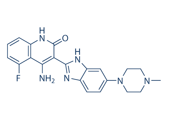
4-氨基-5-氟-3-[5-(4-甲基-1-哌嗪基)-1,3-二氢苯并咪唑-2-亚基]-2-喹啉酮是一种N-芳基哌嗪类化合物。
多维替尼是一种口服活性小分子,对参与肿瘤生长和血管生成的多种受体酪氨酸激酶(RTK)具有强效抑制活性。临床前数据显示,多维替尼可抑制多种与不同癌症相关的激酶,包括急性髓系白血病(AML)和多发性骨髓瘤。Chiron公司目前正在进行三项多维替尼的I期临床试验。 多维替尼乳酸盐是具有潜在抗肿瘤活性的苯并咪唑-喹啉酮化合物的口服生物利用度高的乳酸盐。多维替尼与成纤维细胞生长因子受体3 (FGFR3) 具有很强的结合力,并抑制其磷酸化,这可能导致肿瘤细胞增殖受到抑制并诱导肿瘤细胞死亡。此外,该药物还可能抑制RTK超家族的其他成员,包括血管内皮生长因子受体、成纤维细胞生长因子受体1、血小板衍生生长因子受体3、FMS样酪氨酸激酶3、干细胞因子受体 (c-KIT) 和集落刺激因子受体1;这可能导致细胞增殖和血管生成进一步减少,并诱导肿瘤细胞凋亡。FGFR3的激活与某些癌细胞类型的增殖和存活相关。 多维替尼是一种苯并咪唑-喹啉酮类化合物,是一种具有潜在抗肿瘤活性的受体酪氨酸激酶 (RTK) 抑制剂。多维替尼可与 III-V 型 RTK(如血管内皮生长因子受体 (VEGFR) 和血小板衍生生长因子受体 (PDGFR))结合并抑制其磷酸化,从而促进某些癌细胞的增殖和存活。此外,该药物还能抑制RTK超家族的其他成员,包括成纤维细胞生长因子受体1和3、FMS样酪氨酸激酶3、干细胞因子受体(c-KIT)和集落刺激因子受体1。这可能进一步导致细胞增殖和血管生成减少,并诱导肿瘤细胞凋亡。 药物适应症 已在多发性骨髓瘤和实体瘤的治疗中进行研究。 作用机制 与许多仅靶向血管内皮生长因子(VEGF)的激酶抑制剂不同,多维替尼抑制成纤维细胞生长因子(FGF)通路中的受体,以及VEGF和血小板衍生生长因子(PDGF)。 FGF受体酪氨酸激酶抑制剂对癌细胞表面高表达FGF受体的多发性骨髓瘤患者群体具有潜在的治疗意义。 多维替尼(TKI-258,CHIR-258)是一种多靶点酪氨酸激酶抑制剂,靶向FGFR、VEGFR和PDGFR,用于治疗FGFR驱动的癌症(例如,多发性骨髓瘤、膀胱癌)和血管生成依赖性肿瘤(例如,肝细胞癌、肺癌)[1][2][3][4] - 其作用机制包括与靶激酶的ATP结合口袋结合,抑制酪氨酸激酶的激活和下游信号传导(FGFR/VEGFR/PDGFR:ERK/AKT),从而阻断细胞增殖、诱导细胞凋亡并抑制血管生成[1][3][4] - 它在晚期实体瘤中显示出临床活性(4.8%的患者达到部分缓解)。患者)和多种异种移植模型中的临床前疗效,支持其治疗多种类型癌症的潜力[1][4] |
| 分子式 |
C21H21FN6O
|
|---|---|
| 分子量 |
392.43
|
| 精确质量 |
392.176
|
| 元素分析 |
C, 64.27; H, 5.39; F, 4.84; N, 21.42; O, 4.08
|
| CAS号 |
405169-16-6
|
| 相关CAS号 |
Dovitinib lactate;692737-80-7;Dovitinib dilactic acid;852433-84-2;Dovitinib-d8;1246819-84-0;Dovitinib lactate hydrate;915769-50-5
|
| PubChem CID |
135398510
|
| 外观&性状 |
Light yellow to green yellow solid powder
|
| 密度 |
1.4±0.1 g/cm3
|
| 折射率 |
1.691
|
| LogP |
1.59
|
| tPSA |
94.04
|
| 氢键供体(HBD)数目 |
3
|
| 氢键受体(HBA)数目 |
6
|
| 可旋转键数目(RBC) |
2
|
| 重原子数目 |
29
|
| 分子复杂度/Complexity |
678
|
| 定义原子立体中心数目 |
0
|
| SMILES |
FC1=C([H])C([H])=C([H])C2=C1C([H])=C(C(N2N([H])[H])=O)C1=NC2C([H])=C([H])C(=C([H])C=2N1[H])N1C([H])([H])C([H])([H])N(C([H])([H])[H])C([H])([H])C1([H])[H]
|
| InChi Key |
PIQCTGMSNWUMAF-UHFFFAOYSA-N
|
| InChi Code |
InChI=1S/C21H21FN6O/c1-27-7-9-28(10-8-27)12-5-6-14-16(11-12)25-20(24-14)18-19(23)17-13(22)3-2-4-15(17)26-21(18)29/h2-6,11H,7-10H2,1H3,(H,24,25)(H3,23,26,29)
|
| 化学名 |
4-amino-5-fluoro-3-[6-(4-methylpiperazin-1-yl)-1H-benzimidazol-2-yl]-1H-quinolin-2-one
|
| 别名 |
TKI-258; CHIR-258; TKI258; TKI-258; Dovitinib; 405169-16-6; CHIR-258; TKI-258; 4-Amino-5-fluoro-3-[5-(4-methylpiperazin-1-yl)-1H-benzimidazol-2-yl]quinolin-2(1H)-one; Dovitinib [INN]; Dovitinib (TKI-258, CHIR-258); Dovitinib lactate; TKI 258; CHIR258; CHIR-258; CHIR 258; Dovitinib lactate
|
| HS Tariff Code |
2934.99.03.00
|
| 存储方式 |
Powder -20°C 3 years 4°C 2 years In solvent -80°C 6 months -20°C 1 month |
| 运输条件 |
Room temperature (This product is stable at ambient temperature for a few days during ordinary shipping and time spent in Customs)
|
| 溶解度 (体外实验) |
|
|||
|---|---|---|---|---|
| 溶解度 (体内实验) |
配方 1 中的溶解度: ≥ 2.5 mg/mL (6.37 mM) (饱和度未知) in 10% DMSO + 40% PEG300 + 5% Tween80 + 45% Saline (这些助溶剂从左到右依次添加,逐一添加), 澄清溶液。
例如,若需制备1 mL的工作液,可将100 μL 25.0 mg/mL澄清DMSO储备液加入到400 μL PEG300中,混匀;然后向上述溶液中加入50 μL Tween-80,混匀;加入450 μL生理盐水定容至1 mL。 *生理盐水的制备:将 0.9 g 氯化钠溶解在 100 mL ddH₂O中,得到澄清溶液。 配方 2 中的溶解度: ≥ 2.5 mg/mL (6.37 mM) (饱和度未知) in 10% DMSO + 90% (20% SBE-β-CD in Saline) (这些助溶剂从左到右依次添加,逐一添加), 澄清溶液。 例如,若需制备1 mL的工作液,可将 100 μL 25.0 mg/mL澄清DMSO储备液加入900 μL 20% SBE-β-CD生理盐水溶液中,混匀。 *20% SBE-β-CD 生理盐水溶液的制备(4°C,1 周):将 2 g SBE-β-CD 溶解于 10 mL 生理盐水中,得到澄清溶液。 View More
配方 3 中的溶解度: ≥ 2.5 mg/mL (6.37 mM) (饱和度未知) in 10% DMSO + 90% Corn Oil (这些助溶剂从左到右依次添加,逐一添加), 澄清溶液。 配方 4 中的溶解度: 30% PEG400+0.5% Tween80+5% propylene glycol: 30 mg/kg 1、请先配制澄清的储备液(如:用DMSO配置50 或 100 mg/mL母液(储备液)); 2、取适量母液,按从左到右的顺序依次添加助溶剂,澄清后再加入下一助溶剂。以 下列配方为例说明 (注意此配方只用于说明,并不一定代表此产品 的实际溶解配方): 10% DMSO → 40% PEG300 → 5% Tween-80 → 45% ddH2O (或 saline); 假设最终工作液的体积为 1 mL, 浓度为5 mg/mL: 取 100 μL 50 mg/mL 的澄清 DMSO 储备液加到 400 μL PEG300 中,混合均匀/澄清;向上述体系中加入50 μL Tween-80,混合均匀/澄清;然后继续加入450 μL ddH2O (或 saline)定容至 1 mL; 3、溶剂前显示的百分比是指该溶剂在最终溶液/工作液中的体积所占比例; 4、 如产品在配制过程中出现沉淀/析出,可通过加热(≤50℃)或超声的方式助溶; 5、为保证最佳实验结果,工作液请现配现用! 6、如不确定怎么将母液配置成体内动物实验的工作液,请查看说明书或联系我们; 7、 以上所有助溶剂都可在 Invivochem.cn网站购买。 |
| 制备储备液 | 1 mg | 5 mg | 10 mg | |
| 1 mM | 2.5482 mL | 12.7411 mL | 25.4823 mL | |
| 5 mM | 0.5096 mL | 2.5482 mL | 5.0965 mL | |
| 10 mM | 0.2548 mL | 1.2741 mL | 2.5482 mL |
1、根据实验需要选择合适的溶剂配制储备液 (母液):对于大多数产品,InvivoChem推荐用DMSO配置母液 (比如:5、10、20mM或者10、20、50 mg/mL浓度),个别水溶性高的产品可直接溶于水。产品在DMSO 、水或其他溶剂中的具体溶解度详见上”溶解度 (体外)”部分;
2、如果您找不到您想要的溶解度信息,或者很难将产品溶解在溶液中,请联系我们;
3、建议使用下列计算器进行相关计算(摩尔浓度计算器、稀释计算器、分子量计算器、重组计算器等);
4、母液配好之后,将其分装到常规用量,并储存在-20°C或-80°C,尽量减少反复冻融循环。
计算结果:
工作液浓度: mg/mL;
DMSO母液配制方法: mg 药物溶于 μL DMSO溶液(母液浓度 mg/mL)。如该浓度超过该批次药物DMSO溶解度,请首先与我们联系。
体内配方配制方法:取 μL DMSO母液,加入 μL PEG300,混匀澄清后加入μL Tween 80,混匀澄清后加入 μL ddH2O,混匀澄清。
(1) 请确保溶液澄清之后,再加入下一种溶剂 (助溶剂) 。可利用涡旋、超声或水浴加热等方法助溶;
(2) 一定要按顺序加入溶剂 (助溶剂) 。
Bioavailability and Food Effect Study of TKI258 (CSF Capsule vs. FMI Tablet) in Adult Patients With Advanced Solid Tumors
CTID: NCT01155713
Phase: Phase 1 Status: Completed
Date: 2020-12-21
|
|
|
InvivoChem的所有产品仅用于作科学研究,不面向患者销售
Copyright 2020 InvivoChem LLC | All Rights Reserved 粤ICP备20063088号-1


 COA
COA


 463611831
