| 规格 | 价格 | 库存 | 数量 |
|---|---|---|---|
| 10 mM * 1 mL in DMSO |
|
||
| 5mg |
|
||
| 10mg |
|
||
| 25mg |
|
||
| 50mg |
|
||
| 100mg |
|
||
| 250mg |
|
||
| 500mg |
|
||
| 1g |
|
||
| Other Sizes |
|

| 靶点 |
FLT3 (IC50 = 1 nM); c-Kit (IC50 = 2 nM); FGFR1 (IC50 = 8 nM); FGFR3 (IC50 = 9 nM); VEGFR1 (IC50 = 1 nM); VEGFR3 (IC50 = 8 nM); VEGFR2 (IC50 = 13 nM); PDGFRα (IC50 = 27 nM); PDGFRβ (IC50 = 210 nM)
1. Dovitinib (TKI258; CHIR-258) Lactate is a multi-targeted tyrosine kinase inhibitor with high activity against FGFR, VEGFR, and PDGFR families, with the following IC50 values: FGFR1: 8 nM, FGFR2: 10 nM, FGFR3: 7 nM, VEGFR2 (KDR): 21 nM, PDGFRβ: 26 nM [1] 2. It also inhibits FLT3 with an IC50 of 12 nM and c-Kit with an IC50 of 34 nM; no significant inhibition (IC50 > 1 μM) is observed against EGFR and HER2 [3] 3. For mutant FGFR3 (FGFR3-TACC3 fusion), Dovitinib (TKI258; CHIR-258) Lactate exhibits an IC50 of 9 nM, similar to its activity against wild-type FGFR3 [3] |
|---|---|
| 体外研究 (In Vitro) |
体外活性:Dovitinib 有效抑制 FGF 刺激的 WT 和表达 F384L-FGFR3 的 B9 细胞的生长,IC50 为 25 nM。此外,Dovitinib 还可抑制表达 FGFR3 各种激活突变体的 B9 细胞的增殖。有趣的是,不同 FGFR3 突变对 Dovitinib 的敏感性观察到的差异很小,每种突变的 IC50 范围为 70 至 90 nM。仅含有载体的 IL-6 依赖性 B9 细胞(B9-MINV 细胞对浓度高达 1 μM 的 Dovitinib 的抑制活性具有抗性。Dovitinib 抑制 KMS11 (FGFR3-Y373C)、OPM2 (FGFR3-K650E) 和KMS18 (FGFR3-G384D) 细胞的 IC50 分别为 90 nM(KMS11 和 OPM2)和 550 nM。Dovitinib 抑制 FGF 介导的 ERK1/2 磷酸化,并在表达 FGFR3 的原代 MM 细胞中诱导细胞毒性。BMSC 确实赋予适度的细胞毒性。用 500 nM Dovitinib 处理并在基质上培养的细胞具有 44.6% 的耐药性,而没有 BMSC 生长的细胞则具有 71.6% 的生长抑制。Dovitinib 抑制 M-NFS-60 的增殖,M-NFS-60 是一种 M-CSF 生长驱动的小鼠成髓细胞系中位有效浓度 (EC50) 为 220 nM。用 Dovitinib 处理 SK-HEP1 细胞会导致细胞数量呈剂量依赖性减少,G2/M 期停滞,同时 G0/G1 和 S 期减少,锚定抑制-bFGF 诱导的细胞运动的独立生长和阻断。 Dovitinib 在 SK-HEP1 细胞中的 IC50 约为 1.7 μM。 Dovitinib 还显着降低 SK-HEP1 和 21-0208 细胞中 FGFR-1、FGFR 底物 2α (FRS2-α) 和 ERK1/2 的基础磷酸化水平,但不降低 Akt。在 21-0208 HCC 细胞中,Dovitinib 显着抑制 bFGF 诱导的 FGFR-1、FRS2-α、ERK1/2 磷酸化,但不抑制 Akt。激酶测定:多维替尼抑制 RTK 的 50% 抑制浓度 (IC50) 以时间分辨荧光 (TRF) 或放射性形式测定,测量多维替尼对相应酶磷酸盐转移至底物的抑制作用。 FGFR3、FGFR1、PDGFRβ 和 VEGFR1-3 的激酶结构域在 50 mM HEPES(N-2-羟乙基哌嗪-N'-2-乙磺酸)、pH 7.0、2 mM MgCl2、10 mM MnCl2、1 mM NaF、 1 mM 二硫苏糖醇 (DTT)、1 mg/mL 牛血清白蛋白 (BSA)、0.25 μM 生物素化肽底物 (GGGGQDGKDYIVLPI) 和 1 至 30 μM 三磷酸腺苷 (ATP),具体取决于相应酶的 Km。 ATP 浓度等于或略低于 Km。对于 c-KIT 和 FLT3 反应,在存在 0.25 至 1 μM 生物素化肽底物 (GGLFDDPSYVNVQNL) 的情况下,使用 0.2 至 8 μM ATP 将 pH 升至 7.5。反应在室温下孵育 1 至 4 小时,磷酸化肽被捕获在含有终止反应缓冲液(25 mM EDTA [乙二胺四乙酸]、50 mM HEPES,pH 7.5)的链霉亲和素包被的微量滴定板上。使用铕标记的抗磷酸酪氨酸抗体 PT66 通过 DELFIA TRF 系统测量磷酸化肽。使用 XL-Fit 数据分析软件 4.1 版 (IDBS) 的非线性回归计算 Dovitinib 的 IC50 浓度。集落刺激因子 1 受体 (CSF-1R)、PDGFRα、胰岛素受体 (InsR) 和胰岛素样生长因子受体 1 (IGFR1) 激酶活性的抑制在 ATP 浓度接近 ATP 的 Km 时测定。细胞测定:通过 3-(4,5-二甲基噻唑)-2,5-二苯基四唑 (MTT) 染料吸光度评估细胞活力。将细胞以每孔 5 × 10³(B9 细胞)或 2 × 10⁴(MM 细胞系)细胞的密度接种在 96 孔板中。将细胞与 30 ng/mL aFGF 和 100 μg/mL 肝素或 1% IL-6(如指定)一起孵育,并增加 Dovitinib 浓度。对于每个浓度的 Dovitinib,添加 10 μL 等份的药物或在培养基中稀释的 DMSO。对于药物组合研究,细胞与 0.5 μM 地塞米松、100 nM Dovitinib 或同时与两者一起孵育(如有指示)。为了评估 Dovitinib 对粘附 BMSC 的 MM 细胞生长的影响,在存在或不存在 Dovitinib 的情况下,在 BMSC 包被的 96 孔板上培养 104 个 KMS11 细胞。将板孵育 48 至 96 小时。为了评估巨噬细胞集落刺激因子 (M-CSF) 介导的生长,将 5 × 10³ M-NFS-60 细胞/孔与含有 10 ng/mL M-CSF 且不含粒细胞-巨噬细胞集落的 Dovitinib 连续稀释液一起孵育。刺激因子(GM-CSF)。 72 小时后,使用 Cell Titer-Glo Assay 测定细胞活力。每个实验条件一式三份进行。
1. FGFR3-TACC3阳性的RT112膀胱癌细胞中:多韦替尼乳酸盐(10-100 nM)抑制细胞增殖,IC50为18 nM。50 nM处理72小时后,细胞活力较未处理对照组降低约80% [3] 2. VEGFR2依赖的人脐静脉内皮细胞(HUVECs)中:多韦替尼乳酸盐(20-200 nM)呈剂量依赖性抑制VEGF诱导的管形成和迁移。100 nM浓度下,管长度较VEGF刺激组降低约75%,迁移能力降低约70% [1] 3. MV4-11细胞(FLT3-ITD阳性急性髓系白血病,AML)中:多韦替尼乳酸盐(5-50 nM)诱导凋亡。20 nM处理48小时后,凋亡率(Annexin V阳性细胞)从对照组的约4%升至约42% [1] 4. HepG2肝癌(HCC)细胞Western blot分析:多韦替尼乳酸盐(100 nM)使FGFR1(Tyr653/654)磷酸化水平降低约90%、VEGFR2(Tyr1175)降低约85%,下游p-AKT(Ser473)降低约80% [2] 5. PDGFRβ过表达的NIH3T3细胞中:多韦替尼乳酸盐(30-300 nM)抑制克隆形成。100 nM浓度下,克隆数较未处理组减少约65% [3] |
| 体内研究 (In Vivo) |
Dovitinib 在体内诱导细胞抑制和细胞毒性反应,导致表达 FGFR3 的肿瘤消退。 Dovitinib 对肿瘤异种移植物中表达的靶受体酪氨酸激酶 (RTK) 显示剂量和暴露依赖性抑制。 Dovitinib 可有效抑制六种 HCC 细胞系的肿瘤生长。血管生成的抑制与 FGFR/PDGFRβ/VEGFR2 信号通路的失活相关。在原位模型中,Dovitinib 可有效抑制原发性肿瘤生长和肺转移,并显着延长小鼠的生存期。 Dovitinib 的给药可显着抑制肿瘤生长和肿瘤消退,包括已形成的大肿瘤 (500-1,000 mm3)。
1. 裸鼠RT112膀胱癌异种移植模型:口服多韦替尼乳酸盐(30 mg/kg,每日1次,持续28天)的肿瘤生长抑制率(TGI)为75%,处理组肿瘤重量约为溶媒对照组(0.5%甲基纤维素)的25% [3] 2. SCID小鼠MV4-11 AML静脉移植模型:多韦替尼乳酸盐(50 mg/kg,灌胃,每日1次,持续14天)延长小鼠生存期,中位生存期从对照组的19天延长至36天,7只小鼠中有2只存活超过50天 [1] 3. 裸鼠HepG2肝癌异种移植模型:多韦替尼乳酸盐(40 mg/kg,口服,每日1次,持续35天)使肿瘤体积减少约70%,肿瘤内微血管密度(CD31阳性血管)较溶媒组降低约65% [2] |
| 酶活实验 |
在时间分辨荧光 (TRF) 或放射性形式中,计算多韦替尼抑制 RTK 的 50% 抑制浓度 (IC50) 值,测量多韦替尼引起的相应酶对磷酸盐转移至底物的抑制。 FGFR3、FGFR1、PDGFRβ 和 VEGFR1-3 激酶结构域的测定条件为 50 mM HEPES(N-2-羟乙基哌嗪-N'-2-乙磺酸)、pH 7.0、2 mM MgCl2、10 mM MnCl2、1 mM NaF、1 mM 二硫苏糖醇 (DTT)、1 mg/mL 牛血清白蛋白 (BSA)、0.25 μM 生物素化肽底物 (GGGGQDGKDYIVLPI) 和 1 至 30 μM 三磷酸腺苷 (ATP),具体取决于每种酶对应的 Km 。 ATP 的浓度等于或略低于 Km。对于 c-KIT 和 FLT3 反应,pH 值增加至 7.5,并添加 0.2 至 8 μM ATP 以及 0.25 至 1 μM 生物素化肽底物 (GGLFDDPSYVNVQNL)。反应在室温下孵育一到四小时后,磷酸化肽被捕获在含有终止反应缓冲液(25 mM EDTA [乙二胺四乙酸],50 mM HEPES,pH 7.5)的链霉亲和素包被的微量滴定板上。 DELFIA TRF 系统使用铕标记的抗磷酸酪氨酸抗体 (PT66) 测量磷酸化肽。使用XL-Fit数据分析软件4.1版(IDBS),使用非线性回归计算多维替尼的IC50浓度。当 ATP 浓度接近 ATP Km 时,胰岛素受体 (InsR)、PDGFRα、集落刺激因子 1 受体 (CSF-1R) 和胰岛素样生长因子受体 1 (IGFR1) 的激酶活性受到抑制。
1. 重组FGFR1激酶活性测定:反应缓冲液含50 mM Tris-HCl(pH 7.5)、10 mM MgCl2、1 mM DTT、25 μM ATP及1 μg/well GST-FGFR1激酶结构域。不同浓度多韦替尼乳酸盐(1-50 nM)与激酶在30°C预孵育15分钟,加入1 μg/well肽底物(序列:EAIYAAPFAKKK)启动反应,30°C孵育45分钟。用磷酸酪氨酸特异性抗体和化学发光法检测磷酸化底物,通过非线性回归拟合抑制曲线计算IC50 [3] 2. 重组VEGFR2(KDR)激酶测定:重组VEGFR2激酶(5 ng/well)与多韦替尼乳酸盐(5-100 nM)在含25 mM HEPES(pH 7.4)、5 mM MnCl2、1 mM DTT、10 μM ATP及0.5 μg/well Poly(Glu,Tyr)4:1底物的缓冲液中混合。37°C反应60分钟后,加入3%磷酸终止反应,将混合物转移至P81板,用0.5%磷酸洗涤,通过闪烁计数器检测[γ-32P]ATP的放射性信号以确定IC50 [1] |
| 细胞实验 |
3-(4,5-二甲基噻唑)-2,5-二苯基四唑(MTT)染料吸光度代表细胞活力。每孔 5 × 10³(B9 细胞)或 2 × 10⁴(MM 细胞系)细胞的密度用于在 96 孔板中接种细胞。为了培养细胞,根据需要添加不同浓度的 Dovitinib 以及 30 ng/mL aFGF、100 μg/mL 肝素或 1% IL-6。对于每个浓度的多韦替尼,添加在培养基中稀释的 10 μL 药物或 DMSO 等分试样。药物组合研究涉及将细胞与 100 nM Dovitinib 或 0.5 μM 地塞米松一起孵育,或在必要时同时与两者一起孵育。为了评估Dovitinib对粘附于BMSC的MM细胞生长的影响,在存在或不存在Dovitinib的情况下在涂有BMSC的96孔板上培养104个KMS11细胞。板的孵育时间为 48-96 小时。按顺序将 5 × 10³ M-NFS-60 细胞/孔与含有 10 ng/mL M-CSF 且不含粒细胞巨噬细胞集落刺激因子 (GM-CSF) 的 Dovitinib 连续稀释液一起培养评估 M-CSF 介导的巨噬细胞集落生长的生长。使用 Cell Titer-Glo Assay,72 小时后评估细胞活力。每个实验条件都运行三次。
1. RT112细胞增殖测定(MTT法):RT112细胞以2×10³个/well接种于96孔板,培养过夜。加入多韦替尼乳酸盐(1-100 nM),37°C孵育72小时。每孔加入MTT试剂(5 mg/mL,10 μL),继续孵育4小时。用DMSO(100 μL/well)溶解甲瓒结晶,在570 nm处测吸光度。细胞活力以对照组的百分比表示,从剂量-反应曲线推导IC50 [3] 2. HUVEC管形成实验:Matrigel冰上融化后铺于24孔板(500 μL/well),37°C聚合30分钟。HUVECs(2×10⁴个/well)悬浮于含多韦替尼乳酸盐(20-200 nM)和VEGF(50 ng/mL)的培养基中,接种于Matrigel上。6小时后拍摄管状结构,用图像分析软件定量每孔管总长度,计算相对VEGF对照组的抑制率 [1] 3. HepG2细胞Western blot分析:HepG2细胞(5×10⁵个/well)接种于6孔板,用多韦替尼乳酸盐(100 nM)处理2小时。用含蛋白酶/磷酸酶抑制剂的RIPA裂解液裂解细胞,BCA法测蛋白浓度。等量蛋白(40 μg)经10% SDS-PAGE分离,转移至PVDF膜,用抗p-FGFR1(Tyr653/654)、FGFR1、p-VEGFR2(Tyr1175)、VEGFR2、p-AKT(Ser473)及AKT抗体孵育。HRP偶联二抗和ECL试剂显影,ImageJ定量条带强度 [2] |
| 动物实验 |
溶于 5 mM 柠檬酸缓冲液;10、30 或 60 mg/kg;口服。荷 KMS11 细胞的雌性 BNX 小鼠溶于 5 mM 柠檬酸缓冲液;10、30 或 60 mg/kg;口服。荷 KMS11 细胞的雌性 BNX 小鼠异种移植小鼠模型[1]
异种移植小鼠模型的制备方法如前所述。简而言之,从弗雷德里克癌症研究与发展中心获得的 6 至 8 周龄雌性 BNX 小鼠,将 3 × 10⁷ 个 KMS11 细胞悬浮于 150 μL IMDM 培养基中,并加入 150 μL Matrigel 基底膜基质,皮下注射到小鼠右侧腹部。当肿瘤体积达到 200 mm³ 时开始治疗,此时将小鼠随机分为三组,分别接受 10、30 或 60 mg/kg 的多维替尼 (CHIR-258) 或 5 mM 柠檬酸盐缓冲液灌胃给药。每日一次,连续 21 天。每组包含 8 至 10 只小鼠。每周两次使用游标卡尺测量肿瘤体积,并使用公式:4π/3 × (宽度/2)² × (长度/2) 估算肿瘤体积。采用单因素方差分析比较载体组和 CHIR-258 治疗组之间的差异。使用 21-0208 和 SK-HEP1 细胞以及患者来源的肝细胞癌 (HCC) 模型研究多维替尼的抗肿瘤作用。通过蛋白质印迹法检测与 FGFR/VEGFR/PDGFR 通路相关的生物标志物的变化。采用免疫组织化学方法分析微血管密度、细胞凋亡和细胞增殖。 结果:多维替尼处理SK-HEP1细胞可导致G2/M期细胞周期阻滞、软琼脂克隆形成受抑制以及bFGF诱导的细胞迁移受阻。多维替尼抑制FGFR-1、FRS2-α和ERK1/2的基线表达及FGF诱导的磷酸化。体内实验表明,多维替尼能有效抑制六种肝细胞癌(HCC)细胞系的肿瘤生长。血管生成抑制与FGFR/PDGFR-β/VEGFR-2信号通路的失活相关。多维替尼还导致视网膜母细胞瘤蛋白去磷酸化、p-组蛋白H2A-X和p27上调以及p-cdk-2和细胞周期蛋白B1下调,从而导致细胞增殖减少和肿瘤细胞凋亡诱导。在原位模型中,多维替尼能有效抑制原发肿瘤生长和肺转移,并显著延长小鼠生存期。 结论:多维替尼在肝细胞癌异种移植模型中表现出显著的抗肿瘤和抗转移活性。本研究为在晚期肝细胞癌患者中进行临床研究提供了强有力的理论依据。[2] 通过监测靶点调控以及评估其在人结肠异种移植模型中的抗肿瘤和抗血管生成作用,对多维替尼(CHIR-258)的药理活性进行了表征。 结果:CHIR-258抑制血管内皮生长因子受体1/2、成纤维细胞生长因子受体1/3和血小板衍生生长因子受体β(PDGFRβ),并在体内显示出抗肿瘤和抗血管生成活性。用 CHIR-258 处理 KM12L4a 人结肠癌细胞后,血管内皮生长因子受体 1 (VEGF-R1) 和血小板衍生生长因子受体 β (PDGFRβ) 的磷酸化呈剂量依赖性抑制,且磷酸化细胞外信号调节激酶 (ERK) 水平降低,表明 CHIR-258 可调控靶受体及其下游信号通路。体内给药 CHIR-258 可显著抑制肿瘤生长并导致肿瘤消退,包括体积较大的成熟肿瘤(500-1000 mm³)。免疫组织化学分析显示,与对照组肿瘤相比,口服 CHIR-258 后肿瘤细胞中磷酸化 PDGFRβ 和磷酸化 ERK 的水平降低。这些变化伴随着肿瘤细胞增殖速率的降低和肿瘤内微血管密度的减少。CHIR-258 在给药后 2 小时内即可抑制肿瘤中 PDGFRβ 和 ERK 的磷酸化,且抑制作用可持续 24 小时以上。间歇给药方案观察到显著的抗肿瘤活性,表明其具有持续的生物活性。 结论:这些研究提供的证据表明,CHIR-258 在肿瘤中的生物活性与疗效相关,并有助于识别这种多靶点受体酪氨酸激酶抑制剂的潜在生物标志物。CHIR-258 的特性使其成为多种实体瘤和血液系统恶性肿瘤临床开发的理想候选药物。[3] 1. 裸鼠 RT112 异种移植模型:将 5×10⁶ 个 RT112 细胞(悬浮于 100 μL PBS/Matrigel 1:1 混合液中)皮下注射到 6-8 周龄雌性无胸腺裸鼠的右侧腹部。当肿瘤体积达到约 100 mm³ 时,将小鼠随机分为两组(每组 n=6):载体对照组(0.5% 甲基纤维素 + 0.1% Tween 80)和乳酸多维替尼(TKI258;CHIR-258)(30 mg/kg)。药物通过灌胃给药,每日一次,持续 28 天。每 3 天测量一次肿瘤体积(V = 长×宽²/2),并监测体重以评估毒性[3] 2. SCID 小鼠 MV4-11 AML 模型:将 1×10⁷ 个 MV4-11 细胞静脉注射到 7-9 周龄的雄性 SCID 小鼠体内。三天后,将小鼠分为两组(每组 n=7):载体组(0.5% 甲基纤维素)和乳酸多维替尼(TKI258;CHIR-258)组(50 mg/kg,每日一次灌胃,持续 14 天)。每日记录小鼠存活情况,每周采集外周血检测人 CD45 阳性细胞(疾病负荷)[1] 3. 裸鼠 HepG2 肝细胞癌异种移植模型:将 5×10⁶ 个 HepG2 细胞(悬浮于 100 μL PBS/Matrigel 1:1 混合液中)皮下注射到 6-8 周龄的雌性裸鼠体内。当肿瘤体积达到约 100 mm³ 时,将小鼠随机分为两组(每组 n=6):载体组和乳酸多维替尼(TKI258;CHIR-258)组(40 mg/kg,每日一次灌胃,持续 35 天)。实施安乐死时,切除肿瘤并称重,然后将肿瘤组织固定用于CD31免疫组织化学染色[2] |
| 药代性质 (ADME/PK) |
1. 在小鼠中:口服多维替尼(TKI258;CHIR-258)乳酸盐(30 mg/kg)后,口服生物利用度(F)为52%,血浆峰浓度(Cmax)为1.7 μg/mL,达峰时间(Tmax)为2小时,末端半衰期(t1/2)为7.8小时[1]
2. 在大鼠中:静脉注射多维替尼(TKI258;CHIR-258)乳酸盐(10 mg/kg)后,t1/2为6.9小时,清除率为1.3 mL/min/kg。口服给药(20 mg/kg)显示 F=46% 和 Cmax=1.2 μg/mL [1] 3. 血浆蛋白结合率:在人血浆中,多维替尼(TKI258;CHIR-258)乳酸盐的蛋白结合率 >95%(通过超滤法测定)[3] |
| 毒性/毒理 (Toxicokinetics/TK) |
1. 小鼠急性毒性:单次口服多维替尼(TKI258;CHIR-258)乳酸盐(剂量高达 200 mg/kg)在 7 天内不会导致死亡。150-200 mg/kg 组的小鼠出现短暂的体重减轻(48 小时内减轻 6-9%)和食物摄入量减少,这些症状在 10 天内恢复 [1]
2. 大鼠亚慢性毒性(28 天口服给药):- 25 mg/kg 组:体重、器官重量(肝脏、肾脏)或血清生化指标(ALT、AST、肌酐)均无显著变化 [1] - 50 mg/kg 组:轻度体重减轻(4-6%),肝脏重量略有增加(12-15%),血小板计数下降 20%;主要器官未见组织病理学改变[1] 3. 在裸鼠异种移植研究中(治疗28-35天),多维替尼(TKI258;CHIR-258)乳酸盐(30-40 mg/kg)不会引起超过10%的体重减轻或明显的器官毒性(通过肝脏、肾脏和脾脏的组织病理学评估)[2][3] |
| 参考文献 | |
| 其他信息 |
多维替尼乳酸盐是一种口服生物利用度高的苯并咪唑-喹啉酮类化合物的乳酸盐,具有潜在的抗肿瘤活性。多维替尼能与成纤维细胞生长因子受体3 (FGFR3) 强力结合并抑制其磷酸化,从而抑制肿瘤细胞增殖并诱导肿瘤细胞死亡。此外,该药物还能抑制其他受体酪氨酸激酶 (RTK) 超家族成员,包括血管内皮生长因子受体、成纤维细胞生长因子受体1、血小板衍生生长因子受体3、FMS样酪氨酸激酶3、干细胞因子受体 (c-KIT) 和集落刺激因子受体1;这可能导致细胞增殖和血管生成进一步减少,并诱导肿瘤细胞凋亡。 FGFR3 的激活与某些癌细胞类型的增殖和存活相关。
1. 多维替尼(TKI258;CHIR-258)乳酸盐通过三重机制发挥抗肿瘤作用:抑制 FGFR 以阻断肿瘤细胞增殖,抑制 VEGFR 以抑制血管生成,以及靶向 PDGFR 以破坏肿瘤基质支持 [1][3] 2. 在 FGFR 激活的肿瘤模型(例如,FGFR3-TACC3 融合膀胱癌)中,即使这些肿瘤对传统化疗耐药,多维替尼(TKI258;CHIR-258)乳酸盐也有效,其肿瘤生长抑制率 (TGI) 约为 75%,而顺铂的 TGI 约为 30% [3] 3. 在临床前肝细胞癌 (HCC) 模型中,多维替尼(TKI258;CHIR-258)乳酸盐通过抑制血管生成来降低肿瘤乏氧,从而增强后续放射治疗的疗效 [2] |
| 分子式 |
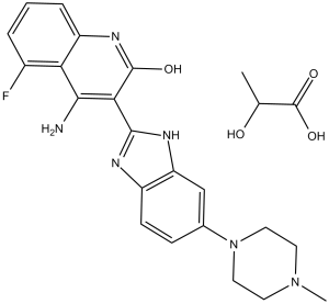
C24H27FN6O4
|
|
|---|---|---|
| 分子量 |
482.51
|
|
| 精确质量 |
500.218
|
|
| 元素分析 |
C, 59.74; H, 5.64; F, 3.94; N, 17.42; O, 13.26
|
|
| CAS号 |
915769-50-5
|
|
| 相关CAS号 |
Dovitinib lactate;692737-80-7;Dovitinib;405169-16-6;Dovitinib dilactic acid;852433-84-2
|
|
| PubChem CID |
135611162
|
|
| 外观&性状 |
white solid powder
|
|
| LogP |
2.516
|
|
| tPSA |
160.8
|
|
| 氢键供体(HBD)数目 |
6
|
|
| 氢键受体(HBA)数目 |
10
|
|
| 可旋转键数目(RBC) |
3
|
|
| 重原子数目 |
36
|
|
| 分子复杂度/Complexity |
737
|
|
| 定义原子立体中心数目 |
0
|
|
| SMILES |
O=C(C(C)O)O.O=C1C(C2NC3C(=CC=C(N4CCN(C)CC4)C=3)N=2)=C(N)C2C(=CC=CC=2F)N1.O
|
|
| InChi Key |
QDPVYZNVVQQULH-UHFFFAOYSA-N
|
|
| InChi Code |
InChI=1S/C21H21FN6O.C3H6O3.H2O/c1-27-7-9-28(10-8-27)12-5-6-14-16(11-12)25-20(24-14)18-19(23)17-13(22)3-2-4-15(17)26-21(18)29;1-2(4)3(5)6;/h2-6,11H,7-10H2,1H3,(H,24,25)(H3,23,26,29);2,4H,1H3,(H,5,6);1H2
|
|
| 化学名 |
4-amino-5-fluoro-3-[6-(4-methylpiperazin-1-yl)-1H-benzimidazol-2-yl]-1H-quinolin-2-one;2-hydroxypropanoic acid;hydrate
|
|
| 别名 |
|
|
| HS Tariff Code |
2934.99.9001
|
|
| 存储方式 |
Powder -20°C 3 years 4°C 2 years In solvent -80°C 6 months -20°C 1 month |
|
| 运输条件 |
Room temperature (This product is stable at ambient temperature for a few days during ordinary shipping and time spent in Customs)
|
| 溶解度 (体外实验) |
|
|||
|---|---|---|---|---|
| 溶解度 (体内实验) |
注意: 如下所列的是一些常用的体内动物实验溶解配方,主要用于溶解难溶或不溶于水的产品(水溶度<1 mg/mL)。 建议您先取少量样品进行尝试,如该配方可行,再根据实验需求增加样品量。
注射用配方
注射用配方1: DMSO : Tween 80: Saline = 10 : 5 : 85 (如: 100 μL DMSO → 50 μL Tween 80 → 850 μL Saline)(IP/IV/IM/SC等) *生理盐水/Saline的制备:将0.9g氯化钠/NaCl溶解在100 mL ddH ₂ O中,得到澄清溶液。 注射用配方 2: DMSO : PEG300 :Tween 80 : Saline = 10 : 40 : 5 : 45 (如: 100 μL DMSO → 400 μL PEG300 → 50 μL Tween 80 → 450 μL Saline) 注射用配方 3: DMSO : Corn oil = 10 : 90 (如: 100 μL DMSO → 900 μL Corn oil) 示例: 以注射用配方 3 (DMSO : Corn oil = 10 : 90) 为例说明, 如果要配制 1 mL 2.5 mg/mL的工作液, 您可以取 100 μL 25 mg/mL 澄清的 DMSO 储备液,加到 900 μL Corn oil/玉米油中, 混合均匀。 View More
注射用配方 4: DMSO : 20% SBE-β-CD in Saline = 10 : 90 [如:100 μL DMSO → 900 μL (20% SBE-β-CD in Saline)] 口服配方
口服配方 1: 悬浮于0.5% CMC Na (羧甲基纤维素钠) 口服配方 2: 悬浮于0.5% Carboxymethyl cellulose (羧甲基纤维素) 示例: 以口服配方 1 (悬浮于 0.5% CMC Na)为例说明, 如果要配制 100 mL 2.5 mg/mL 的工作液, 您可以先取0.5g CMC Na并将其溶解于100mL ddH2O中,得到0.5%CMC-Na澄清溶液;然后将250 mg待测化合物加到100 mL前述 0.5%CMC Na溶液中,得到悬浮液。 View More
口服配方 3: 溶解于 PEG400 (聚乙二醇400) 请根据您的实验动物和给药方式选择适当的溶解配方/方案: 1、请先配制澄清的储备液(如:用DMSO配置50 或 100 mg/mL母液(储备液)); 2、取适量母液,按从左到右的顺序依次添加助溶剂,澄清后再加入下一助溶剂。以 下列配方为例说明 (注意此配方只用于说明,并不一定代表此产品 的实际溶解配方): 10% DMSO → 40% PEG300 → 5% Tween-80 → 45% ddH2O (或 saline); 假设最终工作液的体积为 1 mL, 浓度为5 mg/mL: 取 100 μL 50 mg/mL 的澄清 DMSO 储备液加到 400 μL PEG300 中,混合均匀/澄清;向上述体系中加入50 μL Tween-80,混合均匀/澄清;然后继续加入450 μL ddH2O (或 saline)定容至 1 mL; 3、溶剂前显示的百分比是指该溶剂在最终溶液/工作液中的体积所占比例; 4、 如产品在配制过程中出现沉淀/析出,可通过加热(≤50℃)或超声的方式助溶; 5、为保证最佳实验结果,工作液请现配现用! 6、如不确定怎么将母液配置成体内动物实验的工作液,请查看说明书或联系我们; 7、 以上所有助溶剂都可在 Invivochem.cn网站购买。 |
| 制备储备液 | 1 mg | 5 mg | 10 mg | |
| 1 mM | 2.0725 mL | 10.3625 mL | 20.7250 mL | |
| 5 mM | 0.4145 mL | 2.0725 mL | 4.1450 mL | |
| 10 mM | 0.2072 mL | 1.0362 mL | 2.0725 mL |
1、根据实验需要选择合适的溶剂配制储备液 (母液):对于大多数产品,InvivoChem推荐用DMSO配置母液 (比如:5、10、20mM或者10、20、50 mg/mL浓度),个别水溶性高的产品可直接溶于水。产品在DMSO 、水或其他溶剂中的具体溶解度详见上”溶解度 (体外)”部分;
2、如果您找不到您想要的溶解度信息,或者很难将产品溶解在溶液中,请联系我们;
3、建议使用下列计算器进行相关计算(摩尔浓度计算器、稀释计算器、分子量计算器、重组计算器等);
4、母液配好之后,将其分装到常规用量,并储存在-20°C或-80°C,尽量减少反复冻融循环。
计算结果:
工作液浓度: mg/mL;
DMSO母液配制方法: mg 药物溶于 μL DMSO溶液(母液浓度 mg/mL)。如该浓度超过该批次药物DMSO溶解度,请首先与我们联系。
体内配方配制方法:取 μL DMSO母液,加入 μL PEG300,混匀澄清后加入μL Tween 80,混匀澄清后加入 μL ddH2O,混匀澄清。
(1) 请确保溶液澄清之后,再加入下一种溶剂 (助溶剂) 。可利用涡旋、超声或水浴加热等方法助溶;
(2) 一定要按顺序加入溶剂 (助溶剂) 。
Pharmacokinetic Drug-drug Interaction Study of Dovitinib (TKI258) in Patients With Advanced Solid Tumors
CTID: NCT01596647
Phase: Phase 1 Status: Completed
Date: 2020-12-21
CHIR-258 inhibits the viability of FGFR3-expressing B9 cells but not parental IL-6-stimulated cells. Blood. 2005 Apr 1;105(7):2941-8. |
CHIR-258 inhibits viability of KMS11 cells in the presence of IL-6, IGF-1, and BMSCs. Blood. 2005 Apr 1;105(7):2941-8. |
CHIR-258 inhibits FGFR3 phosphorylation and demonstrates antitumor effects in vivo. Blood. 2005 Apr 1;105(7):2941-8. |
InvivoChem的所有产品仅用于作科学研究,不面向患者销售
Copyright 2020 InvivoChem LLC | All Rights Reserved 粤ICP备20063088号-1


 COA
COA
 Lactate")
 Lactate")
 Lactate")
 463611831
