| 规格 | 价格 | 库存 | 数量 |
|---|---|---|---|
| 10 mM * 1 mL in DMSO |
|
||
| 5mg |
|
||
| 10mg |
|
||
| 50mg |
|
||
| 100mg |
|
||
| 250mg |
|
||
| 500mg |
|
||
| 1g |
|
||
| 2g |
|
||
| 5g |
|
||
| Other Sizes |
|

| 靶点 |
p160ROCK (Ki = 0.33 μM); ROCK2 (IC50 = 0.158 μM); PKA (IC50 = 4.58 μM); PKC (IC50 = 12.30 μM); PKG (IC50 = 1.65 μM)
Fasudil (HA-1077) HCl primarily targets Rho-associated coiled kinase (ROCK) isoforms ROCK1 and ROCK2 (ROCK1 IC50 = 300 nM; ROCK2 IC50 = 150 nM) [3][1] Fasudil (HA-1077) HCl shows weak to moderate inhibition of other kinases (PKC IC50 = 3.3 μM; MLCK IC50 = 5.0 μM; PKA IC50 > 10 μM) [3][1] |
|---|---|
| 体外研究 (In Vitro) |
在大鼠 HSC(肝星状细胞)和人 HSC 衍生的 TWNT-4 细胞中,盐酸Fasudil (100 μM) 通过阻止细胞扩散、应力纤维产生和 α-SMA 表达来抑制细胞发育[4]。在大鼠 HSC 和人 HSC 衍生的 TWNT-4 细胞中,Fasudil Hydrochronide(50-100 μM;24 小时)可抑制 LPA(溶血磷脂酸)引起的 ERK1/2、JNK 和 p38 磷酸化[4]。在人 HSC 衍生的 TWNT-4 细胞中,Fasudil盐酸盐(25–100 μM;24 小时)可促进 MMP-1 转录,同时抑制胶原蛋白和 TIMP 转录[4]。
背景/目的:Rho-ROCK信号通路在肝星状细胞(HSC)的激活中起着重要作用。我们研究了Rho激酶(ROCK)抑制剂Fasudil盐酸盐水合物(FasudilFasudil(100μM)抑制细胞扩散、应力纤维的形成和α-SMA的表达,同时抑制细胞生长,尽管它没有诱导细胞凋亡。Fasudil抑制ERK1/2、JNK和p38的磷酸化。用法舒地尔治疗抑制了胶原蛋白和TIMP的产生和转录,刺激了MMP-1的产生和翻译,并增强了胶原酶活性。 结论:这些发现表明,法舒地尔不仅抑制增殖和胶原蛋白的产生,而且增加胶原酶的活性[4]。 在大鼠肝星状细胞(HSCs)中,法舒地尔(Fasudil, HA-1077)盐酸盐(10 μM)处理72小时后,分别抑制I型和III型胶原蛋白生成62%和58%,蛋白水平下调纤维化标志物α-SMA表达70%。它还使胶原酶活性较对照组提高2.3倍 [4] - 在经历缺氧/复氧(H/R)损伤的新生大鼠心肌细胞中,法舒地尔(Fasudil, HA-1077)盐酸盐(5 μM)使凋亡率降低55%,膜联蛋白V阳性细胞比例从38%降至17%。它抑制JNK磷酸化(降低70%)并阻断凋亡诱导因子(AIF)从线粒体向细胞核转位 [6] - 在大鼠主动脉平滑肌细胞中,法舒地尔(Fasudil, HA-1077)盐酸盐(1 μM)抑制钙敏感性收缩80%,减少ROCK介导的肌球蛋白轻链(MLC)Ser19位点磷酸化75% [3] - 在小鼠小胶质细胞中,法舒地尔(Fasudil, HA-1077)盐酸盐(20 μM)抑制脂多糖(LPS)诱导的促炎细胞因子产生(TNF-α降低68%;IL-1β降低62%),并抑制小胶质细胞活化 [7] - 在人脑微血管内皮细胞(HBMECs)中,法舒地尔(Fasudil, HA-1077)盐酸盐(10 μM)降低通透性52%,减少紧密连接蛋白claudin-5的降解(裂解产物降低45%)[2] |
| 体内研究 (In Vivo) |
在手术前一小时静脉注射时,盐酸Fasudil (10 mg/kg) 已被证明可以预防心血管疾病,抑制 JNK 激活,并减少缺血期间在线粒体和细胞核之间易位的 AIF 量[5]。盐酸Fasudil (50 mg/kg/d; ip) 抑制蛋白脂质蛋白PLP p139-151,该蛋白可引起急性和复发性实验性自身免疫性脑脊髓炎(EAE)。它还可以减少淋巴细胞增殖,下调白细胞介素 (IL)-17,并显着降低 IFN-γ/IL-4 比率 [6]。 Fasudil 盐酸盐(100 mg/kg/d;口服)可抑制小鼠脊髓的炎症、脱髓鞘、轴突损失和 APP 阳性。它还显着降低了SJL/J小鼠实验性自身免疫性脑脊髓炎(EAE)的发生率和病理检查评分[6]。
目前针对中枢神经系统疾病的疗法只能减轻症状,无法延缓或预防疾病进展,迫切需要具有疾病调节活性的新方法。法舒地尔在动物模型和/或中枢神经系统疾病临床应用中的显著作用使其成为克服人类中枢神经系统障碍的有前景的策略。鉴于中枢神经系统疾病的复杂病理,有必要进一步努力开发多功能法舒地尔衍生物或与其他药物的联合策略,以便在对抗中枢神经系统障碍时发挥更强大的作用,同时将不良反应降到最低。[1] 血脑屏障(BBB)和血脊髓屏障(BSCB)功能障碍是多发性硬化症(MS)的主要特征。我们在豚鼠脊髓诱导的实验性自身免疫性脑脊髓炎(EAE)模型中评估了选择性ROCK抑制剂Fasudil的保护作用。此外,我们还研究了Fasudil对BBB和BSCB通透性的影响。我们发现法舒地尔通过降低BBB和BSCB的通透性,部分减轻了EAE依赖性损伤。这些结果为开发Rho激酶选择性抑制剂作为MS的新疗法提供了理论基础。 [2] 缺血再灌注导致Rho激酶、c-Jun NH2末端激酶(JNK)和凋亡诱导因子(AIF)活性显著增加。给予Rho激酶抑制剂Fasudil后,心肌梗死面积从59.89+/-3.83%减少到38.62+/-2.66%(P<0.05),细胞凋亡从32.78+/-5.1%减少到17.05+/-4.2%(P<0.05)。Western blot分析显示,给予法舒地尔可降低JNK的激活,并减轻AIF的线粒体核转位。此外,给予JNK抑制剂SP600125可以减轻AIF的线粒体核转位。 结论:Rho激酶的抑制通过抑制JNK介导的AIF易位来减少体内I/R中的细胞凋亡。[6] 我们研究了选择性Rho激酶抑制剂Fasudil在实验性自身免疫性脑脊髓炎(EAE)中的作用。胃肠外和口服法舒地尔均可预防SJL/J小鼠蛋白脂质蛋白(PLP)p139-151诱导的EAE的发展。淋巴细胞对PLP的特异性增殖显著降低,同时白细胞介素(IL)-17下调,IFN-γ/IL-4比值显著降低。免疫组织化学检查还显示炎症细胞浸润明显减少,脱髓鞘和急性轴突交易减弱。这些结果可能为口服法舒地尔选择性阻断Rho激酶作为多发性硬化症的新疗法提供了理论基础。 在实验性自身免疫性脑脊髓炎(EAE)大鼠模型中,腹腔注射 法舒地尔(Fasudil, HA-1077)盐酸盐(10 mg/kg/天,免疫后0-21天)降低临床评分(中位评分从3.5降至1.2),抑制脱髓鞘(病灶面积减少58%)。它还分别降低血脑屏障(BBB)和血脊髓屏障(BSCB)通透性48%和55% [2][7] - 在大鼠心肌缺血再灌注(I/R)模型中,静脉注射 法舒地尔(Fasudil, HA-1077)盐酸盐(10 mg/kg,再灌注前10分钟给药)减少心肌细胞凋亡60%,缩小梗死面积42%。它抑制JNK活化(磷酸化水平降低70%)和AIF核转位 [6] - 在自发性高血压大鼠(SHRs)中,口服 法舒地尔(Fasudil, HA-1077)盐酸盐(30 mg/kg/天,持续4周)降低收缩压25 mmHg,抑制血管平滑肌细胞肥大(细胞横截面积减少35%)[3] - 在四氯化碳(CCl₄)诱导的大鼠肝纤维化模型中,腹腔注射 法舒地尔(Fasudil, HA-1077)盐酸盐(5 mg/kg/天,持续8周)减少肝脏胶原蛋白含量55%,降低α-SMA阳性肝星状细胞数量60% [4] |
| 酶活实验 |
在最终体积为0.2 mL的反应混合物中,测定环AMP依赖性蛋白激酶的活性,该反应混合物中含有50 mM Tris-HCl (pH 7.0), 10 mM醋酸镁,2 mM EGTA, 1 μM环AMP或不含环AMP, 3.3至20 μM [r-32P] ATP (4×10⁵ c.p.m), 0.5 μg酶,100 μg组蛋白H2B和化合物。在30℃下孵育5 min,加入500 μg牛血清白蛋白作为载体蛋白,加入20%三氯乙酸1mL,终止反应。样品在3000转/分离心15min后,在10%三氯乙酸冰冷溶液中重悬,重复离心-重悬循环3次。最后的颗粒溶解在1ml的1n NaOH中,用液体闪烁计数器测量放射性。
ROCK1/ROCK2激酶活性实验:将纯化的重组大鼠ROCK1或ROCK2与MLC衍生底物肽和 法舒地尔(Fasudil, HA-1077)盐酸盐(0.01 μM-10 μM)在实验缓冲液(50 mM Tris-HCl,pH 7.4,10 mM MgCl₂,1 mM DTT,0.2 mM ATP)中于37°C孵育45分钟。比色法检测磷酸化底物,从剂量-效应曲线计算IC50值 [3][1] - ATP竞争性结合实验:将ROCK2与递增浓度的ATP(0.1-2 mM)和固定浓度的 法舒地尔(Fasudil, HA-1077)盐酸盐(150 nM)孵育。检测激酶活性以证实其与ROCK的ATP结合口袋竞争性结合 [3] - 激酶选择性实验:将 法舒地尔(Fasudil, HA-1077)盐酸盐(10 μM)在各自的底物肽和实验缓冲液中,对一组激酶(PKC、MLCK、PKA、ERK1/2)进行筛选。放射性标记ATP计数法定量激酶活性,测定各脱靶激酶的IC50值 [3][1] |
| 细胞实验 |
Western Blot 分析[4]
细胞类型:大鼠 HSC 和人 HSC 衍生的 TWNT-4 细胞 测试浓度: 50 μM; 100 μM 孵育时间: 24 小时 实验结果: 将 LPA 诱导的 ERK1/2、JNK 和 p38 MAPK 磷酸化抑制 60分别为 %、70% 和 90%。 RT-PCR[4] 细胞类型:大鼠 HSC 和人 HSC 衍生的 TWNT-4 细胞 测试浓度: 25微米; 50μM; 100 μM 24 小时 孵育时间:24 小时 实验结果:降低 I 型胶原蛋白、a-SMA、和TIMP-1。 细胞培养[4] 如前所述,通过依次用胶原酶原位灌注和用链霉蛋白酶消化,然后在双层(17%/11.5%)甲硫酰胺溶液中离心,从雄性Wistar大鼠的肝脏中分离出HSC。HSC在含有10%胎牛血清(FCS)的Dulbecco改良Eagle培养基(DMEM)中培养。本研究中描述的实验是在第二次和第四次连续传代之间的细胞上进行的。由于没有用于测量小鼠基质金属蛋白酶(MMP-1)和TIMP-1的商业试剂盒,我们使用来源于HSC的人细胞系TWNT-4细胞来评估法舒地尔对MMP-1和TIMP-1。如前所述,TWNT-4细胞在含有10%FCS的DMEM中培养。Fasudil由旭化成株式会社捐赠。将法舒地尔溶解在DMEM中并加入培养物中。在24小时的无血清条件下,HSC的细胞存活率超过90% h在100人面前 μM法舒地尔。 免疫细胞化学[4] 在无血清条件下,HSC和TWNT-4细胞在有或没有Fasudil(100μM)的情况下维持24小时 h.免疫细胞化学基本上按照之前的报道进行。用磷酸缓冲盐水(PBS)(137mM NaCl,2.7 mM氯化钾,8.1 mM Na2HPO4和1.5 mM KH2PO4,pH 7.4),细胞固定10 在37°C的3.7%甲醛中浸泡5分钟 在37°C下,在含有0.2%Triton X-100的PBS中浸泡min,用PBS洗涤三次,用含有10%FCS的PBS封闭30分钟 最低温度为37°C。然后将载玻片与抗α-SMA一抗或抗Myc一抗在37°C下孵育60分钟 min。载玻片在PBS中广泛冲洗,然后用罗丹明偶联的鬼笔环肽染色,与Alexa Fluor 488标记的山羊抗小鼠二抗混合。图像用LSM 510共聚焦激光扫描显微镜进行可视化。 BrdU掺入分析[4] 使用细胞增殖ELISA测量BrdU的HSC掺入。简而言之,亚融合造血干细胞在血清饥饿状态下24小时 h.然后用DMEM洗涤并孵育24小时 h在含有10%FCS的DMEM中加入BrdU,在存在或不存在Fasudil(100μM)或Y27632(30μM)(另一种特定的ROCK抑制剂)作为对照的情况下。用BrdU标记细胞后,消化细胞DNA,用过氧化物酶偶联的抗BrdU抗体孵育。通过在450℃下测量上清液的荧光强度来估计BrdU掺入 nm(激发)和690 nm(发射)。 细胞凋亡分析[4] 在无血清条件下,HSC在有或没有Fasudil(100μM)的情况下维持24小时 h.将细胞固定30分钟 在室温下在4%多聚甲醛/PBS中浸泡5分钟 在4°C下,在含有0.2%Triton X-100的PBS中放置至少一分钟。然后用Hoechst 33342对细胞进行染色,并根据制造商的说明使用原位细胞死亡检测试剂盒通过TUNEL法进行分析。样品用LSM 510共聚焦激光扫描显微镜进行可视化。针对每种情况,对来自三个独立实验和三种不同细胞制剂的至少100个细胞进行计数。 磷酸化和非磷酸化MAP激酶(MAPK)的蛋白质印迹分析[4] 蛋白质印迹分析基本上如前所述进行。造血干细胞饿死24天后 h、 用LPA(10μM)刺激它们45 min,然后用或不用100 μM法舒地尔2 h.在100℃下制备含有1×10⁷个TWNT-4细胞的全细胞裂解物 μl SDS-PAGE样品缓冲液。将蛋白质裂解物进行12%SDS-PAGE,转移到聚偏二氟乙烯膜上,并用第一抗体检测细胞外信号相关激酶(ERK)1/2 MAPK、磷酸化ERK1/2 MAPK(Thr202/Tyr204)、JNK、磷酸化JNK(Thr183/Tyr185)、p38 MAPK或磷酸化p38 MAPK(Thr180/Tyr182)。使用过氧化物酶连接的抗兔IgG作为第二抗体检测抗体结合。使用ECL plus绘制印迹,以显示抗体。使用光学扫描系统通过光密度法定量ERK1/2 MAPK、磷酸化ERK1/2 MAPK和JNK、磷酸化JNK、p38 MAPK和磷酸化p38 MAPK的水平。为了进行比较,根据光密度数据分别计算磷酸化ERK1/2、JNK和p38 MAPK与非磷酸化ERK3/2、JNK、p38 MAPK的比率。 使用实时RT-PCR分析基因表达[4] 用Trizol试剂从TWNT-4细胞中制备总RNA,在有或没有Fasudil(25、50或100 μM)在10%FCS中24小时 h.cDNA由1.0合成 μg RNA,使用随机六聚体进行GeneAmp™RNA PCR。根据制造商的说明,使用LightCycler FastStart DNA Master SYBR Green 1(罗氏,日本东京)进行实时PCR。反应混合物(20μl)含有LightCycler FastStart DNA Master SYBR Green 1,4 mM氯化镁,0.5 μM的上游和下游PCR引物,以及2个 μl的第一链cDNA作为模板。为了控制反应的变化,所有PCR都根据甘油醛-3-磷酸脱氢酶(GAPDH)的表达进行了标准化。使用的引物如下:人I型胶原α1链的5′-AGGTGAGACAGGCGAACAG-3′(正向引物)和5′-CTCTGAGTGGGCTGGGGCGGAC-3′(反向引物);人α-SMA的5′-AATGAGTGGGCCACTGCCGC-3′(正向引物)和5′-CAGAGATTTGCGCTCCGGA-3′(反向引物)(GenBank™登录号NM-000088);MMP-1的5′-GATACGGGACAACTCCT-3′(正向引物)和5′-TCCGGGTAGAGAGGATTTGTGTG-3′(反向引物)(GenBank™登录号NM002421);TIMP-1的5′-TTCTGAATTCCGACCTCGT-3′(正向引物)和5′-TCCTGCCACACAGAGT-3′(反向引物)(参考文献3;GenBank™登录号NM003254)。 肝星状细胞(HSC)纤维化实验:大鼠HSCs以2×10⁵个/孔接种到6孔板中,用TGF-β1(10 ng/mL)激活24小时。加入 法舒地尔(Fasudil, HA-1077)盐酸盐(1-50 μM),培养72小时。ELISA法检测胶原蛋白生成,Western blot检测α-SMA表达,酶谱法检测胶原酶活性 [4] - 心肌细胞凋亡实验:新生大鼠心肌细胞以5×10³个/孔接种到96孔板中,培养48小时。用 法舒地尔(Fasudil, HA-1077)盐酸盐(1-20 μM)预处理细胞1小时,再进行H/R(12小时缺氧/6小时复氧)处理。膜联蛋白V-FITC/PI染色检测凋亡,Western blot检测JNK磷酸化,免疫荧光检测AIF定位 [6] - 平滑肌细胞收缩实验:大鼠主动脉平滑肌细胞以1×10⁴个/孔接种到胶原蛋白凝胶中,用 法舒地尔(Fasudil, HA-1077)盐酸盐(0.1-5 μM)处理1小时。去氧肾上腺素(1 μM)诱导钙敏感性收缩,24小时后通过面积减少量量化凝胶收缩。Western blot检测MLC磷酸化 [3] - 小胶质细胞炎症实验:小鼠小胶质细胞以1×10⁶个/孔接种到6孔板中,用 法舒地尔(Fasudil, HA-1077)盐酸盐(5-40 μM)预处理1小时,再用LPS(1 μg/mL)刺激24小时。ELISA法检测TNF-α和IL-1β水平,Iba1免疫染色评估小胶质细胞活化 [7] - HBMEC通透性实验:HBMECs接种到Transwell小室中培养至融合。加入 法舒地尔(Fasudil, HA-1077)盐酸盐(1-30 μM),荧光素异硫氰酸酯(FITC)-葡聚糖渗透法检测通透性。Western blot检测claudin-5表达 [2] |
| 动物实验 |
动物/疾病模型:大鼠心肌缺血再灌注模型(250-300 g)[5]
剂量:10 mg/kg 给药途径:静脉注射;术前1小时 实验结果:激活Rho激酶和JNK,导致AIF易位至细胞核。抑制Rho激酶活性,减少心肌梗死面积和心肌细胞凋亡。 我们研究了选择性Rho激酶抑制剂法舒地尔在实验性自身免疫性脑脊髓炎(EAE)中的作用。法舒地尔的肠外和口服给药均可预防SJL/J小鼠中由蛋白脂蛋白(PLP)p139-151诱导的EAE的发生。淋巴细胞对PLP的特异性增殖显著降低,同时白细胞介素(IL)-17表达下调,IFN-γ/IL-4比值显著降低。免疫组织化学检查也显示炎症细胞浸润显著减少,脱髓鞘和急性轴突损伤减轻。这些结果可能为口服法舒地尔选择性阻断Rho激酶作为多发性硬化症的新疗法提供理论依据。[7] EAE大鼠模型:雌性Lewis大鼠用完全弗氏佐剂乳化的髓鞘碱性蛋白(MBP)免疫以诱导EAE。法舒地尔(HA-1077)盐酸盐溶于生理盐水,于免疫后第0至21天以10 mg/kg/天的剂量腹腔注射。对照组注射生理盐水。每日记录临床评分,并采用伊文思蓝外渗法测定血脑屏障/血脑屏障通透性[2][7] - 心肌缺血/再灌注大鼠模型:雄性Sprague-Dawley大鼠接受30分钟左前降支冠状动脉闭塞,随后进行24小时再灌注。将法舒地尔(HA-1077)盐酸盐(10 mg/kg)溶于生理盐水中,于再灌注前10分钟静脉注射。采用TTC染色法测定梗死面积,采用TUNEL法检测心肌细胞凋亡[6] - 自发性高血压大鼠模型:雄性自发性高血压大鼠口服法舒地尔(HA-1077)盐酸盐(30 mg/kg/天),溶于0.5%羧甲基纤维素钠溶液中,持续4周。对照组给予羧甲基纤维素钠溶液。每周采用尾套法测量收缩压,并通过组织形态计量学分析血管平滑肌细胞肥大[3] - 四氯化碳诱导的肝纤维化大鼠模型:雄性Wistar大鼠腹腔注射四氯化碳(1 mL/kg,1:1 v/v,溶于橄榄油),每周两次,持续8周。法舒地尔(HA-1077)盐酸盐(5 mg/kg/天)溶于生理盐水,腹腔注射,持续8周。收集肝组织进行Masson三色染色(胶原含量)和α-SMA免疫染色[4] - ADME大鼠/犬模型:雄性Sprague-Dawley大鼠和比格犬单次口服法舒地尔(HA-1077)盐酸盐(10 mg/kg)。在特定时间点采集血液、组织(脑、肝、肾、心)、尿液和粪便。采用液相色谱-串联质谱法(LC-MS/MS)测定药物浓度,以确定药代动力学参数[8] |
| 药代性质 (ADME/PK) |
法舒地尔在大鼠体内的药代动力学研究[8]
采用液相色谱-串联质谱法(LC-MS/MS)分析血浆样本中的法舒地尔和羟基法舒地尔。分别在口服(2、4 和 6 mg/kg)和静脉注射(2 mg/kg)法舒地尔后的所有时间点测定血浆样本中的法舒地尔和羟基法舒地尔,并将结果代入标准曲线以获得相应的浓度值。法舒地尔的平均血浆浓度-时间曲线如图 5 所示。使用 DAS 程序计算的法舒地尔和羟基法舒地尔的药代动力学参数列于表 4 和表 5。结果表明,大鼠体内法舒地尔的暴露量在 2-6 mg/kg 剂量范围内呈比例增加。在分别给予低、中、高三种浓度的法舒地尔三次给药后,女性受试者中法舒地尔的消除半衰期(t1/2)分别为1.19 ± 0.51、0.85 ± 0.35、1.09 ± 0.55 h,男性受试者中分别为2.29 ± 0.89、2.74 ± 1.57、2.34 ± 1.83 h。同时,女性受试者中羟基法舒地尔的消除半衰期(t1/2)分别为2.08 ± 0.68、1.84 ± 0.33、1.69 ± 0.41 h,男性受试者中分别为2.40 ± 0.16、2.32 ± 1.02、2.11 ± 0.52 h。结果显示,大鼠灌胃给药后,法舒地尔的药代动力学存在显著的性别差异。 大鼠组织分布[8] 采用LC-MS/MS法分析各组织样本中的法舒地尔和羟基法舒地尔,并将结果代入标准曲线,得到相应的药物浓度值。图6显示了大鼠口服4 mg/kg法舒地尔后0.25、1、3和6 h各组织中法舒地尔和羟基法舒地尔的平均浓度(ng/g)。除胃和小肠外,法舒地尔在所有组织中的浓度均很低;给药后0.5和1 h,胃和小肠中法舒地尔的浓度很高,但6 h后几乎完全消失。然而,羟基法舒地尔在所有组织中的浓度均显著高于法舒地尔。 大鼠体内排泄[8] 采用液相色谱-串联质谱法(LC-MS/MS)分析尿液、粪便和胆汁样本中的法舒地尔和羟基法舒地尔,并将结果代入标准曲线以获得相应的药物浓度值。绘制给药后尿液、粪便和胆汁的累积排泄曲线(图7)。表6显示了雄性和雌性大鼠体内药物排泄差异的统计分析结果。结果表明,给药后48小时内,雌性大鼠尿液中法舒地尔的累积排泄率为0.37%,雄性大鼠为1.08%;而雌性大鼠尿液中羟基法舒地尔的累积排泄率为2.42%,雄性大鼠为16.12%。给药后48小时内,法舒地尔在粪便中的累积排泄率在女性中为0.08%,在男性中为0.36%;羟基法舒地尔在女性中的累积排泄率为0.42%,在男性中为3.82%。结果显示,给药后24小时内,法舒地尔在胆汁中的累积排泄率在女性中为0.46%,在男性中为0.63%;羟基法舒地尔在女性中的累积排泄率为0.40%,在男性中为2.38%。 法舒地尔在犬体内的药代动力学研究[8] 在静脉注射(2 mg/kg)、口服(1、2 和 4 mg/kg)以及多次口服法舒地尔(2 mg/kg)后的所有时间点,测定了血浆样本中法舒地尔和羟基法舒地尔的浓度,并将结果代入标准曲线以获得相应的浓度值。同样,绘制了法舒地尔的平均血浆浓度-时间曲线,如图 8 和图 9 所示。使用 DAS 程序计算的法舒地尔和羟基法舒地尔的药代动力学参数列于表 7 和表 8 中。结果表明,在比格犬中,法舒地尔的暴露量在 1-4 mg/kg 剂量范围内呈比例增加。在分别给予低、中、高浓度三种剂量的法舒地尔后,女性法舒地尔的消除半衰期(t1/2)分别为 2.39 ± 0.95、4.58 ± 2.36、2.69 ± 1.45 小时,男性分别为 1.50 ± 0.64、3.00 ± 0.69、3.22 ± 1.02 小时;而女性羟基法舒地尔的消除半衰期(t1/2)分别为 4.53 ± 1.66、6.89 ± 2.11、8.78 ± 2.96 小时,男性分别为 4.38 ± 1.68、5.16 ± 1.49、6.39 ± 1.03 小时。在分别给予低、中、高浓度三种剂量的法舒地尔后,女性法舒地尔的AUC(0-t)分别为44.63 ± 24.11、123.88 ± 57.81、221.21 ± 108.98 ng/mLh,男性分别为30.32 ± 13.22、115.94 ± 60.18、531.68 ± 199.84 ng/mLh;羟基法舒地尔的AUC(0-t)分别为92.79 ± 30.97、233.58 ± 96.30、345.13 ± 115.31 ng/mLh,男性分别为67.26 ± 24.97、266.12 ± 153.35 ng/mLh。男性为 444.94 ± 190.21 ng/mLh。在分别给予低、中、高浓度三种不同剂量的法舒地尔后,女性的法舒地尔Cmax值分别为17.60 ± 10.31、63.45 ± 28.75、148.51 ± 161.40 ng/mL,男性分别为19.72 ± 11.63、56.84 ± 43.57、304.70 ± 97.36 ng/mL;羟基法舒地尔的Cmax值在女性中分别为18.90 ± 6.48、21.97 ± 6.70、26.68 ± 5.58 ng/mL,男性分别为11.43 ± 4.75、25.04 ± 14.13、34.54 ± 15.52 ng/mL。结果显示,法舒地尔灌胃给药后,犬体内的药代动力学无性别差异。 法舒地尔盐酸盐作为一种细胞内钙离子拮抗剂,具有扩张血管的作用,在治疗心绞痛方面展现出非常显著的药理作用。本研究旨在分别测定法舒地尔在大鼠和比格犬体内的吸收、分布和排泄情况,以阐明其药代动力学模式。我们开发并建立了一种灵敏可靠的液相色谱-串联质谱(LC-MS/MS)方法,并成功将其应用于包括吸收、组织分布和排泄在内的药代动力学研究。结果表明,在2-6 mg/kg剂量范围内,大鼠体内法舒地尔的药代动力学行为(例如AUC和Cmax)呈剂量依赖性。然而,血浆浓度结果表明,法舒地尔在大鼠体内的药代动力学存在显著的性别差异,包括绝对生物利用度和排泄。有趣的是,比格犬的数据表明,单次或重复给药后,盐酸法舒地尔的绝对生物利用度不存在性别差异。总之,本研究通过吸收、组织分布和排泄研究,阐明了法舒地尔在大鼠和比格犬体内的药代动力学特征。这些发现可能具有重要价值,并为进一步研究及其在临床实践中的安全应用提供理论依据。[8]法舒地尔是一种细胞内钙拮抗剂,它通过磷酸化肌球蛋白轻链阻断血管收缩过程,从而扩张血管并抑制血管痉挛(Somlyo & Somlyo, 2003; Fukushima et al., 2010),临床上用于治疗蛛网膜下腔出血(Fu et al., 2018; Kondoh, Mizusawa, Murakami, Nakamichi, & Nagata, 1997)。羟基法舒地尔是盐酸法舒地尔的活性代谢产物,在特异性实验中表现出更高的选择性(Nakamura et al., 2001; Shimokawa & Rashid, 2007)。本研究建立了一种灵敏可靠的液相色谱-串联质谱(LC-MS/MS)方法,用于测定大鼠和比格犬体内法舒地尔和羟基法舒地尔的浓度,并将其应用于给药后的吸收、组织分布和排泄研究,进一步阐明了法舒地尔在动物模型中的药代动力学特性。[8] 分别对大鼠进行静脉注射(4 mg/kg)和口服(2、4 和 6 mg/kg)给药后,在不同时间点测定血浆中法舒地尔和羟基法舒地尔的浓度。结果显示,大鼠体内法舒地尔的药代动力学存在显著的性别差异。此外,在 2~6 mg/kg 的剂量范围内,观察到法舒地尔的药代动力学行为呈剂量依赖性。静脉注射法舒地尔和羟基法舒地尔的半衰期(tl/2)分别为0.6 ± 0.3 h和1.8 ± 0.5 h,与文献报道基本一致(Zhang, Gao, Huang, & Xu, 2009)。口服法舒地尔的半衰期分别为2.3 ± 0.90 h、2.7 ± 1.6 h和2.3 ± 1.8 h,明显长于静脉注射,但羟基法舒地尔的半衰期保持不变。口服盐酸法舒地尔后,雌性大鼠的平均绝对生物利用度为35.8%,而雄性大鼠的平均绝对生物利用度仅为9.46%。结果表明,大鼠口服盐酸法舒地尔后,其绝对生物利用度存在性别差异。 [8] 大鼠口服4.0 mg/kg法舒地尔后,雄性大鼠各组织/器官中法舒地尔的浓度显著高于雌性大鼠,表明法舒地尔在雄性大鼠体内的分布比例更高。尤其值得注意的是,雄性大鼠肝脏中羟基法舒地尔的浓度显著高于雌性大鼠,而其他组织中羟基法舒地尔的浓度差异不大。除胃和小肠外,其他组织中法舒地尔的浓度极低,而各种组织中羟基法舒地尔的浓度则显著较高,表明羟基法舒地尔在大鼠体内分布广泛。[8] 大鼠口服4.0 mg/kg法舒地尔后,采用液相色谱-串联质谱法(LC-MS/MS)定量测定尿液、粪便和胆汁中的法舒地尔和羟基法舒地尔。累积排泄率顺序为:尿液 > 粪便 > 胆汁,表明口服法舒地尔和羟基法舒地尔后主要经尿液排泄。雌性大鼠尿液和粪便中法舒地尔的累积排泄率显著低于雄性大鼠,而雌性大鼠尿液和粪便中羟基法舒地尔的累积排泄率显著高于雄性大鼠,表明大鼠口服盐酸法舒地尔的吸收和排泄存在显著的性别差异。[8] 在比格犬中,分别静脉注射(2 mg/kg)、口服(1、2 和 4 mg/kg)以及多次口服(2 mg/kg)法舒地尔后,测定不同时间点血浆中法舒地尔和羟基法舒地尔的浓度。结果显示,口服法舒地尔(1、2 和 4 mg/kg)后,随着给药剂量的增加,Cmax、AUC(0-48h) 和 AUC(0-∞) 均显著升高,呈良好的比例关系。此外,比格犬体内羟基法舒地尔的 Cmax、AUC(0-48h) 和 AUC(0-∞) 也以类似的方式升高,表明口服法舒地尔后,比格犬体内法舒地尔和羟基法舒地尔的药代动力学过程符合一级动力学。采用非房室模型法计算,比格犬口服法舒地尔(1、2 和 4 mg/kg)后的 t1/2 分别为 1.9 ± 0.9 h、3.8 ± 1.8 h 和 3.0 ± 1.2 h。采用非房室模型法计算的羟基法舒地尔的半衰期分别为4.5 ± 1.6、6.0 ± 1.9和7.6 ± 2.4小时(Yamashita等,2007)。结果表明,法舒地尔在比格犬体内的消除速度快于羟基法舒地尔,且各剂量组间无显著差异(p > 0.05)(Tsounapi等,2012)。口服盐酸法舒地尔片后,雌性比格犬的平均绝对生物利用度为20.5%,雄性比格犬的平均绝对生物利用度为24.5%。结果表明,比格犬口服盐酸法舒地尔后,其绝对生物利用度无性别差异。在比格犬中进行的重复给药实验结果表明,即使间隔24小时,法舒地尔的血药浓度也无法稳定。虽然可以检测到羟基法舒地尔,但其浓度远低于最大浓度,因此建议在临床应用中缩短给药间隔。此外,口服法舒地尔的研究有助于开发新的临床适应症并提高患者的依从性(Zhang et al., 2013)。[8] 采用已建立的LC-MS/MS方法研究了法舒地尔在大鼠和犬体内的药代动力学、吸收、组织分布和排泄。结果表明,大鼠体内盐酸法舒地尔的绝对生物利用度存在性别差异。法舒地尔和羟基法舒地尔主要经尿液排泄,且盐酸法舒地尔的吸收和排泄也存在显著的性别差异。此外,在比格犬中,法舒地尔的清除速度比羟基法舒地尔更快,且各组之间无显著差异。这项在大鼠和犬中进行的研究可能为法舒地尔在临床实践中的安全使用提供支持性信息和理论依据。 吸收:法舒地尔(HA-1077)盐酸盐在大鼠中的口服生物利用度为45%,在犬中的口服生物利用度为38%。口服给药后,大鼠的血浆峰浓度(Cmax)在1.5小时达到,犬的血浆峰浓度在2小时达到[8]。 - 分布:大鼠的分布容积(Vd)为1.8 L/kg,犬的分布容积为2.2 L/kg。该药物分布于多种组织,给药后2小时,大鼠脑/血浆浓度比为0.3,犬脑/血浆浓度比为0.25[8] - 代谢:该药物主要在肝脏通过水解和氧化代谢,已鉴定出两种主要代谢物(M1:去乙酰化法舒地尔;M2:羟基化法舒地尔)[8] - 排泄:在大鼠中,68%的剂量经尿液排泄(32%为原药,36%为代谢物),25%经粪便排泄。在犬中,55%经尿液排泄,38%经粪便排泄。末端消除半衰期(t1/2)在大鼠中为2.8小时,在犬中为3.5小时[8] |
| 毒性/毒理 (Toxicokinetics/TK) |
大鼠口服 LD50 335 mg/kg 感觉器官和特殊感觉:眼睑下垂;行为:震颤;行为:惊厥或对癫痫阈值的影响 Yakuri to Chiryo。药理学与治疗学,20(增刊)
大鼠皮下注射LD50 123 mg/kg 感觉器官和特殊感觉:眼睑下垂;行为:震颤;行为:惊厥或对癫痫阈值的影响。药理学与治疗学,20(增刊) 大鼠静脉注射LD50 59900 ug/kg 感觉器官和特殊感觉:眼睑下垂;行为:惊厥或对癫痫阈值的影响;胃肠道:唾液腺结构或功能的变化。药理学与治疗学, 20(增刊 小鼠口服LD50 274 mg/kg 感觉器官和特殊感觉:眼睑下垂;行为:睡眠时间改变(包括翻正反射改变);行为:惊厥或对癫痫阈值的影响 Yakuri to Chiryo. Pharmacology and Therapeutics., 20(增刊 小鼠皮下注射LD50 124 mg/kg 感觉器官和特殊感觉:眼睑下垂;行为:睡眠时间改变(包括翻正反射改变);行为:惊厥或对癫痫阈值的影响 Yakuri to Chiryo. Pharmacology and治疗学,20(增刊) 体外研究表明,盐酸法舒地尔 (HA-1077)对正常细胞毒性较低(人骨髓内皮细胞 IC50 > 100 μM;新生儿心肌细胞 IC50 > 80 μM)[2][6] -体内研究表明,盐酸法舒地尔 (HA-1077)在测试剂量(5-30 mg/kg,口服/腹腔/静脉注射)下,未引起大鼠和犬的显著体重减轻(<5% vs. 基线)或明显的死亡[3][4][6][8] -与载体对照组相比,盐酸法舒地尔 (HA-1077)治疗组动物的肝功能(ALT、AST)或肾功能(肌酐、BUN)未见显著变化[4][6][8] -血浆蛋白结合率法舒地尔 (HA-1077) 盐酸盐在大鼠体内的血浆结合率为 82-85%,在犬体内为 86-88%(体外血浆结合试验)[8] - 大鼠静脉注射剂量 >50 mg/kg 时观察到轻度低血压,但这种低血压是短暂且可逆的[3] |
| 参考文献 |
|
| 其他信息 |
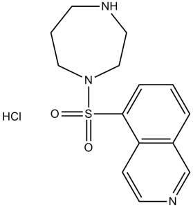
法舒地尔盐酸盐是由法舒地尔与一当量盐酸反应制得的盐酸盐。它具有抗高血压、钙通道阻滞、EC 2.7.11.1(非特异性丝氨酸/苏氨酸蛋白激酶)抑制剂、神经保护、促智和血管扩张等作用。它含有法舒地尔(1+)。
药物适应症 治疗非创伤性蛛网膜下腔出血。 法舒地尔是一种异喹啉类化合物,其5位被(1,4-二氮杂环庚烷-1-基)磺酰基取代。它是一种Rho激酶抑制剂,其盐酸盐水合物已获准用于治疗脑血管痉挛和脑缺血。它具有多种作用,包括抗衰老、抑制EC 2.7.11.1(非特异性丝氨酸/苏氨酸蛋白激酶)、血管扩张、促智、神经保护、降压和钙通道阻滞。它是一种N-磺酰二氮杂环庚烷类化合物,属于异喹啉类。它是法舒地尔(1+)的共轭碱。 法舒地尔已在颈动脉狭窄的治疗中得到研究。 引言:Rho激酶(ROCK)在肌动蛋白细胞骨架的组织中起着关键作用,并参与多种基本细胞功能,例如收缩和基因表达。法舒地尔是一种ROCK抑制剂,自1995年以来,已在日本用于治疗蛛网膜下腔出血(SAH)。越来越多的证据表明,法舒地尔可能对中枢神经系统(CNS)疾病,例如阿尔茨海默病,具有显著的治疗作用。本文总结了支持法舒地尔治疗多种中枢神经系统疾病潜在疗效的证据,并概述了其类似物的特性。专家观点:目前针对中枢神经系统疾病的疗法只能缓解症状,无法延缓或阻止疾病进展,因此迫切需要具有疾病改善作用的新疗法。法舒地尔在动物模型和/或临床应用中对中枢神经系统疾病的显著疗效使其成为治疗人类中枢神经系统疾病的一种有前景的策略。鉴于中枢神经系统疾病的病理机制复杂,需要进一步开发多功能法舒地尔衍生物或与其他药物联合使用,以期在对抗中枢神经系统疾病时发挥更强的疗效并最大限度地减少不良反应。 https://pubmed.ncbi.nlm.nih.gov/23461757/ 血脑屏障 (BBB) 和血脊髓屏障 (BSCB) 功能障碍是多发性硬化症 (MS) 的主要特征。我们评估了选择性 ROCK 抑制剂法舒地尔在豚鼠脊髓诱导的实验性自身免疫性脑脊髓炎 (EAE) 模型中的保护作用。此外,我们还研究了法舒地尔对 BBB 和 BSCB 通透性的影响。我们发现,法舒地尔通过降低 BBB 和 BSCB 通透性,部分缓解了 EAE 引起的损伤。这些结果为开发选择性 Rho 激酶抑制剂作为 MS 的新型疗法提供了理论依据。 https://pubmed.ncbi.nlm.nih.gov/21978848/ 背景/目的:Rho-ROCK信号通路在肝星状细胞(HSCs)的激活中发挥重要作用。我们研究了Rho激酶(ROCK)抑制剂法舒地尔盐酸盐水合物(法舒地尔)对HSCs细胞生长、胶原蛋白生成和胶原酶活性的影响。方法:培养大鼠HSCs和人HSC来源的TWNT-4细胞,用于研究应力纤维形成和α-平滑肌肌动蛋白(α-SMA)表达。采用BrdU掺入法检测细胞增殖,采用TUNEL法检测细胞凋亡。采用Western blot分析评估MAP激酶(MAPKs)、细胞外信号调节激酶1/2(ERK1/2)、c-Jun激酶(JNK)和p38的磷酸化状态。分别采用酶联免疫吸附试验(ELISA)和实时定量PCR检测I型胶原、基质金属蛋白酶-1(MMP-1)和基质金属蛋白酶组织抑制剂-1(TIMP-1)的生成和基因表达。同时检测胶原酶活性(活性MMP-1)。结果:法舒地尔(100 μM)抑制细胞铺展、应力纤维形成和α-SMA表达,并伴随细胞生长抑制,但未诱导细胞凋亡。法舒地尔抑制ERK1/2、JNK和p38的磷酸化。法舒地尔处理抑制胶原和TIMP的生成和转录,刺激MMP-1的生成和转录,并增强胶原酶活性。结论:这些结果表明,法舒地尔不仅抑制细胞增殖和胶原生成,还能增强胶原酶活性。 https://pubmed.ncbi.nlm.nih.gov/15998434/ 法舒地尔 (HA-1077) 盐酸盐 是一种选择性 Rho 相关卷曲激酶 (ROCK) 抑制剂,与其他一些激酶的交叉反应较弱 [1][3] - 其作用机制涉及与 ROCK1/ROCK2 的 ATP 结合口袋竞争性结合,从而抑制激酶活性并阻断下游信号传导(MLC 磷酸化、JNK 激活、细胞骨架重排)[3][6][8] - 法舒地尔 (HA-1077) 盐酸盐 在中枢神经系统疾病(EAE)、心血管疾病(高血压、心肌缺血/再灌注损伤)和肝纤维化模型中均显示出体外和体内疗效 [1][2][3][4][6][7] - 它已在一些国家获得临床批准,用于治疗该药物可用于治疗脑血管痉挛,并具有治疗神经炎症性疾病、纤维化和心血管疾病的潜在应用价值[1] - 该药物能够穿透血脑屏障和血脊髓屏障,支持其在中枢神经系统相关研究和治疗中的应用[2][7][8] |
| 分子式 |
C14H17N3O2S.HCL
|
|
|---|---|---|
| 分子量 |
327.83
|
|
| 精确质量 |
327.0808
|
|
| 元素分析 |
C, 51.29; H, 5.53; Cl, 10.81; N, 12.82; O, 9.76; S, 9.78
|
|
| CAS号 |
105628-07-7
|
|
| 相关CAS号 |
Fasudil;103745-39-7;Fasudil dihydrochloride; 203911-27-7; 105628-07-7 (HCl); 186694-02-0 (hydrochloride hydrate)
|
|
| PubChem CID |
163751
|
|
| 外观&性状 |
Typically exists as White to off-white solids at room temperature
|
|
| 沸点 |
506.2ºC at 760 mmHg
|
|
| 熔点 |
222 °C(dec.)
|
|
| 闪点 |
259.9ºC
|
|
| LogP |
4.17
|
|
| tPSA |
70.68
|
|
| 氢键供体(HBD)数目 |
2
|
|
| 氢键受体(HBA)数目 |
5
|
|
| 可旋转键数目(RBC) |
2
|
|
| 重原子数目 |
21
|
|
| 分子复杂度/Complexity |
421
|
|
| 定义原子立体中心数目 |
0
|
|
| SMILES |
Cl[H].S(C1=C([H])C([H])=C([H])C2C([H])=NC([H])=C([H])C1=2)(N1C([H])([H])C([H])([H])N([H])C([H])([H])C([H])([H])C1([H])[H])(=O)=O
|
|
| InChi Key |
LFVPBERIVUNMGV-UHFFFAOYSA-N
|
|
| InChi Code |
InChI=1S/C14H17N3O2S.ClH/c18-20(19,17-9-2-6-15-8-10-17)14-4-1-3-12-11-16-7-5-13(12)14;/h1,3-5,7,11,15H,2,6,8-10H2;1H
|
|
| 化学名 |
5-(1,4-diazepan-1-ylsulfonyl)isoquinoline;hydrochloride
|
|
| 别名 |
|
|
| HS Tariff Code |
2934.99.9001
|
|
| 存储方式 |
Powder -20°C 3 years 4°C 2 years In solvent -80°C 6 months -20°C 1 month 注意: 请将本产品存放在密封且受保护的环境中,避免吸湿/受潮。 |
|
| 运输条件 |
Room temperature (This product is stable at ambient temperature for a few days during ordinary shipping and time spent in Customs)
|
| 溶解度 (体外实验) |
|
|||
|---|---|---|---|---|
| 溶解度 (体内实验) |
配方 1 中的溶解度: ≥ 2.08 mg/mL (6.34 mM) (饱和度未知) in 10% DMSO + 40% PEG300 + 5% Tween80 + 45% Saline (这些助溶剂从左到右依次添加,逐一添加), 澄清溶液。
例如,若需制备1 mL的工作液,可将100 μL 20.8 mg/mL澄清DMSO储备液加入400 μL PEG300中,混匀;然后向上述溶液中加入50 μL Tween-80,混匀;加入450 μL生理盐水定容至1 mL。 *生理盐水的制备:将 0.9 g 氯化钠溶解在 100 mL ddH₂O中,得到澄清溶液。 配方 2 中的溶解度: ≥ 2.08 mg/mL (6.34 mM) (饱和度未知) in 10% DMSO + 90% (20% SBE-β-CD in Saline) (这些助溶剂从左到右依次添加,逐一添加), 澄清溶液。 例如,若需制备1 mL的工作液,可将 100 μL 20.8 mg/mL澄清DMSO储备液加入900 μL 20% SBE-β-CD生理盐水溶液中,混匀。 *20% SBE-β-CD 生理盐水溶液的制备(4°C,1 周):将 2 g SBE-β-CD 溶解于 10 mL 生理盐水中,得到澄清溶液。 View More
配方 3 中的溶解度: ≥ 2.08 mg/mL (6.34 mM) (饱和度未知) in 10% DMSO + 90% Corn Oil (这些助溶剂从左到右依次添加,逐一添加), 澄清溶液。 配方 4 中的溶解度: Saline: 30 mg/mL 配方 5 中的溶解度: 100 mg/mL (305.04 mM) in PBS (这些助溶剂从左到右依次添加,逐一添加), 澄清溶液; 超声助溶. 1、请先配制澄清的储备液(如:用DMSO配置50 或 100 mg/mL母液(储备液)); 2、取适量母液,按从左到右的顺序依次添加助溶剂,澄清后再加入下一助溶剂。以 下列配方为例说明 (注意此配方只用于说明,并不一定代表此产品 的实际溶解配方): 10% DMSO → 40% PEG300 → 5% Tween-80 → 45% ddH2O (或 saline); 假设最终工作液的体积为 1 mL, 浓度为5 mg/mL: 取 100 μL 50 mg/mL 的澄清 DMSO 储备液加到 400 μL PEG300 中,混合均匀/澄清;向上述体系中加入50 μL Tween-80,混合均匀/澄清;然后继续加入450 μL ddH2O (或 saline)定容至 1 mL; 3、溶剂前显示的百分比是指该溶剂在最终溶液/工作液中的体积所占比例; 4、 如产品在配制过程中出现沉淀/析出,可通过加热(≤50℃)或超声的方式助溶; 5、为保证最佳实验结果,工作液请现配现用! 6、如不确定怎么将母液配置成体内动物实验的工作液,请查看说明书或联系我们; 7、 以上所有助溶剂都可在 Invivochem.cn网站购买。 |
| 制备储备液 | 1 mg | 5 mg | 10 mg | |
| 1 mM | 3.0504 mL | 15.2518 mL | 30.5036 mL | |
| 5 mM | 0.6101 mL | 3.0504 mL | 6.1007 mL | |
| 10 mM | 0.3050 mL | 1.5252 mL | 3.0504 mL |
1、根据实验需要选择合适的溶剂配制储备液 (母液):对于大多数产品,InvivoChem推荐用DMSO配置母液 (比如:5、10、20mM或者10、20、50 mg/mL浓度),个别水溶性高的产品可直接溶于水。产品在DMSO 、水或其他溶剂中的具体溶解度详见上”溶解度 (体外)”部分;
2、如果您找不到您想要的溶解度信息,或者很难将产品溶解在溶液中,请联系我们;
3、建议使用下列计算器进行相关计算(摩尔浓度计算器、稀释计算器、分子量计算器、重组计算器等);
4、母液配好之后,将其分装到常规用量,并储存在-20°C或-80°C,尽量减少反复冻融循环。
计算结果:
工作液浓度: mg/mL;
DMSO母液配制方法: mg 药物溶于 μL DMSO溶液(母液浓度 mg/mL)。如该浓度超过该批次药物DMSO溶解度,请首先与我们联系。
体内配方配制方法:取 μL DMSO母液,加入 μL PEG300,混匀澄清后加入μL Tween 80,混匀澄清后加入 μL ddH2O,混匀澄清。
(1) 请确保溶液澄清之后,再加入下一种溶剂 (助溶剂) 。可利用涡旋、超声或水浴加热等方法助溶;
(2) 一定要按顺序加入溶剂 (助溶剂) 。
|
|---|
|
 HA-100 dihydrochloride
 ROCK-IN-7
 ROCK-IN-9
 ROCK-IN-6
InvivoChem的所有产品仅用于作科学研究,不面向患者销售
Copyright 2020 InvivoChem LLC | All Rights Reserved 粤ICP备20063088号-1


 COA
COA
 HCl")
 HCl")
 463611831
