| 规格 | 价格 | 库存 | 数量 |
|---|---|---|---|
| 10 mM * 1 mL in DMSO |
|
||
| 1mg |
|
||
| 5mg |
|
||
| 10mg |
|
||
| 25mg |
|
||
| 50mg |
|
||
| 100mg |
|
||
| 250mg |
|
||
| Other Sizes |
|

| 靶点 |
PF-03814735 is a potent and orally bioavailable small-molecule inhibitor of Aurora kinases, with high selectivity for Aurora A and Aurora B kinases. The IC50 values are as follows: Aurora A (1.8 nM), Aurora B (4.6 nM). It shows weak inhibitory activity against other kinases (e.g., IC50 >1000 nM for Abl, EGFR, and VEGFR2), confirming its specificity for the Aurora kinase family [1]
|
|---|---|
| 体外研究 (In Vitro) |
由于 PF-03814735 对 Aurora1 和 Aurora2 激酶的抑制作用,完整细胞中磷酸化 Aurora1、磷酸化组蛋白 H3 和磷酸化 Aurora2 的含量均较低。 PF-03814735 会抑制多倍体多核细胞的发育和细胞增殖,因为它会抑制胞质分裂 [1]。小细胞肺癌 (SCLC) 细胞系和结肠癌细胞系均显示出对 PF-03814735 的高敏感性。 PF-03814735 的有效性与视网膜母细胞瘤途径和 Myc 基因家族成员的状态之间存在显着相关性。
抗增殖活性:PF-03814735可抑制多种人类肿瘤细胞系的增殖,包括血液系统恶性肿瘤细胞系(K562、MV4-11、Raji)和实体瘤细胞系(HCT116、A549、MCF-7、PC-3),IC50值范围为9-68 nM。其中,对血液系统肿瘤细胞系的抑制活性更强(IC50:9-22 nM),对部分实体瘤细胞系的活性相对较低[1] - 抑制组蛋白H3磷酸化:Western blot分析显示,用PF-03814735(10-100 nM)处理HCT116细胞4小时,可浓度依赖性降低磷酸化组蛋白H3(p-Histone H3,Ser10,Aurora B激酶的特异性底物)水平,在50 nM浓度下可完全抑制p-Histone H3的表达[1] - 诱导细胞有丝分裂停滞与凋亡:流式细胞术分析表明,25 nM PF-03814735处理HCT116细胞24小时可导致G2/M期停滞(细胞比例从15%升至65%),处理48小时后通过Annexin V-FITC/PI染色检测到凋亡细胞比例达30%[1] - 基于基因组分析的预测生物标志物筛选:在54种人类肿瘤细胞系中,PF-03814735的响应(IC50 <30 nM vs. >30 nM)与特定基因表达特征相关。RNA测序和qPCR验证显示,有丝分裂进程相关基因(如CCNB1、CDK1、AURKA)高表达与细胞对PF-03814735的敏感性增加相关;反之,DNA修复基因(如BRCA1、RAD51)高表达与耐药性相关[2] - Aurora A表达与药物敏感性的相关性:免疫印迹和qPCR实验证实,内源性Aurora A mRNA和蛋白水平高的肿瘤细胞系(如MV4-11、K562)对PF-03814735的IC50值更低(9-12 nM),而Aurora A表达低的细胞系(如MCF-7)IC50值更高(45-68 nM)[2] |
| 体内研究 (In Vivo) |
当每天一次给患有人类异种移植肿瘤的小鼠口服 PF-03814735 时,它通过将磷酸化组蛋白 H3 的水平降低至可控制的水平,显着减缓了肿瘤的生长。在异种移植小鼠肿瘤模型中,多西紫杉醇和PF-03814735的组合显示出协同肿瘤生长抑制作用[1]。当以每周 80 mg/kg 的方案而不是每日 15 mg/kg 的方案给药时,PF-03814735 在 NCI-H82 异种移植物中更成功。 PF-03814735 引起的每周生长延迟 23.5 天相当于整个治疗过程中 0.9 个对数净细胞死亡 [2]。
皮下异种移植模型的抗肿瘤疗效:在携带K562(血液肿瘤)皮下异种移植瘤的裸鼠中,口服PF-03814735(25 mg/kg、50 mg/kg或100 mg/kg,每日一次,连续14天)可剂量依赖性抑制肿瘤生长。其中50 mg/kg和100 mg/kg剂量组的肿瘤生长抑制率(TGI)分别达78%和92%,但未观察到完全肿瘤消退;肿瘤组织分析显示,p-Histone H3水平较溶剂对照组降低60-80%[1] - 原位异种移植模型的抗肿瘤活性:在盲肠原位接种HCT116(结直肠癌)细胞的裸鼠模型中,口服PF-03814735(75 mg/kg,每日一次,连续21天)可使肿瘤体积减少65%,并降低肝脏转移结节数量(从对照组的12±3个/鼠降至3±1个/鼠)[1] - 药效与生物标志物的相关性:在携带MV4-11(Aurora A高表达)异种移植瘤的小鼠中,口服PF-03814735(50 mg/kg)在第14天的TGI达85%;相反,在携带MCF-7(Aurora A低表达)异种移植瘤的小鼠中,相同剂量仅实现32%的TGI,证实了体外Aurora A表达水平与体内药物敏感性的相关性[2] |
| 酶活实验 |
极光激酶活性测定(基于HTRF技术):将重组人源Aurora A(含激活突变T288E)或Aurora B(与INCENP肽形成复合物)与含ATP(10 μM)、生物素化组蛋白H3底物(500 nM)及系列浓度PF-03814735(0.1-100 nM)的反应缓冲液在30°C孵育60分钟。加入链霉亲和素偶联的铕穴状化合物和XL665标记的抗磷酸化组蛋白H3(Ser10)抗体终止反应,测定时间分辨荧光共振能量转移(HTRF)信号(665 nm和620 nm),通过665/620 nm信号比值计算PF-03814735对Aurora A和Aurora B的抑制率及IC50值[1]
- 激酶选择性测定:采用与极光激酶活性测定相同的HTRF方法,测试100 nM PF-03814735对64种非极光激酶(包括Abl、EGFR、VEGFR2、CDK2、PI3K)的抑制活性。通过比较药物组与溶剂组的HTRF信号计算各激酶的抑制率,仅3种激酶(CDK1、PLK1、JAK2)的抑制率>20%,证实该化合物对极光激酶的选择性[1] |
| 细胞实验 |
抗增殖实验(SRB法):将人类肿瘤细胞(K562、HCT116、MCF-7等)以2×10³-5×10³个/孔的密度接种于96孔板,培养24小时后加入系列稀释的PF-03814735(0.1-1000 nM),继续培养72小时。用10%三氯乙酸(TCA)在4°C固定细胞1小时,0.4%磺酰罗丹明B(SRB)(溶于1%乙酸)染色30分钟,1%乙酸洗去未结合染料,10 mM Tris碱溶解结合的SRB,测定510 nm处吸光度,通过非线性回归分析计算IC50值[1]
- G2/M期停滞与凋亡实验:将HCT116细胞以1×105个/孔接种于6孔板,用25 nM PF-03814735分别处理24小时(细胞周期分析)或48小时(凋亡分析)。细胞周期分析:收集细胞,-20°C下70%乙醇固定过夜,加入含RNase A的碘化丙啶(PI)染色,流式细胞术检测G0/G1、S、G2/M期细胞比例;凋亡分析:用Annexin V-FITC和PI染色,流式细胞术定量早期(Annexin V+/PI-)和晚期(Annexin V+/PI+)凋亡细胞百分比[1] - 生物标志物筛选的基因表达分析:采用标准酚-氯仿法从54种肿瘤细胞系中提取总RNA,逆转录为cDNA后,用基因特异性引物通过qPCR检测候选基因(AURKA、CCNB1、BRCA1等)的表达水平。RNA测序实验中,从总RNA构建文库,在Illumina平台测序,通过差异基因表达分析筛选与PF-03814735 IC50值相关的基因(Pearson相关系数>0.6或<-0.6)[2] - Aurora A蛋白表达实验(免疫印迹法):用含蛋白酶抑制剂的RIPA缓冲液提取肿瘤细胞总蛋白,取30 μg蛋白进行SDS-PAGE电泳,转印至PVDF膜,5%脱脂牛奶封闭1小时。膜与抗Aurora A一抗(1:1000稀释)在4°C孵育过夜,再与HRP偶联的二抗(1:5000稀释)室温孵育1小时,ECL化学发光显影,ImageJ软件定量条带强度,分析其与PF-03814735 IC50值的相关性[2] |
| 动物实验 |
溶于 Cremophor EL [Cremophor/乙醇/0.9% 生理盐水 (12.5%/12.5%/75%)];剂量分别为 10、20 和 30 mg/kg;灌胃法。将 HCT116 肿瘤皮下植入裸鼠右侧腹部。
皮下异种移植模型(抗肿瘤疗效):将 5×10⁶ 个 K562 细胞(悬浮于 50% Matrigel 中)皮下注射到 6-8 周龄雌性裸鼠的右侧腹部。当肿瘤体积达到 100-150 mm³ 时,将小鼠随机分为 4 组(每组 n=6):载体对照组(0.5% 甲基纤维素 + 0.1% Tween 80)、PF-03814735 25 mg/kg 组、50 mg/kg 组和 100 mg/kg 组。该化合物每日口服一次,连续给药14天。每2天使用游标卡尺测量肿瘤体积(V = L×W²/2,其中L为最长直径,W为最短直径),并记录体重以监测毒性。研究结束时,切除肿瘤,液氮速冻,用于p-组蛋白H3的Western blot分析[1] - 原位异种移植模型(结肠癌):雌性裸鼠用异氟烷麻醉,腹部做一个小切口以暴露盲肠。将1×10⁶个HCT116细胞(悬浮于20 μL PBS中)注射到盲肠壁内,然后用缝线缝合切口。细胞注射两周后(肿瘤形成后),将小鼠分为两组(每组 n=8):一组为载体对照组,另一组为 PF-03814735 75 mg/kg 口服组(每日一次,连续 21 天)。治疗结束后处死小鼠;称量盲肠肿瘤重量,并将肝组织固定于 4% 多聚甲醛溶液中,用于计数转移结节 [1]。 - 生物标志物验证异种移植模型:将 5×10⁶ 个 MV4-11(高 Aurora A 表达)或 MCF-7(低 Aurora A 表达)细胞皮下注射到裸鼠体内。当肿瘤体积达到 100 mm³ 时,给予小鼠 PF-03814735 50 mg/kg 口服治疗,每日一次,连续 14 天。每 2 天测量一次肿瘤体积,并计算肿瘤生长指数 (TGI)。切除肿瘤组织,通过免疫印迹法确认 Aurora A 的表达,从而验证 Aurora A 水平与药物反应之间的相关性 [2] |
| 药代性质 (ADME/PK) |
口服吸收:在CD-1小鼠中,口服PF-03814735(10 mg/kg)后,血浆峰浓度(Cmax)为85±12 ng/mL,曲线下面积(AUC0-24h)为320±45 ng·h/mL。通过比较AUC0-24h与静脉给药(2 mg/kg,AUC0-24h = 152±20 ng·h/mL)[1]计算得出,口服生物利用度(F)为42±5%。
- 分布:在Sprague-Dawley (SD)大鼠中,静脉注射PF-03814735(5 mg/kg)后,稳态分布容积(Vss)为3.8±0.6 L/kg,表明其组织分布广泛。小鼠组织分布研究表明,该化合物会在肿瘤中蓄积(口服给药后 4 小时,肿瘤/血浆浓度比为 3.2±0.4)[1] - 代谢:在人肝微粒体中,PF-03814735 的代谢半衰期 (t1/2) 为 3.6±0.5 小时。主要代谢酶为 CYP3A4(占代谢的 60%)和 CYP2D6(25%),这是通过与选择性 CYP 抑制剂(例如,CYP3A4 的酮康唑,CYP2D6 的奎尼丁)孵育测定的。主要代谢产物是 N-氧化产物,不具有 Aurora 激酶抑制活性 [1] - 排泄:在 SD 大鼠中,静脉注射 PF-03814735 (5 mg/kg) 后,48 小时内,18±3% 的剂量以原形药物形式从粪便中排出,3±1% 从尿液中排出,表明粪便排泄是主要途径 [1] - 大鼠药代动力学参数:静脉注射 (5 mg/kg):Cmax = 520±65 ng/mL,AUC0-24h = 480±70 ng·h/mL,消除半衰期 (t1/2) = 2.8±0.3 小时,清除率 (CL) = 8.5±1.2 mL/min/kg。口服(10 mg/kg):Cmax = 190±25 ng/mL,AUC0-24h = 840±95 ng·h/mL,t1/2 = 3.1±0.4 小时 [1] |
| 毒性/毒理 (Toxicokinetics/TK) |
小鼠急性毒性:单次口服剂量高达 200 mg/kg 的 PF-03814735 不会导致死亡或严重毒性(例如,惊厥、嗜睡)。小鼠重复口服给药(14 天)的最大耐受剂量 (MTD) 为 150 mg/kg/天,因为超过此剂量会导致体重减轻 10% 以上 [1]。肝肾毒性:SD 大鼠口服 PF-03814735(50 mg/kg/天,持续 28 天)后,血清丙氨酸氨基转移酶 (ALT)、天冬氨酸氨基转移酶 (AST)、血尿素氮 (BUN) 和肌酐 (Cr) 水平与溶剂对照组无显著差异。肝肾组织病理学分析显示无坏死或炎症迹象[1]
- 血液毒性:裸鼠连续14天接受PF-03814735(100 mg/kg/天)治疗后,外周血细胞计数(白细胞、血小板、红细胞)均在正常范围内,表明无明显的骨髓抑制[1] - 血浆蛋白结合率:采用平衡透析法测定人、小鼠和大鼠血浆中PF-03814735的血浆蛋白结合率分别为95±2%(人)、93±3%(小鼠)和94±2%(大鼠),表明其与血浆蛋白具有较高的结合率[1] - 药物相互作用:体外研究表明,PF-03814735不抑制人CYP酶(CYP1A2、2C9、2C19、2D6、 3A4)浓度高达 100 μM(所有化合物的 IC50 > 100 μM),表明与 CYP 代谢药物相互作用的风险较低 [1] |
| 参考文献 |
|
| 其他信息 |
PF-03814735 已用于实体瘤治疗研究的临床试验。
Aurora 激酶抑制剂 PF-03814735 是一种具有潜在抗肿瘤活性的 Aurora 激酶抑制剂。PF-03814735 可结合并抑制 Aurora 激酶,Aurora 激酶是一种丝氨酸/苏氨酸激酶,在有丝分裂过程中发挥着重要的有丝分裂检查点控制作用。抑制 Aurora 激酶可能导致过度表达 Aurora 激酶的肿瘤细胞的细胞分裂和增殖受到抑制。 作用机制:PF-03814735 通过抑制 Aurora A 和 Aurora B 激酶发挥抗肿瘤作用。Aurora A 调节有丝分裂过程中纺锤体极的形成;抑制 Aurora A 会导致纺锤体组装缺陷。Aurora B 对染色体排列和胞质分裂至关重要;抑制作用导致染色体分离异常和 G2/M 期阻滞,最终导致细胞凋亡 [1] - 口服制剂的原理:PF-03814735 的设计具有较高的口服生物利用度(小鼠为 42%,大鼠约为 40%),以便癌症患者能够方便地进行门诊给药,避免许多抗癌药物需要静脉输注 [1] - 临床应用预测性生物标志物:对 54 种肿瘤细胞系的基因组分析发现,Aurora A mRNA/蛋白质表达和有丝分裂基因特征(CCNB1、CDK1)是 PF-03814735 疗效的潜在预测性生物标志物。这些生物标志物可用于临床试验,以筛选可能从药物中获益的患者,从而提高治疗效果并减少不必要的药物暴露[2] - 血液肿瘤的临床前研究:PF-03814735 对血液肿瘤细胞系表现出更高的效力(IC50:9-22 nM),并且在 K562/MV4-11 异种移植模型中具有更高的体内疗效(TGI:85-92%),与某些实体瘤相比,这表明它可能对治疗血液恶性肿瘤(例如白血病、淋巴瘤)特别有效[1,2] |
| 分子式 |
C23H25F3N6O2
|
|
|---|---|---|
| 分子量 |
474.48
|
|
| 精确质量 |
474.199
|
|
| CAS号 |
942487-16-3
|
|
| 相关CAS号 |
|
|
| PubChem CID |
51346455
|
|
| 外观&性状 |
White to off-white solid powder
|
|
| 密度 |
1.4±0.1 g/cm3
|
|
| 折射率 |
1.642
|
|
| LogP |
2.11
|
|
| tPSA |
99.25
|
|
| 氢键供体(HBD)数目 |
3
|
|
| 氢键受体(HBA)数目 |
9
|
|
| 可旋转键数目(RBC) |
6
|
|
| 重原子数目 |
34
|
|
| 分子复杂度/Complexity |
778
|
|
| 定义原子立体中心数目 |
2
|
|
| SMILES |
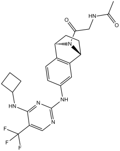
CC(=O)NCC(=O)N1[C@H]2CC[C@@H]1C3=C2C=CC(=C3)NC4=NC=C(C(=N4)NC5CCC5)C(F)(F)F
|
|
| InChi Key |
RYYNGWLOYLRZLK-RBUKOAKNSA-N
|
|
| InChi Code |
InChI=1S/C23H25F3N6O2/c1-12(33)27-11-20(34)32-18-7-8-19(32)16-9-14(5-6-15(16)18)30-22-28-10-17(23(24,25)26)21(31-22)29-13-3-2-4-13/h5-6,9-10,13,18-19H,2-4,7-8,11H2,1H3,(H,27,33)(H2,28,29,30,31)/t18-,19+/m0/s1
|
|
| 化学名 |
N-(2-((1S,4R)-6-((4-(cyclobutylamino)-5-(trifluoromethyl)pyrimidin-2-yl)amino)-1,2,3,4-tetrahydro-1,4-epiminonaphthalen-9-yl)-2-oxoethyl)acetamide
|
|
| 别名 |
PF-03814735; PF 03814735; PF03814735
|
|
| HS Tariff Code |
2934.99.9001
|
|
| 存储方式 |
Powder -20°C 3 years 4°C 2 years In solvent -80°C 6 months -20°C 1 month |
|
| 运输条件 |
Room temperature (This product is stable at ambient temperature for a few days during ordinary shipping and time spent in Customs)
|
| 溶解度 (体外实验) |
|
|||
|---|---|---|---|---|
| 溶解度 (体内实验) |
配方 1 中的溶解度: ≥ 2.5 mg/mL (5.27 mM) (饱和度未知) in 10% DMSO + 40% PEG300 + 5% Tween80 + 45% Saline (这些助溶剂从左到右依次添加,逐一添加), 澄清溶液。
例如,若需制备1 mL的工作液,可将100 μL 25.0 mg/mL澄清DMSO储备液加入到400 μL PEG300中,混匀;然后向上述溶液中加入50 μL Tween-80,混匀;加入450 μL生理盐水定容至1 mL。 *生理盐水的制备:将 0.9 g 氯化钠溶解在 100 mL ddH₂O中,得到澄清溶液。 配方 2 中的溶解度: ≥ 2.5 mg/mL (5.27 mM) (饱和度未知) in 10% DMSO + 90% (20% SBE-β-CD in Saline) (这些助溶剂从左到右依次添加,逐一添加), 澄清溶液。 例如,若需制备1 mL的工作液,可将 100 μL 25.0 mg/mL澄清DMSO储备液加入900 μL 20% SBE-β-CD生理盐水溶液中,混匀。 *20% SBE-β-CD 生理盐水溶液的制备(4°C,1 周):将 2 g SBE-β-CD 溶解于 10 mL 生理盐水中,得到澄清溶液。 View More
配方 3 中的溶解度: ≥ 2.5 mg/mL (5.27 mM) (饱和度未知) in 10% DMSO + 90% Corn Oil (这些助溶剂从左到右依次添加,逐一添加), 澄清溶液。 配方 4 中的溶解度: 2% Cremophor EL, 2% N,N-dimethylacetamide, pH 5.0: ~30mg/mL 1、请先配制澄清的储备液(如:用DMSO配置50 或 100 mg/mL母液(储备液)); 2、取适量母液,按从左到右的顺序依次添加助溶剂,澄清后再加入下一助溶剂。以 下列配方为例说明 (注意此配方只用于说明,并不一定代表此产品 的实际溶解配方): 10% DMSO → 40% PEG300 → 5% Tween-80 → 45% ddH2O (或 saline); 假设最终工作液的体积为 1 mL, 浓度为5 mg/mL: 取 100 μL 50 mg/mL 的澄清 DMSO 储备液加到 400 μL PEG300 中,混合均匀/澄清;向上述体系中加入50 μL Tween-80,混合均匀/澄清;然后继续加入450 μL ddH2O (或 saline)定容至 1 mL; 3、溶剂前显示的百分比是指该溶剂在最终溶液/工作液中的体积所占比例; 4、 如产品在配制过程中出现沉淀/析出,可通过加热(≤50℃)或超声的方式助溶; 5、为保证最佳实验结果,工作液请现配现用! 6、如不确定怎么将母液配置成体内动物实验的工作液,请查看说明书或联系我们; 7、 以上所有助溶剂都可在 Invivochem.cn网站购买。 |
| 制备储备液 | 1 mg | 5 mg | 10 mg | |
| 1 mM | 2.1076 mL | 10.5379 mL | 21.0757 mL | |
| 5 mM | 0.4215 mL | 2.1076 mL | 4.2151 mL | |
| 10 mM | 0.2108 mL | 1.0538 mL | 2.1076 mL |
1、根据实验需要选择合适的溶剂配制储备液 (母液):对于大多数产品,InvivoChem推荐用DMSO配置母液 (比如:5、10、20mM或者10、20、50 mg/mL浓度),个别水溶性高的产品可直接溶于水。产品在DMSO 、水或其他溶剂中的具体溶解度详见上”溶解度 (体外)”部分;
2、如果您找不到您想要的溶解度信息,或者很难将产品溶解在溶液中,请联系我们;
3、建议使用下列计算器进行相关计算(摩尔浓度计算器、稀释计算器、分子量计算器、重组计算器等);
4、母液配好之后,将其分装到常规用量,并储存在-20°C或-80°C,尽量减少反复冻融循环。
计算结果:
工作液浓度: mg/mL;
DMSO母液配制方法: mg 药物溶于 μL DMSO溶液(母液浓度 mg/mL)。如该浓度超过该批次药物DMSO溶解度,请首先与我们联系。
体内配方配制方法:取 μL DMSO母液,加入 μL PEG300,混匀澄清后加入μL Tween 80,混匀澄清后加入 μL ddH2O,混匀澄清。
(1) 请确保溶液澄清之后,再加入下一种溶剂 (助溶剂) 。可利用涡旋、超声或水浴加热等方法助溶;
(2) 一定要按顺序加入溶剂 (助溶剂) 。
| NCT Number | Recruitment | interventions | Conditions | Sponsor/Collaborators | Start Date | Phases |
| NCT00424632 | Completed Has Results | Drug: PF-03814735 | Solid Tumors | Pfizer | November 2006 | Phase 1 |
 SNS-314 Mesylate
 BI-847325
 MK-8745
 MLN8054
InvivoChem的所有产品仅用于作科学研究,不面向患者销售
Copyright 2020 InvivoChem LLC | All Rights Reserved 粤ICP备20063088号-1


 COA
COA
 463611831
