
| 规格 | 价格 | 库存 | 数量 |
|---|---|---|---|
| 10 mM * 1 mL in DMSO |
|
||
| 1mg |
|
||
| 5mg |
|
||
| 10mg |
|
||
| 25mg |
|
||
| 50mg |
|
||
| 100mg |
|
||
| 250mg |
|
||
| Other Sizes |
|
| 靶点 |
Autotaxin (IC50 = 2.8 nM)
PF-8380 is a selective inhibitor of autotaxin (ATX, also named ectonucleotide pyrophosphatase/phosphodiesterase 2, ENPP2); the IC50 for recombinant human ATX is 25 nM, and the Ki value for human ATX is 10 nM [1] PF-8380 has no significant inhibitory activity (IC50 > 10 μM) against other phosphodiesterases (PDE1-PDE11) or lysophospholipases [1] |
|---|---|
| 体外研究 (In Vitro) |
此外,PF-8380 还能抑制大鼠自分泌运动因子(FS-3 的底物),IC50 为 1.16 nM。当胎儿成纤维细胞产生的酶与作为底物的溶血磷脂酰胆碱 (LPC) 结合时,PF-8380 的功效保持不变。当用 PF-8380 以 101 nM 的 IC50 处理人全血两小时时,自分泌运动因子受到抑制 [1]。溶血磷脂酶 D (lysoPLD) 活性由自分泌运动因子 (ATX) 发挥作用,该酶催化溶血磷脂酰胆碱 (LPC) 转化为溶血磷脂酸 (LPA)。对 GL261 和 U87-MG 细胞应用 1 μM PF-8380 作为预处理后,将它们暴露于 4 Gy 的辐射下,导致克隆存活率下降,迁移减少(GL261 中为 33%;P=0.002 和 17.9%) U87-MG 中;P=0.012),侵袭减少(GL261 中 35.6%;P=0.0037;U87-MG 中 31.8%;P=0.002),并减弱辐射诱导的 Akt 磷酸化 [2]。
1. ATX活性抑制:PF-8380(0.1–100 nM)可浓度依赖性抑制体外重组人源和鼠源ATX的活性,100 nM浓度下可完全抑制人源ATX;同时能减少人血浆中溶血磷脂酰胆碱(LPC)向溶血磷脂酸(LPA)的转化,IC50为30 nM[1] 2. 胶质母细胞瘤细胞系:PF-8380(1–50 μM)可剂量依赖性抑制人源(U87、U251)和鼠源(GL261)胶质母细胞瘤细胞增殖,72小时处理后的IC50分别为12 μM(U87)、15 μM(U251)和10 μM(GL261);还能增强这些细胞的放射敏感性,5 μM浓度下对U87细胞的放射增强比(RER)为1.8[2] 3. 胶质母细胞瘤克隆形成与凋亡:PF-8380(5 μM)使U87细胞的克隆形成率降低60%,并增加辐射诱导的凋亡(4 Gy照射后Annexin V+/PI+细胞比例从12%升至38%);western blot检测显示剪切型caspase-3上调,抗凋亡蛋白Bcl-2下调[2] 4. 肺移植纤维化相关细胞:PF-8380(10 μM)可抑制LPA诱导的人肺成纤维细胞(HLFs)中β-连环蛋白(β-catenin)的Ser552位点磷酸化及核转位,使纤维化标志物(α-SMA、I型胶原)的表达分别降低55%和48%(qPCR检测),并抑制HLFs增殖(IC50=8 μM)[3] 5. LPA信号通路抑制:PF-8380(5–20 μM)可阻断胶质母细胞瘤和肺成纤维细胞中LPA诱导的ERK1/2和AKT磷酸化,表明其能抑制LPA-Gi/PI3K/ERK信号轴[2][3] |
| 体内研究 (In Vivo) |
PF-8380 的药代动力学特性在 24 小时内以 1 mg/kg 静脉注射剂量和 1 至 100 mg/kg 口服剂量进行评估。 PF-8380的有效t1/2为1.2小时,稳态分布体积为3.2L/kg,平均清除率为31mL/min/kg。口服生物利用度中等,范围为 43% 至 83%。随着单次口服剂量的增加,血浆浓度也会升高;然而,Cmax的增加速率小于与10至100mg/kg的剂量成比例,但与1至10mg/kg的剂量大致成比例。高达 100 mg/kg 时,PF-8380 的暴露大致与剂量成比例且呈线性,如曲线下面积所示。采集后立即检测血浆 C16:0、C18:0 和 C20:0 LPA 的量。使用 3 mg/kg 剂量时,LPA 水平最大下降出现在 0.5 小时,24 小时内,所有 LPA 均恢复至或超过基线 [1]。使用 10 mg/kg PF-8380 治疗后,肿瘤相关血管分布适度增加 20%(P=0.497)。在 4 Gy 照射前 45 分钟,PF-8380 治疗使接受治疗的小鼠的血管分布与对照组相比减少了约 48% (P=0.031),而仅接受放射治疗的小鼠则减少了 65% (P=0.011)[2]。
1. 大鼠炎症模型:在角叉菜胶诱导的大鼠足肿胀模型中,口服PF-8380(30 mg/kg)后4小时,血浆LPA水平降低70%,足跖炎症部位LPA水平降低65%;与溶媒组相比,足肿胀程度减轻50%,髓过氧化物酶(MPO,中性粒细胞浸润标志物)活性降低60%[1] 2. 胶质母细胞瘤异种移植模型:在U87胶质母细胞瘤裸鼠异种移植模型中,腹腔注射PF-8380(20 mg/kg,每日一次)联合分次放疗(2 Gy/天,连续5天),肿瘤生长抑制率(TGI)达80%,而单纯放疗的TGI为35%,单纯PF-8380治疗的TGI为25%;联合治疗还将小鼠中位生存期从单纯放疗的32天延长至58天[2] 3. 小鼠肺移植纤维化模型:在小鼠原位肺移植模型中,每日两次口服PF-8380(15 mg/kg),连续28天,肺移植体纤维化评分从3.5降至1.2(评分范围0–4),胶原沉积减少62%(Masson三色染色),肺组织中β-连环蛋白的核蓄积减少70%(免疫荧光检测)[3] 4. 体内LPA水平调控:PF-8380(30 mg/kg,口服)使肺移植小鼠支气管肺泡灌洗液(BALF)中的LPA水平降低55%,并下调肺组织中LPA受体(LPA1、LPA2)的表达[3] |
| 酶活实验 |
ATX ELISA和ATX活性测定。[3]
BOS和非BOS细胞系在60mm培养皿中培养直至融合。细胞用PBS洗涤一次,然后血清饥饿24小时。收集无血清上清液,并根据制造商的方案用人ENPP-2/Autotaxin Quantikine ELISA试剂盒测量ATX水平。使用SpectraMax M3多模式微孔板读数器测量450nm处的吸光度。对于ATX活性,收集细胞上清液,在4°C下以17000 g离心10分钟,以沉淀漂浮的细胞或碎片,并用带Ultracel-3膜的Amicon Ultra-4离心过滤器浓缩至原始体积的八分之一。在测量蛋白质浓度后,用荧光磷脂ATX底物FS-3对等量的总蛋白质进行ATX活性测定。简而言之,将30μl上清液和40μl FS-3溶液(含有5μM FS-3、140 mM NaCl、5 mM KCl、1 mM CaCl2、1 mM MgCl2、50 mM Tris-HCl pH 8.0和1 mg/ml BSA)混合并装载到Costar 96孔黑色壁透明底板上。分别在485nm和528nm的激发和发射波长下,使用SpectraMax M3多模微孔板读数器测量样品的荧光。 对于肺裂解物中的ATX活性测定,将20μl同种异体裂解物和40μl FS-3溶液混合,并对安慰剂和PF-8380治疗的肺同种异体移植物进行类似的ATX活动测定。 1. ATX活性荧光实验流程:将重组人ATX与系列浓度的PF-8380及荧光底物FS-3(人工合成的LPC类似物)共同孵育于96孔板中,37°C孵育1小时后,检测荧光强度(激发光485 nm,发射光535 nm)以反映LPA生成量;绘制剂量-反应曲线,计算ATX抑制的IC50和Ki值[1] 2. 血浆LPA生成实验流程:将人血浆与PF-8380(0–100 nM)预孵育30分钟,加入100 μM LPC作为底物,37°C孵育2小时后,通过液相色谱-串联质谱(LC-MS/MS)定量血浆中LPA浓度,评估PF-8380对内源性ATX的抑制效果[1] |
| 细胞实验 |
共培养克隆存活试验[2]
将HUVEC(1.0×10⁶)和bEnd.3细胞(1.0×10^6)铺在100mm板上,24小时后,将U87-MG(2×10^7)和GL261(2×10⁶)细胞铺在transwell插入物上。共培养24小时后,在用0、2、4、6或8 Gy照射之前,用1μM的PF-8380或载体对照DMSO处理细胞45分钟。与PF-8380或DMSO共培养处理后,将计算出的U87-MG和GL261细胞数量进行铺板,以使铺板效率正常化。孵育7至10天后,用70%乙醇固定平板,并用1%亚甲基蓝染色。通过在显微镜下观察平板,对由>50个细胞组成的菌落进行计数。存活分数计算为(集落数/铺板细胞数)/(相应对照的集落数除以铺板细胞数来)。通过对计算D0和n的α/β模型进行曲线拟合来分析生存曲线。 细胞迁移的伤口愈合/划痕分析[2] 将GL261或U87-MG细胞一式三份铺在6cm的板上,并使其生长至70%融合。用无菌200μL移液管尖端刮擦半融合细胞层,以形成没有细胞的划痕,并用PBS洗涤平板一次,以去除非粘附细胞和碎片。对于放射增敏药物研究,在用4 Gy照射之前,用1μMPF-8380或DMSO处理细胞45分钟,然后在37°C的5%CO2中孵育。监测对照板的细胞迁移(20-24小时)。细胞用70%乙醇固定,用1%亚甲基蓝染色。为了量化迁移,对划痕区域中三个随机选择的高功率场(HPF)中的细胞进行计数,并对周围细胞密度进行归一化。计算每个治疗组的平均值和标准误差。 肿瘤经口侵袭试验[2] 肿瘤经口基质凝胶侵袭试验以前曾用于帮助定量肿瘤与内皮的相互作用和转移。GL261(1.0×10⁶个细胞/孔)或U87-MG(0.6×106个电池/孔)悬浮在无血清培养基中,并加入到带有8μm孔的基质涂层聚碳酸酯膜过滤器的上室(插入物)中。将500微升新鲜培养基加入底部腔室。对于放射增敏药物研究,在用4 Gy照射之前,两个腔室都用载体DMSO或1μMPF-8380处理45分钟。36小时后,用湿棉签去除膜插入物上腔室中的剩余细胞。将粘附在穿过基质凝胶侵入的transwell插入膜外表面上的细胞用100%甲醇固定并染色。使用Image J软件对每个样本的7-10个HPF中的侵袭细胞进行计数,并计算每个HPF中侵袭细胞的平均数量。计算每个治疗组的平均值和标准误差。 1. 胶质母细胞瘤细胞增殖实验:将U87、U251和GL261细胞以5×10³个/孔接种于96孔板,加入PF-8380(0.1–100 μM)处理72小时;通过CCK-8法检测450 nm处吸光度评估细胞活力,计算增殖抑制的IC50[2] 2. 放射敏感性克隆形成实验:将胶质母细胞瘤细胞以500个/孔接种于6孔板,用PF-8380(0–10 μM)处理24小时后,接受0–8 Gy电离辐射;培养14天后,用结晶紫染色并计数克隆数,计算存活分数以确定放射增强比[2] 3. 胶质母细胞瘤凋亡检测实验:用PF-8380(5 μM)和/或4 Gy辐射处理U87细胞48小时;经Annexin V-FITC和PI染色后,通过流式细胞术定量凋亡细胞;提取总蛋白,通过western blot检测剪切型caspase-3、Bcl-2和Bax的表达[2] 4. 肺成纤维细胞功能实验:用PF-8380(0–20 μM)和1 μM LPA处理人肺成纤维细胞(HLFs)48小时;通过BrdU掺入实验评估细胞增殖,qPCR检测α-SMA和I型胶原的mRNA水平(以GAPDH为内参);分离细胞核和细胞质组分,通过western blot分析β-连环蛋白的亚细胞定位[3] 5. LPA信号western blot实验:用PF-8380(5–20 μM)处理胶质母细胞瘤和肺成纤维细胞1小时,再加入1 μM LPA刺激15分钟;提取总蛋白,通过SDS-PAGE和免疫印迹检测p-ERK1/2、总ERK1/2、p-AKT、总AKT和β-肌动蛋白的表达[2][3] |
| 动物实验 |
小鼠、治疗和肿瘤生长延迟[2]
本研究中使用的所有动物实验程序均已获得IACUC批准。动物的饲养和处理均遵循DCM指南。将GL261细胞(1 × 10⁶)注射到裸鼠的右后肢。待肿瘤可触及后,将小鼠按蛇形分选法分成6至7只一组,每组代表肿瘤大小分布相似(范围 = 240 mm³)。荷瘤小鼠腹腔注射赋形剂(DMSO)或PF-8380(10 mg/kg体重),每日一次,连续5天。给药45分钟后,用异氟烷麻醉小鼠,并将其置于RS2000照射器中。随后,小鼠每天接受2 Gy的照射,连续5天,总剂量为10 Gy。使用铅块(10 mm厚)屏蔽小鼠的头部、胸部和腹部。使用外部可溯源数字游标卡尺对肿瘤大小进行纵向监测。 在动物房的隔离室内进行灌胃给药。PF-8380和AM095溶解于PEG 400中,浓度为6 mg/ml。每日测量动物体重。从肺移植后第14天开始,每天两次通过灌胃给予PF-8380或AM095,剂量为30 mg/kg体重。安慰剂组小鼠通过灌胃给予载体(PEG 400)。肺移植后第 40 天,处死小鼠,并收集肺同种异体移植物用于蛋白质印迹、羟脯氨酸测定或免疫组织化学分析。 1. 大鼠角叉菜胶诱导的爪水肿模型:将雄性 Sprague-Dawley 大鼠随机分为载体组和 PF-8380 组(每组 n=6);将 PF-8380 溶解于 10% DMSO、40% PEG400 和 50% 生理盐水中,并以 10、30 和 50 mg/kg 的剂量口服给药;1 小时后,向右后爪注射 100 μL 1% 角叉菜胶以诱导炎症;在注射角叉菜胶后 1、4 和 8 小时测量爪体积,并收集血浆/爪组织进行 LPA 和 MPO 活性分析 [1] 2. U87 胶质母细胞瘤异种移植模型:将 U87 细胞 (1×10⁷) 皮下接种到 6-8 周龄雌性 BALB/c 裸鼠的侧腹部;当肿瘤体积达到 100 mm³ 时,将小鼠随机分为四组:载体组、PF-8380 单药治疗组(20 mg/kg,腹腔注射,每日一次)、放射单药治疗组(2 Gy/天,连续 5 天)和联合治疗组;PF-8380 溶解于 5% DMSO、20% Cremophor EL 和 75% 生理盐水中;每周测量两次肿瘤体积,并监测生存期80天[2] 3. 小鼠肺移植纤维化模型:C57BL/6小鼠(受体)接受BALB/c小鼠(供体)的原位左肺移植;PF-8380配制于0.5% CMC-Na溶液中,从移植当天开始,以15 mg/kg的剂量每日两次灌胃给药,持续28天;载体对照组小鼠仅接受0.5% CMC-Na溶液;研究结束时,收集肺组织进行组织病理学评分、Masson三色染色和β-catenin免疫荧光检测[3] |
| 药代性质 (ADME/PK) |
1. 口服生物利用度:大鼠口服 30 mg/kg PF-8380 后,其口服生物利用度为 45% [1]
2. 血浆药代动力学:大鼠口服 30 mg/kg PF-8380 后,1.5 小时达到最大血浆浓度 (Cmax) 1.2 μM,血浆半衰期 (t1/2) 为 4.2 小时;曲线下面积 (AUC0-24h) 为 8.5 μM·h [1] 3. 组织分布:口服给药 4 小时后,PF-8380 在炎症组织(大鼠爪:2.8 μM)和肺组织(小鼠肺:3.1 μM)中蓄积,组织/血浆比值分别为 2.3(爪)和 2.6(肺)[1][3] 4. 代谢和排泄:PF-8380 主要在肝脏通过葡萄糖醛酸化代谢;约 60% 的药物在 24 小时内经粪便排泄,30% 经尿液排泄,原形药物占总排泄量的 15% [1] |
| 毒性/毒理 (Toxicokinetics/TK) |
1. 急性毒性:在小鼠和大鼠中,口服剂量高达 200 mg/kg 和腹腔注射剂量高达 100 mg/kg 时,PF-8380 耐受性良好,未观察到死亡或严重临床症状(体重减轻、嗜睡)[1][2]
2. 亚慢性毒性:在一项为期 28 天的大鼠研究中,口服 PF-8380(10、30、100 mg/kg/天)仅在 100 mg/kg 剂量下引起轻度腹泻,血液学(红细胞、白细胞、血小板)或血清生化(ALT、AST、肌酐)参数未见显著变化[1] 3. 血浆蛋白结合率:PF-8380 在人血浆中的血浆蛋白结合率为 92%,在大鼠血浆中的血浆蛋白结合率为 90%,在小鼠血浆中的血浆蛋白结合率为 88%。 (通过超滤法测定)[1] 4. 器官毒性:对经PF-8380治疗的动物的肝脏、肾脏和肺组织进行组织学分析,未发现炎症、坏死或纤维化的迹象[1][3] 5. 药物相互作用:体外研究表明,PF-8380在治疗浓度(最高达1 μM)下不抑制CYP450同工酶(CYP3A4、CYP2C9、CYP2D6)[1] |
| 参考文献 |
|
| 其他信息 |
自泌素是负责将溶血磷脂酰胆碱 (LPC) 转化为溶血磷脂酸 (LPA) 的酶,在包括但不限于癌症、关节炎和多发性硬化症在内的多种炎症性疾病中表达上调。LPA 信号通路可促进血管生成、有丝分裂、细胞增殖和细胞因子分泌。抑制自泌素可能在多种疾病中具有抗炎作用;然而,由于缺乏有效的抑制剂,这一假设尚未得到药理学验证。本文报道了一种高效自泌素抑制剂 PF-8380 [6-(3-(哌嗪-1-基)丙酰基)苯并[d]恶唑-2(3H)-酮] 的研制,其在分离酶活性测定中的 IC50 为 2.8 nM,在人全血中的 IC50 为 101 nM。 PF-8380 具有足够的口服生物利用度和体内暴露量,可用于自泌素抑制的体内试验。本研究采用大鼠气囊模型,检测了自泌素在血浆和炎症部位产生溶血磷脂酸 (LPA) 的作用。特异性抑制剂 PF-8380 以 30 mg/kg 的剂量口服给药,3 小时内即可使血浆和气囊中的 LPA 水平降低 95% 以上,表明自泌素是炎症期间 LPA 的主要来源。30 mg/kg 的 PF-8380 可减轻炎症性痛觉过敏,其疗效与 30 mg/kg 的萘普生相当。血浆自泌素活性的抑制与炎症部位和离体全血中自泌素活性的抑制呈正相关。此外,还观察到密切的药代动力学/药效学关系,提示 LPA 在体内生成和降解迅速。PF-8380 可作为阐明 LPA 在炎症中作用的工具化合物。 [1]
目的:多形性胶质母细胞瘤(GBM)是一种侵袭性强的原发性脑肿瘤,具有放射抗性,即使采用积极的手术、化疗和放疗也容易复发。自泌素(ATX)在包括GBM在内的多种癌症中过表达,并与肿瘤进展、侵袭和血管生成有关。我们使用ATX特异性抑制剂PF-8380,研究了ATX作为增强GBM放射敏感性的潜在靶点。方法和材料:我们使用小鼠GL261细胞和人U87-MG细胞作为GBM细胞模型。通过PF-8380进行克隆形成存活实验和肿瘤Transwell侵袭实验,以评估ATX在细胞存活和侵袭中的作用。通过免疫印迹分析放射依赖性Akt激活。使用GL261背侧皮肤褶皱模型研究肿瘤诱导的血管生成。本研究采用异位小鼠GL261肿瘤模型评估PF-8380作为放射增敏剂的疗效。结果显示:用1 μM PF-8380预处理GL261和U87-MG细胞,随后进行4 Gy照射,可降低细胞克隆形成能力、迁移能力(GL261细胞降低33%,P = 0.002;U87-MG细胞降低17.9%,P = 0.012)、侵袭能力(GL261细胞降低35.6%,P = 0.0037;U87-MG细胞降低31.8%,P = 0.002)以及放射诱导的Akt磷酸化水平。在肿瘤窗口模型中,抑制ATX可消除放射诱导的肿瘤新生血管形成(降低65%,P = 0.011)。在异位小鼠GL261肿瘤模型中,未经治疗的小鼠肿瘤体积达到7000 mm³需要11.2天,而PF-8380(10 mg/kg)联合放射治疗(5次,每次2 Gy)则需要超过32天才能达到7000 mm³的肿瘤体积。结论:PF-8380抑制ATX可降低GBM细胞的侵袭性并增强其放射敏感性。ATX抑制剂可阻断放射诱导的Akt激活。此外,ATX抑制剂还可降低肿瘤血管生成并延缓肿瘤生长。这些结果表明,抑制ATX可能改善GBM对放射治疗的反应。[2] 组织纤维化是器官移植后长期移植物功能衰竭的主要原因。在肺移植中,进行性终末气道纤维化会导致肺功能不可逆性下降,即闭塞性细支气管炎综合征(BOS)。本研究发现了一条自分泌通路,该通路连接活化T细胞核因子2(NFAT1)、自分泌素(ATX)、溶血磷脂酸(LPA)和β-catenin,促进肺移植中纤维化的进展。源自纤维化肺移植的间充质细胞(BOS MCs)表现出组成型核β-catenin表达,且该表达依赖于自分泌ATX和LPA信号传导。我们发现,NFAT1位于ATX的上游,调控ATX和β-catenin的表达。在BOS MCs中沉默NFAT1可抑制ATX表达,而NFAT1的持续过表达则可增加非纤维化MCs中ATX的表达和活性。 LPA信号通路诱导NFAT1核转位,提示自分泌LPA合成通过正反馈环路促进NFAT1转录激活和ATX分泌。在小鼠原位肺移植诱导的支气管闭塞综合征(BOS)体内模型中,LPA受体(LPA1)拮抗剂或ATX抑制剂可降低同种异体移植肺纤维化,并与活性β-catenin和去磷酸化NFAT1表达降低相关。来自β-catenin报告基因小鼠的肺同种异体移植在LPA1拮抗剂存在下表现出β-catenin转录激活降低,证实了LPA信号通路在β-catenin激活中的体内作用。[3] 1. PF-8380是由辉瑞公司开发的第一代小分子ATX抑制剂,旨在通过抑制LPA信号通路中的关键酶ATX来阻断LPA的产生。[1] 2. PF-8380 的作用机制涉及与 ATX 的催化结构域竞争性结合,阻止 LPC 水解为 LPA,从而抑制 LPA 介导的信号通路(Gi/PI3K/ERK、β-catenin),这些通路参与炎症、癌症进展和纤维化 [1][2][3]。PF-8380 正在被研究用于治疗炎症性疾病、胶质母细胞瘤(与放射治疗联合使用)和肺移植纤维化;目前处于临床前开发阶段,尚未有临床试验或 FDA 警告信息报告 [1][2][3]。PF-8380 表现出组织特异性的 LPA 减少作用,在炎症/纤维化部位的疗效优于全身循环,表明其具有良好的治疗前景 [1][3]。 |
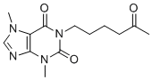
| 分子式 |
C22H21CL2N3O5
|
|---|---|
| 分子量 |
478.3252
|
| 精确质量 |
477.085
|
| 元素分析 |
C, 55.24; H, 4.43; Cl, 14.82; N, 8.78; O, 16.72
|
| CAS号 |
1144035-53-9
|
| 相关CAS号 |
PF-8380 hydrochloride;2070015-01-7
|
| PubChem CID |
25265312
|
| 外观&性状 |
White to light brown solid powder
|
| 密度 |
1.4±0.1 g/cm3
|
| 折射率 |
1.616
|
| LogP |
3.63
|
| tPSA |
95.85
|
| 氢键供体(HBD)数目 |
1
|
| 氢键受体(HBA)数目 |
6
|
| 可旋转键数目(RBC) |
7
|
| 重原子数目 |
32
|
| 分子复杂度/Complexity |
693
|
| 定义原子立体中心数目 |
0
|
| SMILES |
ClC1C([H])=C(C([H])=C(C=1[H])C([H])([H])OC(N1C([H])([H])C([H])([H])N(C([H])([H])C([H])([H])C(C2C([H])=C([H])C3=C(C=2[H])OC(N3[H])=O)=O)C([H])([H])C1([H])[H])=O)Cl
|
| InChi Key |
JMSUDQYHPSNBSN-UHFFFAOYSA-N
|
| InChi Code |
InChI=1S/C22H21Cl2N3O5/c23-16-9-14(10-17(24)12-16)13-31-22(30)27-7-5-26(6-8-27)4-3-19(28)15-1-2-18-20(11-15)32-21(29)25-18/h1-2,9-12H,3-8,13H2,(H,25,29)
|
| 化学名 |
3,5-dichlorobenzyl 4-(3-oxo-3-(2-oxo-2,3-dihydrobenzo[d]oxazol-6-yl)propyl)piperazine-1-carboxylate
|
| 别名 |
PF-8380; PF 8380; PF8380; 1144035-53-9; 3,5-Dichlorobenzyl 4-(3-oxo-3-(2-oxo-2,3-dihydrobenzo[d]oxazol-6-yl)propyl)piperazine-1-carboxylate; (3,5-Dichlorophenyl)methyl 4-[3-oxo-3-(2-oxo-2,3-dihydro-1,3-benzoxazol-6-yl)propyl]piperazine-1-carboxylate; T582DIM5A4; 1-Piperazinecarboxylic acid, 4-[3-(2,3-dihydro-2-oxo-6-benzoxazolyl)-3-oxopropyl]-, (3,5-dichlorophenyl)methyl ester; UNII-T582DIM5A4;
|
| HS Tariff Code |
2934.99.9001
|
| 存储方式 |
Powder -20°C 3 years 4°C 2 years In solvent -80°C 6 months -20°C 1 month |
| 运输条件 |
Room temperature (This product is stable at ambient temperature for a few days during ordinary shipping and time spent in Customs)
|
| 溶解度 (体外实验) |
DMSO : ~100 mg/mL (~209.06 mM)
|
|---|---|
| 溶解度 (体内实验) |
配方 1 中的溶解度: ≥ 0.67 mg/mL (1.40 mM) (饱和度未知) in 10% DMSO + 40% PEG300 + 5% Tween80 + 45% Saline (这些助溶剂从左到右依次添加,逐一添加), 澄清溶液。
例如,若需制备1 mL的工作液,可将100 μL 6.7 mg/mL澄清DMSO储备液加入400 μL PEG300中,混匀;然后向上述溶液中加入50 μL Tween-80,混匀;加入450 μL生理盐水定容至1 mL。 *生理盐水的制备:将 0.9 g 氯化钠溶解在 100 mL ddH₂O中,得到澄清溶液。 配方 2 中的溶解度: ≥ 0.67 mg/mL (1.40 mM) (饱和度未知) in 10% DMSO + 90% (20% SBE-β-CD in Saline) (这些助溶剂从左到右依次添加,逐一添加), 澄清溶液。 例如,若需制备1 mL的工作液,可将 100 μL 6.7mg/mL澄清的DMSO储备液加入到900μL 20%SBE-β-CD生理盐水中,混匀。 *20% SBE-β-CD 生理盐水溶液的制备(4°C,1 周):将 2 g SBE-β-CD 溶解于 10 mL 生理盐水中,得到澄清溶液。 View More
配方 3 中的溶解度: ≥ 0.67 mg/mL (1.40 mM) (饱和度未知) in 10% DMSO + 90% Corn Oil (这些助溶剂从左到右依次添加,逐一添加), 澄清溶液。 配方 4 中的溶解度: 10 mg/mL (20.91 mM) in 50% PEG300 50% Saline (这些助溶剂从左到右依次添加,逐一添加), 悬浊液; 超声助溶。 *生理盐水的制备:将 0.9 g 氯化钠溶解在 100 mL ddH₂O中,得到澄清溶液。 1、请先配制澄清的储备液(如:用DMSO配置50 或 100 mg/mL母液(储备液)); 2、取适量母液,按从左到右的顺序依次添加助溶剂,澄清后再加入下一助溶剂。以 下列配方为例说明 (注意此配方只用于说明,并不一定代表此产品 的实际溶解配方): 10% DMSO → 40% PEG300 → 5% Tween-80 → 45% ddH2O (或 saline); 假设最终工作液的体积为 1 mL, 浓度为5 mg/mL: 取 100 μL 50 mg/mL 的澄清 DMSO 储备液加到 400 μL PEG300 中,混合均匀/澄清;向上述体系中加入50 μL Tween-80,混合均匀/澄清;然后继续加入450 μL ddH2O (或 saline)定容至 1 mL; 3、溶剂前显示的百分比是指该溶剂在最终溶液/工作液中的体积所占比例; 4、 如产品在配制过程中出现沉淀/析出,可通过加热(≤50℃)或超声的方式助溶; 5、为保证最佳实验结果,工作液请现配现用! 6、如不确定怎么将母液配置成体内动物实验的工作液,请查看说明书或联系我们; 7、 以上所有助溶剂都可在 Invivochem.cn网站购买。 |
| 制备储备液 | 1 mg | 5 mg | 10 mg | |
| 1 mM | 2.0906 mL | 10.4530 mL | 20.9061 mL | |
| 5 mM | 0.4181 mL | 2.0906 mL | 4.1812 mL | |
| 10 mM | 0.2091 mL | 1.0453 mL | 2.0906 mL |
1、根据实验需要选择合适的溶剂配制储备液 (母液):对于大多数产品,InvivoChem推荐用DMSO配置母液 (比如:5、10、20mM或者10、20、50 mg/mL浓度),个别水溶性高的产品可直接溶于水。产品在DMSO 、水或其他溶剂中的具体溶解度详见上”溶解度 (体外)”部分;
2、如果您找不到您想要的溶解度信息,或者很难将产品溶解在溶液中,请联系我们;
3、建议使用下列计算器进行相关计算(摩尔浓度计算器、稀释计算器、分子量计算器、重组计算器等);
4、母液配好之后,将其分装到常规用量,并储存在-20°C或-80°C,尽量减少反复冻融循环。
计算结果:
工作液浓度: mg/mL;
DMSO母液配制方法: mg 药物溶于 μL DMSO溶液(母液浓度 mg/mL)。如该浓度超过该批次药物DMSO溶解度,请首先与我们联系。
体内配方配制方法:取 μL DMSO母液,加入 μL PEG300,混匀澄清后加入μL Tween 80,混匀澄清后加入 μL ddH2O,混匀澄清。
(1) 请确保溶液澄清之后,再加入下一种溶剂 (助溶剂) 。可利用涡旋、超声或水浴加热等方法助溶;
(2) 一定要按顺序加入溶剂 (助溶剂) 。
Inhibition of ATX reduces Akt Phosphorylation in GBM cells grown in co-culture.Front Oncol.2013 Sep 17;3:236. |
|---|
 Inhibition of ATX abrogates radiation induced tumor neovascularization.Front Oncol.2013 Sep 17;3:236. |
 Inhibition of ATX in combination with irradiation delays tumor growth in a heterotopic tumor model of GL261.Front Oncol.2013 Sep 17;3:236. |
 FCPR-16
 Mardepodect盐酸盐
 盐酸阿那格雷
 己酮可可碱
己酮可可碱
InvivoChem的所有产品仅用于作科学研究,不面向患者销售
Copyright 2020 InvivoChem LLC | All Rights Reserved 粤ICP备20063088号-1


 COA
COA

 463611831
