| 规格 | 价格 | 库存 | 数量 |
|---|---|---|---|
| 1mg |
|
||
| 2mg |
|
||
| 5mg |
|
||
| 10mg |
|
||
| 25mg |
|
||
| 50mg |
|
||
| 100mg |
|
||
| 250mg |
|
||
| Other Sizes |
|
| 靶点 |
Coccidia; antibiotic; Wnt/β-catenin
Salinomycin (Procoxacin) targets Wnt signaling pathway, inhibiting β-catenin nuclear translocation (IC50=1 μM in primary CLL cells) [1] Salinomycin (Procoxacin) induces reactive oxygen species (ROS) accumulation in cancer cells [2] Salinomycin (Procoxacin) selectively targets cancer stem cells (CSCs) by disrupting their self-renewal signaling [6] |
|---|---|
| 体外研究 (In Vitro) |
Salinomycin 是一种强效 Wnt 信号级联剂。盐霉素恶唑啉的平均 IC50 为 230 nM,细胞可在 48 小时内产生。另一种抗菌钾离子载体是盐霉素。据最近报道,它是一种针对乳腺癌干细胞的新型有效抗癌药物。 SW620细胞和Cisp抗性SW620细胞受到盐霉素的抑制,IC50值分别为1.54±0.23 μM和0.32±0.05 μM。人们发现盐霉素具有破坏癌症干细胞(CSC)及其承载能力的能力。连续盐霉素处理 48 小时后,在显微镜下检查染色细胞,每个视野内至少随机计数 100 个细胞。 Cisp抗性SW620细胞中Hoechst33342染色的细胞量(20.20±3.72)与SW620细胞中的9.40±2.07)/100个细胞有显着差异(p<0.05)。在用盐霉素处理细胞 48 小时后,使用流式细胞术分析发现了 Cisp 抗性细胞和 SW620 细胞。与SW620细胞(16.78±2.56%)相比,Cisp的消毒率(37.82±3.63%)要高得多(p<0.05)。[2]。
在原代慢性淋巴细胞白血病(CLL)细胞及 CLL 细胞系(MEC-1、HG-3)中,Salinomycin (Procoxacin) 抑制细胞增殖(IC50=1 μM),诱导凋亡(2 μM 时 annexin V+/PI- 细胞增加 35–45%),并通过降低核内 β-catenin 水平和下调 Wnt 靶基因(c-Myc、cyclin D1)表达抑制 Wnt 信号通路 [1] 在顺铂耐药结直肠癌细胞(HCT116/DDP、SW480/DDP)中,Salinomycin (Procoxacin) 抑制细胞活力(HCT116/DDP 的 IC50=2.3 μM,SW480/DDP 的 IC50=2.7 μM),诱导凋亡(剪切型 caspase-3/caspase-9 上调 2.5–3 倍),并增加细胞内 ROS 水平(2 μM 时 DCFH-DA 荧光强度升高 40–50%);ROS 清除剂 NAC 可逆转上述效应 [2] 在结直肠癌细胞(HCT116、HT29)及结直肠癌干细胞(CR-CSCs)中,Salinomycin (Procoxacin) 抑制细胞增殖(HCT116 的 IC50=1.8 μM,HT29 的 IC50=2.1 μM),降低球形成效率(1 μM 时从 12% 降至 3%),并在 mRNA 和蛋白水平下调 CSC 标志物(CD44、CD133、ALDH1)[3] 在人肝癌(HCC)细胞(HepG2、SMMC-7721)中,Salinomycin (Procoxacin) 抑制细胞活力(HepG2 的 IC50=2.5 μM,SMMC-7721 的 IC50=2.9 μM),诱导 G2/M 期细胞周期阻滞(2 μM 时 G2/M 期细胞从 15% 增至 42%),并通过线粒体通路促进凋亡(Bax/Bcl-2 比值上调 3.2 倍,细胞色素 c 释放增加)[4] 在膀胱癌细胞 T24 中,Salinomycin (Procoxacin) 抑制细胞侵袭(Transwell 实验:1 μM 时侵袭细胞减少 60%)和迁移(划痕愈合实验:1 μM 时闭合率从 85% 降至 30%),下调基质金属蛋白酶(MMP)-2 和 MMP-9 的表达 [5] 在多种癌细胞系(乳腺癌、前列腺癌、结直肠癌)中,Salinomycin (Procoxacin) 选择性清除 CSCs(CSCs 的 IC50=0.5–2 μM,非 CSCs 的 IC50=5–10 μM),抑制 CSC 自我更新,并通过下调 Snail、Slug、Twist 阻断上皮间质转化(EMT)[6] |
| 体内研究 (In Vivo) |
接受4mg/kg盐霉素(Sal)、8mg/kg盐霉素和10μL/g盐水后,六周后处死小鼠。与对照组相比,盐霉素治疗组的肝脏肿瘤尺寸更小。平均肿瘤直径(从12.17 mm降至3.67 mm;p<0.05)和平均肿瘤体积(V=长×宽2×0.5)显着下降,从819 mm3降至25.25 mm3。为了评估盐霉素的抗肿瘤活性,取出肿瘤并进行免疫组织化学、TUNEL 测定和 HE 染色。 HE染色可显示肝癌的组织结构,显示不同大小的细胞核以及被破坏的肝索结构。盐霉素治疗后,免疫组织化学显示 PCNA 表达降低。 HE染色和TUNEL实验显示盐霉素处理组的细胞凋亡率高于对照组。此外,免疫组织化学表明盐霉素治疗增加了 Bax/Bcl-2 比率。盐霉素治疗组的β-连环蛋白表达水平低于对照组[4]。白色链霉菌发酵产生盐霉素,一种单羧酸聚醚抗生素。其独特的环状结构使其能够与球虫和病原微生物的胞外阳离子,特别是 K+、Na+ 和 Rb+ 形成复合物,从而改变细胞内外的离子浓度 [5]。
在荷 HCT116 结直肠癌异种移植瘤裸鼠中,腹腔注射 Salinomycin (Procoxacin)(5 mg/kg,每周 3 次,持续 4 周),肿瘤体积较对照组减少 65%,肿瘤重量减少 70%;瘤内 ROS 水平升高 2.8 倍,CSC 标志物 CD44+CD133+ 细胞从 18% 降至 4% [3] 在荷 HepG2 肝癌异种移植瘤 BALB/c 裸鼠中,Salinomycin (Procoxacin) 经腹腔注射给药(4 mg/kg,每周 2 次,持续 5 周),抑制肿瘤生长(肿瘤体积减少 62%),诱导肿瘤细胞凋亡(TUNEL 阳性细胞增加 3.5 倍),并抑制肿瘤组织中 Wnt/β-catenin 信号通路(核内 β-catenin 下调 60%)[4] 在荷乳腺癌 CSC 来源异种移植瘤 SCID 鼠中,口服 Salinomycin (Procoxacin)(10 mg/kg,每日 1 次,持续 3 周)可清除 CSCs(ALDH1+ 细胞减少 80%),且较紫杉醇更能预防肿瘤复发(治疗后 8 周复发率 10% vs. 60%)[6] |
| 酶活实验 |
盐霉素是一种抗生素钾离子载体,最近被报道作为一种选择性的乳腺癌症干细胞抑制剂,但其抗癌作用的生化基础尚不清楚。Wnt/β-catenin信号传导通路在干细胞发育中起着核心作用,其异常激活可导致癌症。在这项研究中,我们发现盐霉素是Wnt信号级联的强效抑制剂。在Wnt转染的HEK293细胞中,盐霉素阻断Wnt辅助受体脂蛋白受体相关蛋白6(LRP6)的磷酸化并诱导其降解。另一种具有抗癌症干细胞活性的钾离子载体Nigericin也发挥了类似的作用。在其他未经处理的慢性淋巴细胞白血病细胞中,具有组成性Wnt激活的盐霉素纳摩尔浓度下调了Wnt靶基因如LEF1、细胞周期蛋白D1和纤维连接蛋白的表达,降低了LRP6水平,限制了细胞存活。正常人外周血淋巴细胞抵抗盐霉素毒性。这些结果表明,盐霉素和相关药物诱导的离子变化通过干扰LPR6磷酸化来抑制近端Wnt信号传导,从而损害依赖质膜Wnt信号的细胞的存活[1]。
评估 Wnt 信号抑制作用:构建 β-catenin 响应性荧光素酶报告质粒(TOPflash),转染至 CLL 细胞系(MEC-1)。转染 24 h 后,用系列浓度(0.1–5 μM)的 Salinomycin (Procoxacin) 处理细胞 18 h。裂解细胞后检测荧光素酶活性,评估 Wnt 通路抑制效率 [1] MMP 活性检测:收集经 Salinomycin (Procoxacin)(0.5–2 μM)处理 48 h 的 T24 膀胱癌细胞培养上清液。将上清液与 MMP-2/MMP-9 特异性底物在 37°C 孵育 2 h,在 405 nm 处检测吸光度,定量 MMP 酶活性 [5] |
| 细胞实验 |
结直肠癌癌症患者术后化疗并非完全有效,主要原因可能在于癌症干细胞(CSCs)。新出现的研究表明,CSC过度表达一些与耐药性相关的蛋白质,这些蛋白质有效地将化疗药物输送出癌症细胞。盐霉素被认为是一种新型有效的抗癌药物,具有杀死肿瘤干细胞和耐药癌症细胞的能力。为了探讨盐霉素特异性靶向结直肠癌癌症耐药细胞的潜在机制,我们首先从原始结直肠癌癌症细胞系中重复暴露于5μmol/l的顺铂,获得了顺铂耐药(顺铂耐药)SW620细胞。这些Cisp抗性SW620细胞保持相对静止状态(G0/G1期阻滞)并显示出干样特征(Sox2、Oct4、Nanog、Klf4、Hes1、CD24、CD26、CD44、CD133、CD166、Lgr5、ALDH1A1和ALDH1A3 mRNA表达上调)(p<0.05),对盐霉素敏感(p<0.05)。盐霉素对Cisp抗性SW620细胞的细胞周期没有影响(p>0.05),但可以诱导细胞死亡过程(p<0.05),增加LDH释放和MDA含量,降低SOD和GSH-PX活性(p<0.05)。我们的数据还显示,在Cisp抗性SW620细胞中,促凋亡基因(Caspase-3、Caspase-8、Caspase-9和Bax)上调,抗凋亡基因Bcl-2下调(p<0.05)。积累的活性氧和一些凋亡相关基因的失调可能最终导致Cisp抗性SW620细胞的凋亡。这些发现将为对顺铂耐药的结直肠癌癌症细胞进行新的选择性化疗提供新的线索[2]。
体外培养癌症细胞株T24。在体内建立大鼠膀胱肿瘤模型。将大鼠随机分为两组,实验组腹腔注射盐霉素,对照组腹腔注射生理盐水。观察两组肿瘤细胞的变化。Transwell用于检测细胞迁移和侵袭能力,Real-time PCR用于检测mRNA的表达,Western blot用于测定E-cadherin和波形蛋白的表达。 结果:实验组经盐霉素治疗后,血清膀胱癌症细胞株T24的转移和侵袭能力较对照组显著降低,肿瘤转移灶由平均1.59处降至0.6处(P<0.05)。实验组T24细胞增殖逐渐减少。T24细胞48h增殖明显低于12h和24h(P<0.05)。T24细胞24小时增殖明显低于12小时(P<0.05)。实验组各时间点T24细胞增殖明显低于对照组(P<0.05)。实验组血清mRNA水平和肿瘤组织E-cadherin表达明显高于对照组,而波形蛋白表达水平明显低于对照组(P<0.05)。 结论:盐霉素能抑制膀胱癌症细胞的转移和侵袭,其机制可能与抑制肿瘤细胞EMT有关。[5] CLL 细胞活力及凋亡实验:将原代 CLL 细胞或 MEC-1 细胞(1×10⁵ 个细胞/孔)接种于 96 孔板,用 Salinomycin (Procoxacin)(0.5–4 μM)处理 48 h。采用 MTT 法检测细胞活力, Annexin V-FITC/PI 染色后通过流式细胞术检测凋亡细胞。Wnt 信号分析:处理 24 h 后提取核蛋白和胞质蛋白,Western blot 检测 β-catenin 水平 [1] 结直肠癌细胞 ROS 检测:将 HCT116/DDP 细胞(2×10⁵ 个细胞/孔)接种于 6 孔板,用 Salinomycin (Procoxacin)(1–3 μM)处理 24 h。加入 DCFH-DA 探针(10 μM)负载 30 min,流式细胞术分析 ROS 水平。凋亡相关蛋白检测:处理 48 h 后提取总蛋白,Western blot 检测剪切型 caspase-3、caspase-9、Bax 及 Bcl-2 [2] CSC 球形成实验:从结直肠癌组织中分离 CR-CSCs,将单细胞(100 个细胞/孔)接种于超低吸附 96 孔板,加入 Salinomycin (Procoxacin)(0.5–2 μM)处理。培养 7 天后,计数球状体数量(直径 >50 μm)评估自我更新能力。处理 48 h 后采用 qPCR 检测 CD44、CD133、ALDH1 的 mRNA 表达 [3] 肝癌细胞周期分析:将 HepG2 细胞(3×10⁵ 个细胞/孔)接种于 6 孔板,用 Salinomycin (Procoxacin)(1–3 μM)处理 24 h。70% 乙醇固定细胞,碘化丙啶染色,流式细胞术分析细胞周期分布(G0/G1、S、G2/M 期)[4] 膀胱癌细胞侵袭实验:Transwell 小室上室包被 Matrigel,加入含 Salinomycin (Procoxacin)(0.5–2 μM)的 T24 细胞(5×10⁴ 个细胞/小室),下室加入含 10% 胎牛血清的培养基。孵育 24 h 后,固定并染色下室表面的侵袭细胞,显微镜下计数 [5] |
| 动物实验 |
小鼠:4 和 8 mg/kg,腹腔注射;大鼠:8 mg/kg,腹腔注射
小鼠:使用裸鼠(nu/nu;4-6 周龄)。将 HepG2 细胞悬浮于 100 mL 1:1 无血清 DMEM 和 Matrigel 混合液中。小鼠用氯胺酮/甲苯噻嗪麻醉,开腹手术后,将 HepG2 细胞接种到肝实质内,每 3 天观察一次,持续 35 天。最后,将 18 只裸鼠随机分为三组,每天腹腔注射,持续 6 周:两组盐霉素治疗组(4 mg/kg 盐霉素组、8 mg/kg 盐霉素组)和对照组(生理盐水组)。 大鼠:实验共使用 10 只雄性大鼠。常规麻醉后,打开腹腔。将膀胱移行细胞癌细胞系T24接种于大鼠膀胱实质内,接种前先用无血清高糖DMEM培养基和基质胶进行悬浮,然后缝合腹腔。术后,将大鼠随机分为实验组和对照组,每组5只。实验组大鼠术后立即腹腔注射盐霉素(8 mg/kg),对照组大鼠腹腔注射生理盐水。给药期间密切观察。 15天后,将大鼠颈椎脱臼处死,完整剥离肿瘤组织,观察肿瘤生长和转移情况。 结直肠癌异种移植模型:将HCT116细胞(5×10⁶个细胞/只)皮下注射到6-8周龄的裸鼠体内,建立异种移植瘤模型。当肿瘤体积达到100 mm³时,将小鼠随机分为对照组和治疗组(每组n=6)。盐霉素(普罗昔沙星)溶于DMSO,并用PBS稀释(最终DMSO浓度<5%),以5 mg/kg的剂量腹腔注射,每周3次,持续4周。每3天测量一次肿瘤体积,并在治疗结束后处死小鼠,收集肿瘤组织用于ROS检测和CSC标志物分析[3] HCC异种移植模型:将HepG2细胞(2×10⁶个细胞/只)皮下植入BALB/c裸鼠(6-8周龄)。当肿瘤生长至80-100 mm³时,将小鼠随机分为对照组(载体组)和治疗组(每组n=5)。盐霉素(普罗昔沙星)配制成1 mg/mL的0.5%羧甲基纤维素悬浮液,以4 mg/kg的剂量腹腔注射,每周两次,持续5周。记录肿瘤重量和体积,并收集肿瘤组织进行TUNEL检测和Wnt信号蛋白的Western blot分析[4] CSC异种移植模型:将乳腺癌CSC(1×10⁵个细胞/只)皮下注射到6-8周龄的SCID小鼠体内。肿瘤形成(50 mm³)后,小鼠每天灌胃给予盐霉素(Procoxacin)(溶于0.9% NaCl溶液),剂量为10 mg/kg,持续3周。治疗后监测肿瘤复发情况8周,并分析肿瘤组织中ALDH1⁺ CSC的含量[6] |
| 药代性质 (ADME/PK) |
吸收、分布和排泄
本研究采用口服和静脉注射两种途径给鸡服用盐霉素,以测定其血药浓度、药代动力学、生物利用度和组织残留。药物以20 mg/kg体重的单次剂量通过嗉囊内注射和静脉注射给药。口服给药后半小时达到血清盐霉素最高浓度,吸收半衰期(t0.5(ab))为3.64小时,消除半衰期(t0.5(beta))为1.96小时。嗉囊内注射给药后,系统生物利用度为73.02%,表明鸡通过该途径对盐霉素的吸收率较高。静脉注射后,盐霉素的药代动力学可用二室开放模型描述,其半衰期(t1/2(α))为0.48小时,分布容积(Vd ss)为3.28升/千克,总清除率(Cl(β))为27.39毫升/千克/分钟。体外计算的盐霉素血清蛋白结合率为19.78%。连续两周给予盐霉素预混剂(60 ppm)的禽类,其血清和组织中的盐霉素浓度低于单次嗉囊内注射纯盐霉素(20毫克/千克体重)后的浓度。盐霉素残留浓度最高的组织是肝脏,其次是肾脏、肌肉、脂肪、心脏和皮肤。 48小时后,除肝脏外,其他组织中均未检测到沙利霉素残留,且肝脏中的沙利霉素残留在72小时内完全消失。 代谢/代谢物 ……沙利霉素(SAL)是一种广谱抗生素和抗球虫药,研究发现其对抗肿瘤耐药性和杀伤癌干细胞的疗效优于现有的化疗药物紫杉醇和阿霉素。这重新凸显了其在人类癌症治疗中的重要性。本研究探讨了沙利霉素的体外药物代谢和药代动力学参数。沙利霉素在肝微粒体中代谢迅速,具有较高的固有清除率。沙利霉素的代谢主要由CYP酶介导,其中CYP3A4是主要的代谢酶。与小鼠和大鼠血浆相比,沙利霉素在人体血浆中的蛋白结合率显著降低。我们通过化学抑制和重组酶实验研究了CYP抑制情况。研究发现,沙利铂(SAL)是CYP2D6和CYP3A4的中度抑制剂。由于CYP3A4是SAL代谢的主要酶,因此进行了大鼠体内药代动力学研究,以检验同时服用酮康唑(KTC)对SAL药代动力学的影响。KTC作为一种选择性CYP3A4抑制剂,显著增加了SAL的全身暴露量,在同时服用KTC的大鼠中,SAL的AUC0-a增加了7倍,Cmax增加了3倍。 生物半衰期 ……该药物以20 mg/kg体重的单次剂量,通过嗉囊内和静脉途径给药。口服沙利霉素后半小时血清浓度达到最高值,吸收半衰期(t0.5(ab))为3.64小时,消除半衰期(t0.5(beta))为1.96小时。 |
| 毒性/毒理 (Toxicokinetics/TK) |
毒性概述
鉴定和用途:盐霉素是一种兽药,用于预防肉鸡、烤鸡和后备鸡的球虫病,这些球虫病由柔嫩艾美球虫(Eimeria tenella)、坏死艾美球虫(E. necatrix)、堆型艾美球虫(E. acervulina)、巨型艾美球虫(E. maxima)、布氏艾美球虫(E. brunetti)和米氏艾美球虫(E. mivati)引起。它也用于预防鹌鹑的球虫病,这些球虫病由分散艾美球虫(Eimeria dispersa)和莱氏艾美球虫(E. lettyae)引起。人体暴露和毒性:本研究探讨了盐霉素对人类非恶性细胞的细胞毒性和遗传毒性。使用来自10名受试者的原代人鼻黏膜细胞(单层细胞培养和微型器官培养)和外周血淋巴细胞,通过膜联蛋白-碘化丙啶-MTT法研究了盐霉素(0.1-175 μM)的细胞毒性作用。采用彗星试验评估DNA损伤。此外,采用ELISA法分析白细胞介素-8的分泌。流式细胞术和MTT试验显示,低浓度(10-20 μM)的盐霉素对鼻黏膜细胞和淋巴细胞具有显著的细胞毒性作用。未观察到基因毒性作用。5 μM浓度下IL-8分泌升高。在抗癌治疗相关浓度下,观察到盐霉素诱导的细胞毒性和促炎作用。动物研究:已有大量报道指出,误食盐霉素会导致多种动物死亡。在一个拥有五个鸡舍的火鸡养殖场中,一个鸡舍内600只48周龄雄性种火鸡突然死亡,怀疑与饲料有关。这些火鸡出现喘息和倒地症状;21.7%的受影响火鸡死亡。组织学病变仅限于骨骼肌,表现为变性和坏死,符合离子载体中毒的特征。对受影响鸡舍的饲料样品进行分析,结果显示每吨饲料中含有13.4至18.4克盐霉素。为了进一步研究盐霉素对火鸡的影响,分别在7、11、15、27和32周龄时,对336、24、24、40和40只雄性火鸡进行了为期7天的五次试验。结果表明,随着火鸡年龄的增长,盐霉素的毒性也随之增强。当7周龄火鸡饲喂含44或66 ppm盐霉素的日粮时,84只火鸡中仅有1只死亡;而当27或32周龄火鸡饲喂相同浓度的盐霉素时,20只火鸡中有13只死亡。浓度为 22 ppm 的盐霉素会抑制幼马的生长速度,并阻碍或减缓老年马的生长,同时增加死亡率。另有报道称,六匹马因误食盐霉素而发生意外中毒。这些中毒症状包括厌食、腹痛、虚弱和共济失调,与误食相关离子载体莫能菌素的马的症状相似。在另一起中毒事件中,马匹被喂食了含有 61 mg/kg 盐霉素的浓缩饲料,该饲料因生产商配制不当而存在缺陷。所有马匹均出现严重的临床中毒症状。尽管进行了治疗,仍有八匹马在三到六天内死亡。另有十匹马卧地不起,不得不实施安乐死。最终只有六匹马存活。主要的实验室检测结果显示酶水平极高和碱中毒。最典型的临床表现是后肢瘫痪。此外,还有报道称,猫因食用被盐霉素污染的干猫粮而爆发了中毒性多发性神经病。研究人员收集了823只猫的流行病学和临床数据,约占高危猫总数的1%。其中21只患病猫进行了尸检。患病猫均表现为急性跛行和后肢瘫痪,随后累及前肢。临床和病理检查表明,这些猫患有累及感觉神经和运动神经的远端多发性神经病。此外,研究人员还报告了一起育肥牛中毒事件的临床症状和病理学表现,该事件是由于牛在长达11周的时间里摄入了毒性剂量的盐霉素所致。380头牛中有39头出现与心力衰竭相符的症状,其中8头死亡。临床症状包括呼吸困难、呼吸急促、心动过速和运动不耐受。对两头牛进行了尸检,其中一头牛的肉眼可见病变,提示存在充血性心力衰竭,包括肺水肿、胸腔积液和肝肿大。组织病理学检查显示,慢性心肌病的主要特征是广泛的心肌纤维萎缩,伴有多灶性肥大以及间质和替代性纤维化。肝脏和肺部病变与充血性心力衰竭的病变相符。最后,据报道,一群饲喂含盐霉素饲料的绵羊死亡率达100%。饲喂后的第二天早上,发现78只羊死亡,其中一只出现抽搐。尸检显示肺充血和水肿、皱胃出血、肾脏肿大苍白以及心肌出现白色条纹。 相互作用 肝细胞癌(HCC)是少数几种发病率在过去几年持续上升的癌症之一。耐药性是HCC治疗中的一个主要问题。在本研究中,我们采用沙利霉素(Sal)和5-氟尿嘧啶(5-FU)联合治疗肝癌细胞系Huh7、LM3和SMMC-7721以及裸鼠皮下肿瘤模型,研究Sal是否能提高肝癌细胞对传统化疗药物(如5-FU)的敏感性。结果表明,Sal与5-FU联合用药在体外和体内均表现出对肝肿瘤的协同抗肿瘤作用。Sal逆转了5-FU诱导的CD133(+)EPCAM(+)细胞增多、上皮-间质转化以及Wnt/β-catenin信号通路的激活。Sal与5-FU的联合用药可能为逆转肝癌患者的耐药性提供一种新的治疗策略。 由于软组织肉瘤的化疗敏感性低,其化疗效果仍不尽如人意。即使是一线化疗药物阿霉素的有效率也仅为18-29%。抗生素沙利霉素是一种钾离子载体,最近的研究表明,它能有效清除腺癌中的化疗耐药细胞,例如癌干细胞样细胞(CSC)。本研究评估了沙利霉素对肉瘤细胞系的影响,分析了沙利霉素单药治疗以及与阿霉素联合治疗的效果。为了评估沙利霉素对纤维肉瘤、横纹肌肉瘤和脂肪肉瘤细胞系的影响,我们分别对细胞进行了单药和联合治疗。通过细胞活力检测、细胞周期分析以及caspase 3/7和9活性检测来监测相应治疗的效果。此外,我们还分析了NF-κB活性。 p53、p21 和 PUMA 的转录水平,以及 p53 的表达和丝氨酸 15 位点的磷酸化,均有所变化。盐霉素与阿霉素联用可增强 caspase 的激活并增加亚 G1 期细胞比例。联合治疗可提高 NF-κB 的活性以及 p53、p21 和 PUMA 的转录水平,而盐霉素单药治疗则未引起任何显著变化。盐霉素即使在亚致死浓度下也能提高肉瘤细胞系对细胞抑制剂阿霉素的化疗敏感性。这些发现支持降低盐霉素与阿霉素联用浓度的策略,以减少毒副作用。 采用析因设计(2×3)评估黄曲霉毒素(0、2.5 和 5 mg/kg)与盐霉素(1 和 60 g/吨(909 kg))之间的相互作用。每个处理组设置4个重复,每个重复包含10只雏鸡。……在所有测量的参数中,黄曲霉毒素和盐霉素之间均未观察到显著的交互作用。 本研究旨在探讨盐霉素联合长春新碱对Jurkat细胞增殖和凋亡的影响及其可能的机制。采用CKK-8法检测Jurkat细胞的增殖。采用流式细胞术评估细胞凋亡。采用Western blot法检测BCL-2、caspase-3和caspase-8的表达水平。盐霉素或长春新碱单独或联合使用均能以剂量依赖的方式抑制Jurkat细胞的增殖。盐霉素联合长春新碱对细胞增殖的抑制作用比单独使用任何一种化合物都更为显著(P<0.05)。 Western blot分析显示,盐霉素(Sal)与长春新碱(VCR)联合使用可显著降低BCL-2蛋白的表达,并显著增加caspase 3和caspase 8蛋白的表达。此外,盐霉素与长春新碱联合使用可协同促进Jurkat细胞的凋亡(P<0.05)。盐霉素与长春新碱联合使用可协同抑制T细胞急性淋巴细胞白血病Jurkat细胞的增殖并促进其凋亡。 在用盐霉素(普罗昔沙星)(5 mg/kg,腹腔注射,每周3次,持续4周)治疗的裸鼠中,未观察到体重(变化<10%)或血液学参数(白细胞、红细胞、血小板)的显著变化;肝肾组织病理学检查未见明显的药物相关病变[3] 在接受盐霉素(普罗昔沙星)(4 mg/kg,腹腔注射,每周两次,持续5周)治疗的BALB/c裸鼠中,血清ALT、AST、肌酐和BUN水平均保持在正常范围内,表明未见明显的肝毒性或肾毒性[4] 体外实验表明,盐霉素(普罗昔沙星)对正常外周血单核细胞(PBMCs)的毒性较低,IC50 >10 μM,而对慢性淋巴细胞白血病(CLL)细胞则表现出选择性毒性(IC50=1 μM)[1] 在口服盐霉素(普罗昔沙星)(10 mg/kg,每日一次,持续3周)治疗的SCID小鼠中,20%的小鼠出现轻度腹泻,但随后症状消退。无需治疗中断即可自发进行[6] |
| 参考文献 |
|
| 其他信息 |
治疗用途
抗菌剂;抗球虫药 说明:疟疾药物研发联盟(Medicines for Malaria Venture)提出的疟疾消除/根除政策的药物靶点谱侧重于对疟原虫无性期和有性期均有活性的分子,从而兼具治疗和阻断传播的双重作用。本研究旨在探讨一类单价离子载体(包括兽药和近期被提议作为人类抗癌药物的药物)是否符合这些要求。本文报道了沙利霉素、莫能菌素和尼日利亚菌素对恶性疟原虫无性期和有性红细胞期以及伯氏疟原虫和恶性疟原虫蚊媒期发育的活性。在体外诱导恶性疟原虫3D7株的配子体发生,并将处于II期和III期或IV期和V期的配子体用离子载体处理不同时间,并通过寄生虫乳酸脱氢酶(pLDH)测定法检测其活力。单价离子载体能有效杀灭无性期寄生虫和配子体,其半数抑制浓度(IC50)为纳摩尔级。与标准药物相比,盐霉素的杀灭速度更快,且对IV期和V期配子体的效力高于II期和III期配子体。离子载体抑制了蚊子中肠内动合子的发育和后续卵囊的形成,证实了其阻断传播的活性。由于离子载体仅损伤了受感染的红细胞而非正常红细胞,因此排除了溶血毒性的可能性。我们的数据有力地支持对单价离子载体进行下游探索,以期将其重新定位为新型抗疟疾和阻断传播的先导化合物。 EXPL:盐霉素已被引入作为一种新型的传统抗癌药物替代品。本研究旨在测试一种将盐霉素递送至体外胶质母细胞瘤细胞的策略。我们制备了盐霉素包封的聚山梨醇酯80包覆的聚乳酸-羟基乙酸共聚物纳米颗粒(P80-SAL-PLGA),并对其粒径、形貌、热性能、药物包封率和可控盐霉素释放行为进行了表征。我们评估了P80-SAL-PLGA(5和10 μM)或未包覆纳米颗粒在T98G人胶质母细胞瘤细胞中的体外细胞摄取情况,并研究了其对细胞活力和抗增殖活性的影响。 SAL通过P80包覆的纳米颗粒成功递送至T98G胶质母细胞瘤细胞(60分钟内递送量约为14%),显著降低了T98G细胞的活力(p < 0.01)。T98G细胞的形态发生了显著变化,肌动蛋白细胞骨架受损。因此,P80-SAL-PLGA纳米颗粒诱导细胞死亡,表明这种沙利霉素递送系统在治疗人类胶质母细胞瘤方面具有潜在的治疗作用。 兽药:使用Sacox 60……预防鹌鹑由散布艾美球虫(Eimeria dispersa)和莱氏艾美球虫(E. lettyae)引起的球虫病。 兽药:使用Sacox 60……预防肉鸡、烤鸡和后备鸡由柔嫩艾美球虫(Eimeria tenella)、坏死艾美球虫(E. necatrix)、堆型艾美球虫(E. acervulina)、巨型艾美球虫(E. maxima)、布氏艾美球虫(E. brunetti)和米瓦蒂艾美球虫(E. mivati)引起的球虫病。 药物警告 请勿喂给产蛋供人食用的母鸡。 如果误喂给成年火鸡或马,可能致命。 沙利霉素(普罗昔沙星)通过抑制Wnt信号通路来阻止…… β-catenin 核转位,从而阻断 CLL 细胞中 Wnt 依赖性癌基因(c-Myc、cyclin D1)的转录[1] 沙利霉素(Procoxacin)在顺铂耐药性结直肠癌细胞中的抗肿瘤活性依赖于 ROS 的积累,ROS 可触发线粒体功能障碍和 caspase 依赖性细胞凋亡[2] 与传统化疗药物(例如紫杉醇、顺铂)相比,沙利霉素(Procoxacin)对癌症干细胞具有更高的选择性,使其成为靶向 CSC 驱动的肿瘤复发和转移的潜在候选药物[6] 在肝细胞癌 (HCC) 细胞中,沙利霉素(Procoxacin)与索拉非尼具有协同作用,可增强抗肿瘤疗效,联合用药时可使索拉非尼的 IC50 降低 50%。亚有效浓度[4] 盐霉素(普罗昔沙星)通过下调MMP-2和MMP-9抑制膀胱癌细胞侵袭,MMP-2和MMP-9是参与细胞外基质降解的关键酶[5] |
| 分子式 |
C42H70O11
|
|
|---|---|---|
| 分子量 |
751.00
|
|
| 精确质量 |
750.49
|
|
| 元素分析 |
C, 67.17; H, 9.40; O, 23.43
|
|
| CAS号 |
53003-10-4
|
|
| 相关CAS号 |
Salinomycin sodium salt;55721-31-8
|
|
| PubChem CID |
3085092
|
|
| 外观&性状 |
brown solid powder
|
|
| 密度 |
1.2±0.1 g/cm3
|
|
| 沸点 |
839.2±65.0 °C at 760 mmHg
|
|
| 熔点 |
112.5-113.5 °C(lit.)
|
|
| 闪点 |
243.2±27.8 °C
|
|
| 蒸汽压 |
0.0±0.6 mmHg at 25°C
|
|
| 折射率 |
1.547
|
|
| LogP |
6.1
|
|
| tPSA |
164.04
|
|
| 氢键供体(HBD)数目 |
4
|
|
| 氢键受体(HBA)数目 |
11
|
|
| 可旋转键数目(RBC) |
12
|
|
| 重原子数目 |
53
|
|
| 分子复杂度/Complexity |
1320
|
|
| 定义原子立体中心数目 |
18
|
|
| SMILES |
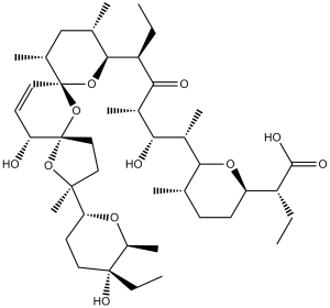
O1[C@@]2([C@@]([H])(C([H])=C([H])[C@@]3([C@]([H])(C([H])([H])[H])C([H])([H])[C@]([H])(C([H])([H])[H])[C@@]([H])([C@@]([H])(C([H])([H])C([H])([H])[H])C([C@@]([H])(C([H])([H])[H])[C@]([H])([C@]([H])(C([H])([H])[H])[C@@]4([H])[C@@]([H])(C([H])([H])[H])C([H])([H])C([H])([H])[C@]([H])([C@]([H])(C(=O)O[H])C([H])([H])C([H])([H])[H])O4)O[H])=O)O3)O2)O[H])C([H])([H])C([H])([H])[C@@]1(C([H])([H])[H])[C@@]1([H])C([H])([H])C([H])([H])[C@](C([H])([H])C([H])([H])[H])([C@]([H])(C([H])([H])[H])O1)O[H]
|
|
| InChi Key |
KQXDHUJYNAXLNZ-XQSDOZFQSA-N
|
|
| InChi Code |
InChI=1S/C42H70O11/c1-11-29(38(46)47)31-15-14-23(4)36(50-31)27(8)34(44)26(7)35(45)30(12-2)37-24(5)22-25(6)41(51-37)19-16-32(43)42(53-41)21-20-39(10,52-42)33-17-18-40(48,13-3)28(9)49-33/h16,19,23-34,36-37,43-44,48H,11-15,17-18,20-22H2,1-10H3,(H,46,47)/t23-,24-,25+,26-,27-,28-,29+,30-,31+,32+,33+,34+,36+,37-,39-,40+,41-,42-/m0/s1
|
|
| 化学名 |
(2R)-2-[(2R,5S,6R)-6-[(2S,3S,4S,6R)-6-[(3S,5S,7R,9S,10S,12R,15R)-3-[(2R,5R,6S)-5-ethyl-5-hydroxy-6-methyloxan-2-yl]-15-hydroxy-3,10,12-trimethyl-4,6,8-trioxadispiro[4.1.57.35]pentadec-13-en-9-yl]-3-hydroxy-4-methyl-5-oxooctan-2-yl]-5-methyloxan-2-yl]butanoic acid
|
|
| 别名 |
|
|
| HS Tariff Code |
2934.99.9001
|
|
| 存储方式 |
Powder -20°C 3 years 4°C 2 years In solvent -80°C 6 months -20°C 1 month |
|
| 运输条件 |
Room temperature (This product is stable at ambient temperature for a few days during ordinary shipping and time spent in Customs)
|
| 溶解度 (体外实验) |
|
|||
|---|---|---|---|---|
| 溶解度 (体内实验) |
配方 1 中的溶解度: ≥ 2.5 mg/mL (3.33 mM) (饱和度未知) in 10% DMSO + 40% PEG300 + 5% Tween80 + 45% Saline (这些助溶剂从左到右依次添加,逐一添加), 澄清溶液。
例如,若需制备1 mL的工作液,可将100 μL 25.0 mg/mL澄清DMSO储备液加入到400 μL PEG300中,混匀;然后向上述溶液中加入50 μL Tween-80,混匀;加入450 μL生理盐水定容至1 mL。 *生理盐水的制备:将 0.9 g 氯化钠溶解在 100 mL ddH₂O中,得到澄清溶液。 配方 2 中的溶解度: ≥ 2.5 mg/mL (3.33 mM) (饱和度未知) in 10% DMSO + 90% Corn Oil (这些助溶剂从左到右依次添加,逐一添加), 澄清溶液。 例如,若需制备1 mL的工作液,可将 100 μL 25.0 mg/mL 澄清 DMSO 储备液加入到 900 μL 玉米油中并混合均匀。 View More
配方 3 中的溶解度: 2.5 mg/mL (3.33 mM) in 5% DMSO + 40% PEG300 + 5% Tween80 + 50% Saline (这些助溶剂从左到右依次添加,逐一添加), 悬浊液; 超声助溶。 1、请先配制澄清的储备液(如:用DMSO配置50 或 100 mg/mL母液(储备液)); 2、取适量母液,按从左到右的顺序依次添加助溶剂,澄清后再加入下一助溶剂。以 下列配方为例说明 (注意此配方只用于说明,并不一定代表此产品 的实际溶解配方): 10% DMSO → 40% PEG300 → 5% Tween-80 → 45% ddH2O (或 saline); 假设最终工作液的体积为 1 mL, 浓度为5 mg/mL: 取 100 μL 50 mg/mL 的澄清 DMSO 储备液加到 400 μL PEG300 中,混合均匀/澄清;向上述体系中加入50 μL Tween-80,混合均匀/澄清;然后继续加入450 μL ddH2O (或 saline)定容至 1 mL; 3、溶剂前显示的百分比是指该溶剂在最终溶液/工作液中的体积所占比例; 4、 如产品在配制过程中出现沉淀/析出,可通过加热(≤50℃)或超声的方式助溶; 5、为保证最佳实验结果,工作液请现配现用! 6、如不确定怎么将母液配置成体内动物实验的工作液,请查看说明书或联系我们; 7、 以上所有助溶剂都可在 Invivochem.cn网站购买。 |
| 制备储备液 | 1 mg | 5 mg | 10 mg | |
| 1 mM | 1.3316 mL | 6.6578 mL | 13.3156 mL | |
| 5 mM | 0.2663 mL | 1.3316 mL | 2.6631 mL | |
| 10 mM | 0.1332 mL | 0.6658 mL | 1.3316 mL |
1、根据实验需要选择合适的溶剂配制储备液 (母液):对于大多数产品,InvivoChem推荐用DMSO配置母液 (比如:5、10、20mM或者10、20、50 mg/mL浓度),个别水溶性高的产品可直接溶于水。产品在DMSO 、水或其他溶剂中的具体溶解度详见上”溶解度 (体外)”部分;
2、如果您找不到您想要的溶解度信息,或者很难将产品溶解在溶液中,请联系我们;
3、建议使用下列计算器进行相关计算(摩尔浓度计算器、稀释计算器、分子量计算器、重组计算器等);
4、母液配好之后,将其分装到常规用量,并储存在-20°C或-80°C,尽量减少反复冻融循环。
计算结果:
工作液浓度: mg/mL;
DMSO母液配制方法: mg 药物溶于 μL DMSO溶液(母液浓度 mg/mL)。如该浓度超过该批次药物DMSO溶解度,请首先与我们联系。
体内配方配制方法:取 μL DMSO母液,加入 μL PEG300,混匀澄清后加入μL Tween 80,混匀澄清后加入 μL ddH2O,混匀澄清。
(1) 请确保溶液澄清之后,再加入下一种溶剂 (助溶剂) 。可利用涡旋、超声或水浴加热等方法助溶;
(2) 一定要按顺序加入溶剂 (助溶剂) 。
 Sal inhibits HCC cell proliferationin vitro.PLoS One.2012;7(12):e50638. |
|---|
 Sal causes cell cycle arrest and induces apoptosis of HCC cellsin vitro.PLoS One.2012;7(12):e50638. |
 Sal increases intracellular Calcium levelsin vitro.PLoS One.2012;7(12):e50638. |
 Anti-tumor activity of Salin vivo.A. HE staining showed the structure of the liver cancer tissue: nuclei of different sizes, hepatic cord structure was destroyed. B. Immunohistochemistry indicates that PCNA expression is down-regulated after Sal.PLoS One.2012;7(12):e50638. |
|---|
 A. Gross observation of HepG2 cell orthotopic tumors in nude mice from the saline group or Sal groups (4 mg/kg or 8 mg/kg). B. Tendency of tumor mean diameter after injection in nude mice (*p<0.05).PLoS One.2012;7(12):e50638. |
 A. Real-time PCR was performed to examine mRNA expression of the Wnt pathway.PLoS One.2012;7(12):e50638. |
InvivoChem的所有产品仅用于作科学研究,不面向患者销售
Copyright 2020 InvivoChem LLC | All Rights Reserved 粤ICP备20063088号-1


 COA
COA
 463611831
